3807
Free-breathing MRI enabled by FD-UNet with ground truth from repeated k-t-subsampling and greedy randomized adaptive search procedure1Department of Diagnostic Radiology, University of Hong Kong, Hong Kong, China, 2Department of Biomedical Engineering, Chinese University of Hong Kong, Hong Kong, China
Synopsis
Deep learning solutions have been proposed to correct motion-induced artefact in free-breathing abdominal MRI that can overcome some challenges in conventional methods, such as requiring respiratory modeling, external motion monitoring devices, or extremely long computation time. However, ground truth data for training can only be acquired with breath-holding or using those conventional methods. In this study, we proposed to use FD-UNet for the motion correction of free-breathing abdominal MRI. The high-quality and artifact-free ground truth data were produced from repeated k-t-subsampling and greedy randomized adaptive search procedure (ReK-GRASP), without relying on respiratory modeling, external devices, or long computation time.
Introduction
Abdominal magnetic resonance imaging (MRI) suffers from respiratory motion artifacts, including blurring and ghosting1. Conventional methods can partially address the motion artifacts, but they often require respiratory modeling, external motion monitoring devices, or extremely long computation time.2,3,4 Recently, several deep learning solutions have been proposed to enable free-breathing abdominal MRI, based on 1) ground truth data acquisition (i.e., motion-artifact-free data) and 2) subsequent network training using the acquired ground truth.5,6 However, acquiring ground truth data with breath-holding can only achieve limited data quality and still shows various minor motion artifacts. In addition to conventional methods, ReKAM was proposed to minimize the respiratory motion artifact in free-breathing MRI by firstly sorting a few repeated k-t-subsampling datasets based on exhaustive attack method (e.g., bootstrapping), and then reconstructing final images using POCSMUSE for further reduction of ghosting. Thus, ReKAM may be able to produce ground truth data, at the expense of computation cost (4.4 hours for 4 repetitions, 3.1 days for 5 repetitions, with a matrix size of 256x256).7 In this study, we propose to improve the performance of ReKAM using the greedy randomized adaptive search procedure (ReK-GRASP)8,9 for producing ground truth data to train the FD-UNet10, and form a complete FD-UNet-ReK-GRASP framework to correct motion artifacts present in free-breathing fast-spin-echo (FSE) images based on deep learning.Materials and methods
Framework of FD-UNet-ReK-GRASPFig.1 shows the framework of FD-UNet-ReK-GRASP technique, which consists of two main stages: 1) ground truth data reconstruction by ReK-GRASP and 2) FD-UNet training with ground truth data and testing. Ground truth data reconstruction includes repeated k-space data acquisition, initial datasets combination, GRASP and final image selection. GRASP is used to accelerate the selection process of k-space data combination, and background ghost energy is used for the main cost of each iteration with the constraint as {snm | g(snm) < min(g) + αmax(g)} , where α controls the weight of greediness and randomness. FD-UNet consists of an encoder and decoder with the training parameters as follows: loss function=L2 loss, learning rate=10-3, batch size=16.
Data acquisition
Repeated k-space data were acquired from 4 subjects on an 1.5T MRI (Explore, GE Healthcare). 5 repetitions of the 16-shot free-breathing FSE datasets (i.e., 5 full k-space data) were acquired with the following parameters: TR=3500ms, TE=100ms, slice thickness=8mm, FOV=40x40cm, ETL=16, matrix size=256x256, number of slices=14, scan time=360s. In addition, for each subject, breath-holding FSE dataset (total scan time = 20s x 4 acquisitions) was also acquired for comparison.
Image reconstruction
For each slice location of each subject, 120 full k-space datasets were generated by randomly regrouping the 16 segments from 5 repeated k-space data acquired under free-breathing, and then Fourier transformed to 120 images (resulting in total 1680 images for 14 different slice locations). Afterward, 20 images (i.e., 17% of generated data) were used to reconstruct a single artifact-free image using ReK-GRASP based on the minimization of ghost energy (Fig.1), and other 100 images with motion artifacts were used for FD-UNet training and testing.
Training and testing
The complete training dataset consisted of 4200 paired real images from 3 subjects, and each pair has one ground truth (produced by ReK-GRASP) and an image with motion artifacts. The testing dataset consisted of total 1400 paired real images generated from the other subject.
Evaluation
For quantitative assessment, ghost-to-signal ratio (GSR) and signal-to-noise ratio (SNR) were measured on the FSE abdominal images produced from i) free-breathing acquisition, ii) breath-holding acquisition, and iii) ReK-GRASP. The computation time for proposed methods was also compared with ReKAM method.
Results
Fig.2 shows the representative images produced from different methods for 4 subjects with quantitative assessment shown at bottom. Fig.3 shows statistic comparison in the quantitative assessment of abdominal FSE image produced from different methods. Fig.4 shows the comparison in computation time between proposed method and ReKAM. Fig.5 shows the image produced by FD-UNet-ReK-GRASP with free-breathing FSE as input.Discussion
The proposed FD-UNet-ReK-GRASP (Fig.5) shows improved performance in motion artifact reduction for free-breathing FSE images compared with breath-holding images. It is partly because ReK-GRASP can generally produce better ground truth data than the data acquired with breath-holding for the network training (Fig.2). The free-breathing image produced by ReK-GRASP shows less residual ghost than breathing-holding, and the superiority of ReK-GRASP was also verified through statistical analysis (Fig.3). In addition, the proposed ReK-GRASP may be a better alternative for the subjects who have difficulty in performing breath-holding, without relying on either external monitoring device or exhaustive attack method (Fig.4). Although high quality and low artifact level images generated by ReK-GRASP can serve as better ground truth for network training, however, it only selects a single image from different k-space segment combinations regardless of their respiratory phases. That may lead to certain geometric discrepancy between the paired input and label images, thereby possibly degrading the performance of FD-UNet-ReK-GRASP framework and requiring further investigation.Conclusion
In conclusion, FD-UNet-ReK-GRASP can enable high-quality free-breathing abdominal FSE imaging using a single acquisition, with better motion reduction than breath-holding strategy and less computation cost than data driven method.Acknowledgements
The work was in part supported by grants from Hong Kong Research Grant Council (GRFs 17121517, 17106820, and 17125321).References
1. Zaitsev, M., Maclaren, J., & Herbst, M. (2015). Motion artifacts in MRI: A complex problem with many partial solutions. Journal of Magnetic Resonance Imaging, 42(4), 887-901.2. Cheng, J. Y., Zhang, T., Ruangwattanapaisarn, N., Alley, M. T., Uecker, M., Pauly, J. M., ... & Vasanawala, S. S. (2015). Free‐breathing pediatric MRI with nonrigid motion correction and acceleration. Journal of Magnetic Resonance Imaging, 42(2), 407-420.
3. Kellman, P., Chefd'hotel, C., Lorenz, C. H., Mancini, C., Arai, A. E., & McVeigh, E. R. (2009). High spatial and temporal resolution cardiac cine MRI from retrospective reconstruction of data acquired in real time using motion correction and resorting. Magnetic Resonance in Medicine: An Official Journal of the International Society for Magnetic Resonance in Medicine, 62(6), 1557-1564.
4. Pipe, J. G. (1999). Motion correction with PROPELLER MRI: application to head motion and free‐breathing cardiac imaging. Magnetic Resonance in Medicine: An Official Journal of the International Society for Magnetic Resonance in Medicine, 42(5), 963-969.
5. Jiang, W., Liu, Z., Lee, K. H., Chen, S., Ng, Y. L., Dou, Q., ... & Kwok, K. W. (2019). Respiratory motion correction in abdominal MRI using a densely connected U-Net with GAN-guided training. arXiv preprint arXiv:1906.09745.
6. Lv, J., Yang, M., Zhang, J., & Wang, X. (2018). Respiratory motion correction for free-breathing 3D abdominal MRI using CNN-based image registration: a feasibility study. The British journal of radiology, 91(xxxx), 20170788.
7. Chu, M. L., Chang, H. C., Chung, H. W., Bashir, M. R., Cai, J., Zhang, L., ... & Chen, N. K. (2018). Free‐breathing abdominal MRI improved by repeated k‐t‐subsampling and artifact‐minimization (Re KAM). Medical physics, 45(1), 178-190.
8. Feo, T. A., & Resende, M. G. (1995). Greedy randomized adaptive search procedures. Journal of global optimization, 6(2), 109-133.
9. Resende, M. G., & Ribeiro, C. C. (2014). GRASP: Greedy randomized adaptive search procedures. In Search methodologies (pp. 287-312). Springer, Boston, MA.
Guan, S., Khan, A. A., Sikdar, S., & Chitnis, P. V. (2019).
10. Fully dense UNet for 2-D sparse photoacoustic tomography artifact removal. IEEE journal of biomedical and health informatics, 24(2), 568-576.
Figures
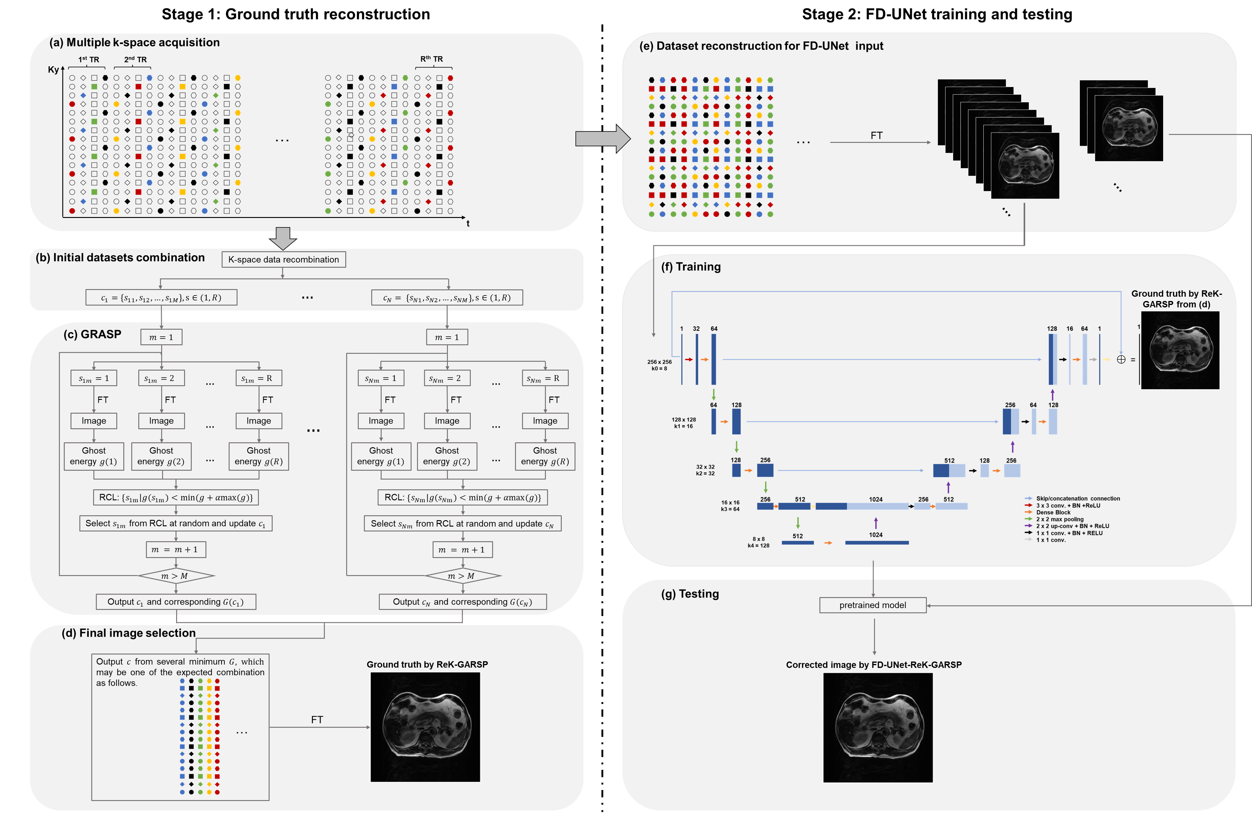
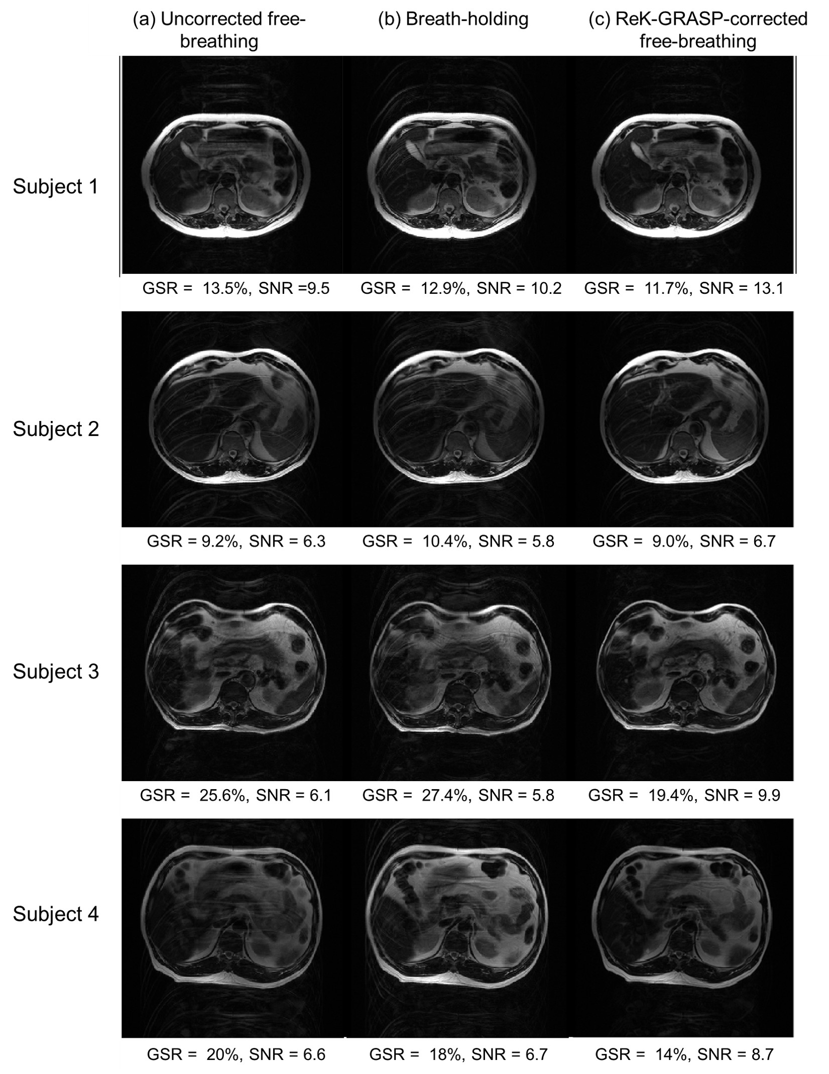
Fig.2 Abdominal FSE images acquired from 4 subjects with (a) uncorrected free-breathing method, (b) breath-holding method, and (c) ReK-GRASP method. The resulted images show that ReK-GRASP can achieve best performance in motion artifact reduction.
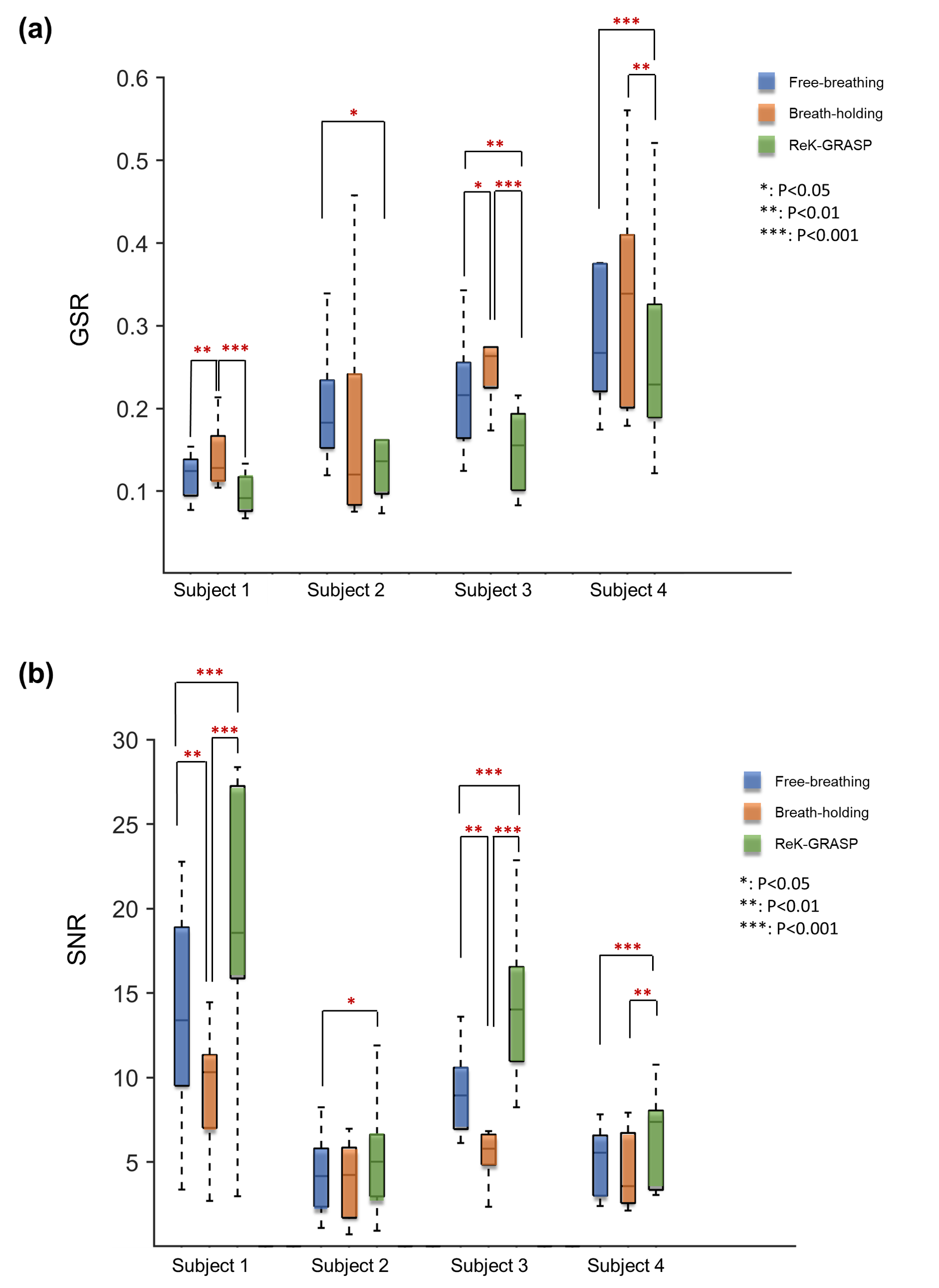
Fig.3 The measured (a) mean GSR (ghost-to-signal) and (b) mean SNR (signal-to-noise) of the images acquired from uncorrected free-breathing method, breath-holding method and ReK-GRASP method, respectively. It shows that the images produced by ReK-GRASP generally have lower GSR and higher SNR than those acquired with either free-breathing or breath-holding methods, except for the subject 2 that shows no significant differences. From the overall assessments of GSR and SNR, ReK-GRASP method shows better performance than breath-holding method in teams of motion artifact reduction.
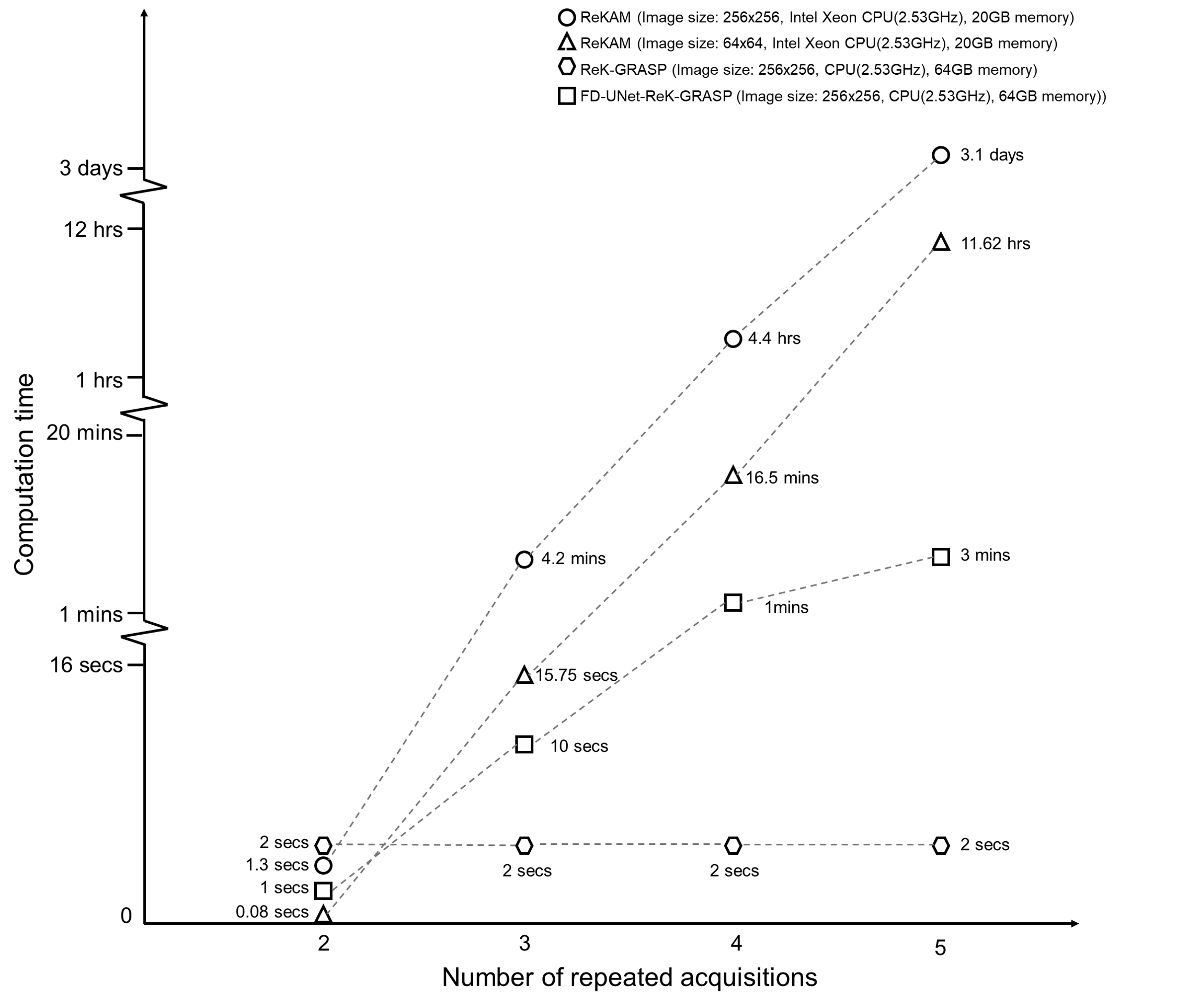
Fig.4 The computation time corresponding to different numbers of repeated acquisitions for producing free-breathing FSE images with motion reduction, using ReKAM for 256x256 image (circle marker), ReKAM for 64x64 image (triangle marker), ReK-GRASP for 256x256 image (square marker), and FD-UNet-ReK-GRASP for 256x256 image (hexagon marker). Comparison shows that ReK-GRASP requires less computation cost than ReKAM, especially when number of repeated acquisitions increases, and FD-UNet-ReK-GRASP can reduce motion artifacts regardless of the number of repetitions.
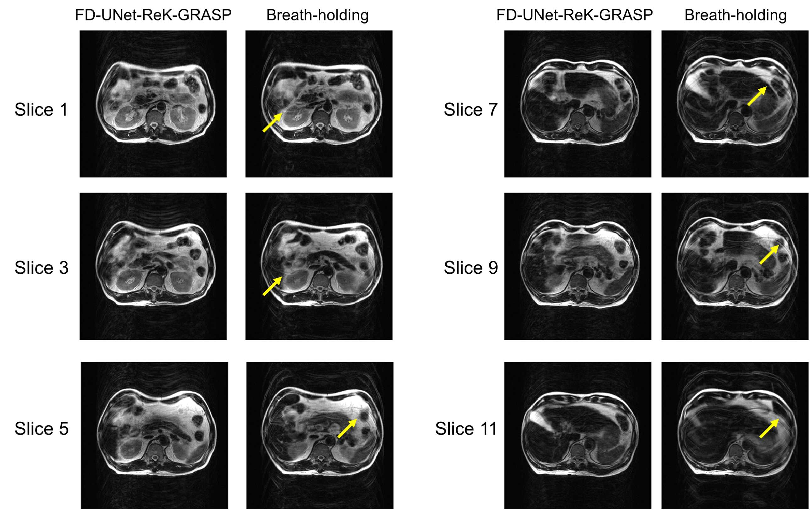
Fig.5 FSE images of six selected slice locations produced from FD-UNet-ReA-GRASP framework and conventional breath-holding method. FD-UNet-ReK-GRASP shows better performance in motion artifact reduction than the breath-holding method, suggesting that FD-UNet-ReK-GRASP may be a better alternative for the subjects who cannot perform breath-holding during routine MRI exam.