3796
Effect of varying intensity discretization and normalization parameters on first order radiomics features.
Abhilasha Indoria1, Sachin Patalasingh1, subhas Konar1, and Jitender Saini1
1NIMHANS, Bengaluru, India
1NIMHANS, Bengaluru, India
Synopsis
This study assessed the impact of normalization scale and intensity discretization on first order radiomics features. Features were extracted from T2W images by varying the normalization parameter and bin width parameter of Pyradiomics library; Un-normalized (Without_normalization), normalized to scale1, normalized to scale 100 while keeping the bin width constant; and Bin width set to 20, bin width set to 40, bin width set to 60 while keeping the normalization scale set to 1. We found that the radiomics features were highly dependent on normalization scale and independent of bin width parameter.
INTRODUCTION
Radiomics is dependent on the extraction of a variety of quantitative image-based features to provide decision support1. The premise of radiomics is that these features can serve as biomarkers characterizing lesions. These features extracted from magnetic resonance images or computed tomography images suffer from being highly dependent on acquisition, reconstruction as well as pre-processing and feature extraction configurations2. There are no guidelines for the feature extraction configuration and pre-processing of MR images before extracting the radiomics features, which is important for the generalization of image-based unique signatures. So, for such biomarkers to be useful, repeatability is a basic requirement which implies that its value must remain stable between two scans, if the conditions remain stable. This study aims to assess the impact of different feature extraction configurations such as intensity normalization scales, intensity discretization on first order radiomics features extracted from T2 weighted (T2W) images of meningioma subjects and hemangiopericytoma (HPC) subjects.METHODS
This retrospective study included 14 HPC and 17 Meningioma subjects, classified with tumor histopathological characterization done using WHO 2016 classification. MRI data included T2W MRI performed on 3T MR scanner (Philips Achieva) using a 15-channel head coil. Nifti Images were subjected to voxel reshaping to 1X1X1 followed by brain extraction using FSL BET. Segmentation wizard extension of 3D slicer was used for lesion segmentation. Semi-automatic method of intensity thresholding was applied to generate the binary label for region of interest (ROI). All ROIs were cross-validated by an experienced neuroradiologist. 18 first order features were extracted from all ROIs using pyradiomics library. features were recursively extracted by; 1. varying the normalization scale parameter (values=1, 60, 100, no normalization) while keeping the bin width parameter constant, and, 2. By varying the bin width parameter (values=20, 40, 60) while keeping the normalization scale parameter constant. Since the features are highly correlated, feature weight (importance score to differentiate between HPC and meningioma) was calculated using random forest estimator from the scikit learn library. Random forest algorithm naturally ranks the features based on gini impurity. Change in feature importance score was assessed by varying normalization and bin width parameters. Kolmogorov Smirnov test was applied to the features to test the distribution of features. Repeated measures ANOVA (rm-anova) was applied to compare means across the normally distributed features to assess the effect of above-mentioned feature extraction configurations. Friedman test was applied for the features which were not normally distributed. Boxplots of were also calculated for certain features which were common in top 5 feature rankings (based on importance score) between normal to 100 and unnormalized groups.RESULTS
All subjects were age and gender matched. There was no effect of changing bin width variable on any first order statistical feature as well as on the feature importance score. Whereas it was found that the feature importance score/ weight changes when the normalization parameter is varied figure2. Normalization parameter had significant effect on first order statistical features. However, there were 6 features-MeanAbsoluteDeviation, Range, InterquartileRange, kurtosis and variance which were not statistically significantly different between unnormalized and normalized to scale 100 group. Rest 10 features found to be statistically significantly different between the two normalized and not normalized group. Similarly, for comparison between normalization to scale1 and normalization to scale 100 we found 14 features to be statistically significantly different. While 4 features- kurtosis, MeanAbsoluteDeviation, variance and minimum were found to have no effect of normalization scale. P values for statistical comparisons are reported in Table1 and Table2. Boxplots generated for four unnormalized and normalized group is shown in figure3.DISCUSSION
We investigated the effect of feature configuration parameters on first order statistical features extracted from T2W images of HPC subjects and Meningioma subjects. Along with machine learning techniques, radiomics is becoming an increasingly popular computer-aided diagnostic tool in the field of medical research3,4. We found that there is very little to no effect of changing bin width parameters on radiomics features. This finding is in line with previously published literature5. This outcome was expected because changing the bin width parameter should only change the appearance of intensity histogram and not the intensity values. Whereas changing the normalization parameter had a big impact on the extracted features. Feature weightage also changed as the scale of normalization was changed. This implies that normalization scale will affect the classification accuracies of different machine learning algorithms. However, we also found certain features that don’t change by changing the normalization scale. Using the features that remain stable for different normalization scales may increase the accuracy and reproducibility of various methods/algorithms used for lesion characterization. For future studies effect of normalization can also be assessed on quantitative maps such as diffusion maps or fractional anisotropy maps.CONCLUSION
First order radiomics features are dependent on normalization scale and independent of bin width parameter. Radiomics quantifies imaging features that can help in lesion characterization. Identifying features that remain stable after normalization and the features that are affected majorly by the normalization scale will help in increasing the radiomics feature reproducibility, thereby helping in the standardization of radiomics pipeline.Acknowledgements
No acknowledgement found.References
- Gillies, R. J., Kinahan, P. E. & Hricak, H. Radiomics: images are more than pictures, they are data. Radiology 278, 563–577 (2016).
- Simmons, A., Tofts, P. S., Barker, G. J. & Arridge, S. R. Sources of intensity nonuniformity in spin echo images at 1.5 T. Magn.Reson. Med. 32, 121–128 (1994).
- Lambin, P. et al. Radiomics: the bridge between medical imaging and personalized medicine. Nat. Rev. Clin. Oncol. 14, 749–762(2017).
- Limkin, E. J. et al. Promises and challenges for the implementation of computational medical imaging (radiomics) in oncology.Ann. Oncol. 28, 1191–1206 (2017).
- Alexandre Carré et. Al. Standardization of brain MR images across machines and protocols: bridging the gap for MRI‑based radiomics. Sci Rep. 2020; 10: 12340
Figures
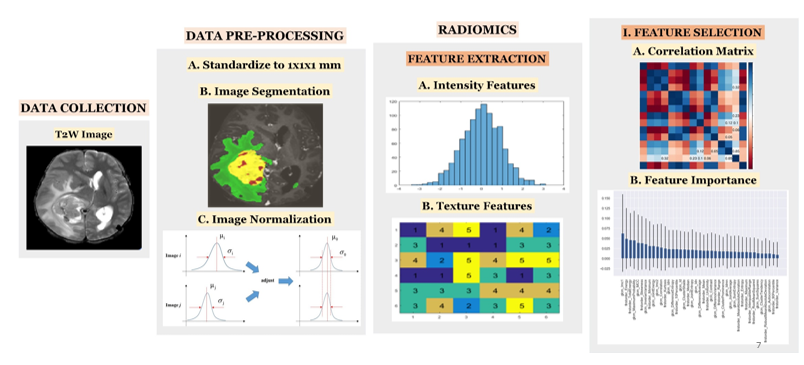
Figure1:Radiomics workflow to extract features and compute feature importance
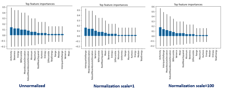
Figure2: Effect of normalization parameter, changing the normalization parameter changes the order of feature importance
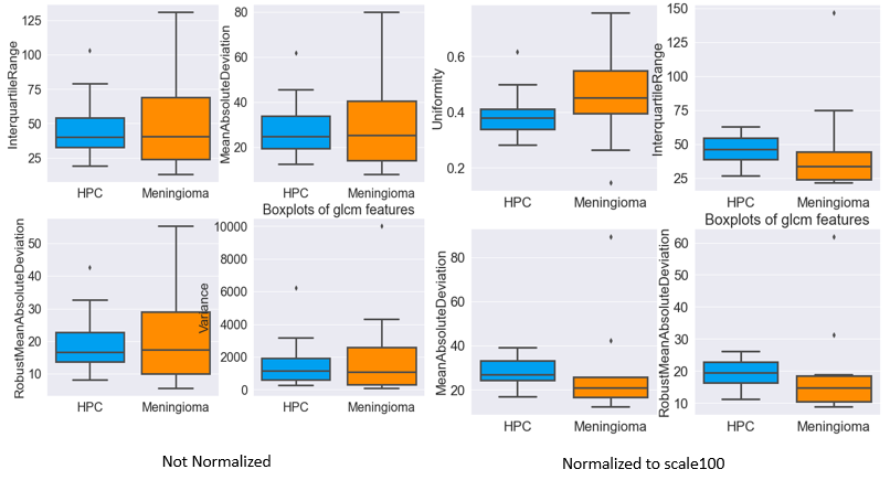
Figure3: Boxplots of few features from un-normalized (left) and normalized(Right) T2W images
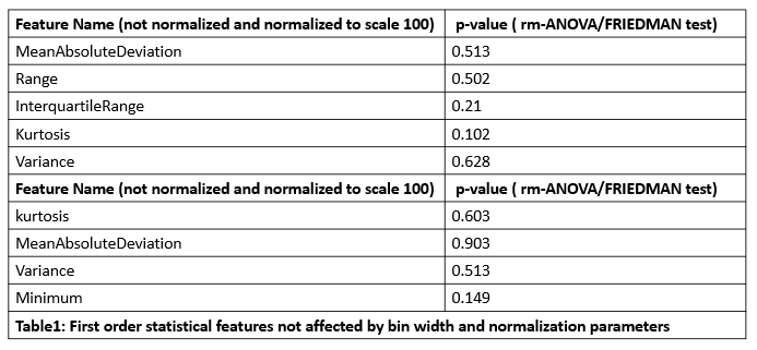
Table1: First order features not affected by varying normalization parameter
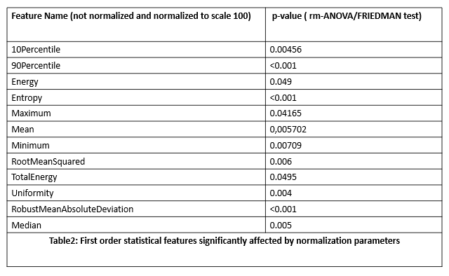
Table2: Features affected by varying the normalization parameters
DOI: https://doi.org/10.58530/2022/3796