3792
Comparison of fat quantification in abdomen and vertebrae by IDEAL-IQ and mDIXON Quant1the First Affiliated Hospital of Dalian Medical University, Da Lian, China, 2Philips Healthcare, Da Lian, China, 3Philips Healthcare, Bei Jing, China
Synopsis
IDEAL-IQ by GE and mDixon Quant by Philips are two MR fat quantification techniques that have been widely used for clinical disease evaluation and diagnosis in recent years. This study aims to compare performance of the two similar methods for fat quantification in the liver, pancreatic and lumbar vertebral. Results showed no significant difference between IDEAL-IQ and mDixon Quant on the liver, pancreas, and lumbar vertebral fat quantification in the same cohort of heathy volunteers
Synopsis
IDEAL-IQ by GE and mDixon Quant by Philips are two MR fat quantification techniques that have been widely used for clinical disease evaluation and diagnosis in recent years. This study aims to compare performance of the two similar methods for fat quantification in the liver, pancreatic and lumbar vertebral. Results showed no significant difference between IDEAL-IQ and mDixon Quant on the liver, pancreas, and lumbar vertebral fat quantification in the same cohort of heathy volunteers.Summary of Main Findings
The IDEAL-IQ and mDixon Quant techniques provided by different vendors showed a high degree of repeatability in fat quantification in human liver, pancreas, and lumbar vertebral, proving the potential these two methods for multicenter research.Introduction
Adipose tissue is one of the largest compartments in the human body which is widely distributed in the subcutaneous, internal organs and surrounding and vertebrae. IDEAL-IQ and mDixon Quant are methods provided by two different vendors for clinical evaluation of tissue fat contents. Both of two methods employ the 3D gradient echo acquisition and obtain the proton density fat fractions (PDFF) by water-fat separation based on chemical shift encoding [1]. In addition, both techniques use a small flip angle (3 degrees) to minimize T1 bias, and multiple echo signals (6 echoes) to correct the T2* effect and reduce noise bias, enabling accurate quantification of fat [2]. While the two methods may differ in the post-processing stage when modeling the water and fat signals. The purpose of this study is to explore the differences between the two techniques in fat quantification in liver, pancreas and lumbar vertebra.Materials and methods
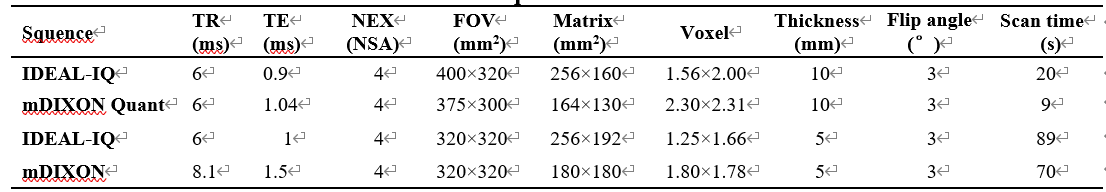
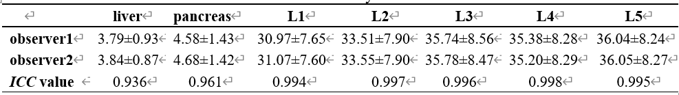
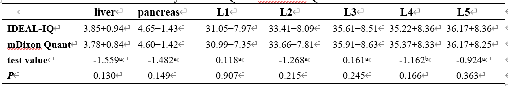
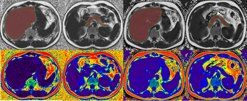
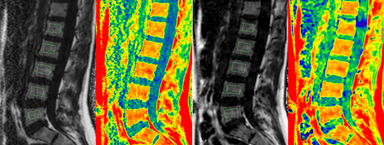
Data were collected from 30 healthy volunteers (12 males), aged 21-29 years (24.40± 2.43 years), BMI of 18.07-29.59 kg/m2 (21.84±2.83kg/m2). All volunteers completed MR scans of the upper abdominal axis position and the lumbar vector with both IDEAL-IQ (GE Signa HDxt 3.0T MR) and mDixon Quant (Philips Ingenia CX 3.0T MR, Philips Healthcare, Best, the Netherlands) sequences within 1 to 4 h of the same day of examination. Scan parameters are shown in Table 1-2. The FF maps were automatically reconstructed on the console of MR scanners. Two observers measured the FF values of the liver, pancreas, and lumbar vertebrae in the fat fraction images by the two sequences. The FF images by IDEAL-IQ and mDixon Quant were transferred to the ISP (Intelli Space Portall Version7) workstation for data measurements. For liver and pancreas FF measurements: the liver and pancreas volumes were semi-automatically split by 3D volume extraction tumor tracking software, and manually adjusted by two observers (Figure 1). For lumbar spine FF measurements, two observers manually place rectangular ROIs on the L1- L5 vertebral vector maximum level with area of 200-230 mm2, avoid the vertebrae head and tail side of the final plate, cartilage and vertebral front and back edge bone cortex. Measurement consistency by the two observers was tested by intra-class correlation coefficients (ICC). The mean FF values by the two observers were compared between IDEAL-IQ and mDIXON Quant methods using the paired sample t test.Results
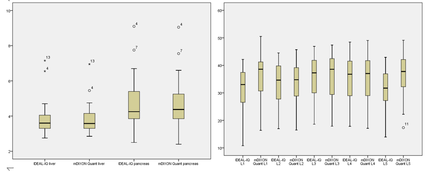
The interobserver measurement consistency was good (all ICC > 0.93). The FF values of liver, pancreas, and lumbar vertebra (L1 to L5) obtained by IDEAL-IQ and mDIXON Quant showed no significant difference (p>0.05,Table 4).Conclusions
There is no significant difference on FF values of the liver, pancreas and lumbar vertebra measured by IDEAL-IQ and mDixon Quant on two different 3.0T platforms, indicating the high repeatability between the two methods and proving their potential in multicenter research. Both IDEAL-IQ and mDixon Quant are non-invasive and radiation-free methods that can be used for precise fat quantifications in more clinical situations.Acknowledgements
No acknowledgement found.References
1. Yidi Chen, Liling Long, Zijian Jiang. Quantification of pancreatic proton density fat fraction in diabetic pigs using MR imaging and IDEAL-IQ sequence. BioMed Central,2019,19(1): 38.
2. Hines Catherine D G, Yu Huanzhou, Shimakawa Ann. Quantification of hepatic steatosis with 3-T MR imaging: validation in ob/ob mice. Radiology,2010,254(1): 119-28.
Figures