3789
Hyperintense globus pallidus rim sign could be a novel hallmark of Wilson disease at 7T MRI: a pilot case-control study1Department of Neurology, Beijing Tiantan Hospital,Captital Medical University, Beijing, China, 2China National Clinical Research Center for Neurological Diseases, Beijing, China, 3Tiantan Neuroimaging Center of Excellence, Beijing Tiantan Hospital, Capital Medical University, Beijing, China, 4MR Collaboration, Siemens Healthineers Ltd., Beijing, China
Synopsis
To explore specific neuroimaging characteristics of iron deposition in WD, we conducted a small sample, prospective case-control study to compare the 7T MRI manifestation of iron deposition in the basal ganglia among PD, WD patients and healthy controls. The hyperintense globus pallidus rim sign could be a specific neuroimaging characteristic of WD in 7T T2*-weighted and SWI images, which may be related to the unique iron deposition pattern in the striatum in WD patients. Internal capsule appears to be hyperintensity with a clear border only observed in WD patients. These findings have the potential to provide diagnostic biomarkers for WD.
Introduction
Wilson disease (WD), also referred to as hepatolenticular degeneration, is a genetic disorder related to the mutation of ATP7B copper transporter. WD is characterized by ataxia and other movement disorders caused by disturbances of the extrapyramidal system. Previous pathological studies have shown that iron deposition instead of copper deposition is related to T2/T2*w in the basal ganglia hypointensity1. Similar to WD, PD is also a movement disorder with iron deposition in the basal ganglia. Compared with 3T MRI, 7T MRI could provide higher sensitivity for detecting iron concentration changes. It is unknown whether there exist distinct image charateristics on 7T MRI between the two diseases. To address this question, we conducted a small sample, prospective case-control study to compare the 7T MRI manifestation of iron deposition in the basal ganglia among PD, WD patients and healthy controls, aimed to explore specific neuroimaging characteristics of iron deposition in WD.Methods
Four WD patients, four PD patients and four age-matched healthy controls were recruited. The study was approved by the local Ethical Standards Committee, and written informed consent was obtained from all participants. All the subjects underwent 7T MR (MAGNETOM Terra, Siemens Healthcare, Erlangen, Germany) scans with three-dimensional (3D) T2*-weighted Imaging (transverse scan, 4-echo 3D GRE, acquisition voxel = 0.4×0.4×0.5mm3, 88 slices, TE = 8/15/22/29 ms, TR = 42ms, flip angle = 16°, acceleration = 3, scan time = 8:24), two-dimensional (2D) T2*-weighted Imaging (transverse scan, 2-echo 2D GRE, acquisition voxel = 0.2×0.2×1.2mm3, 29 axial slices, TE = 15/30 ms, TR = 1400ms, flip angle = 50°, acceleration = 2, scan time = 7:20) and 3D SWI (transverse scan, acquisition voxel = 0.3×0.3×1.2mm3, 104 slices, TE = 12 ms, TR = 19 ms, flip angle = 14°, acceleration = 3, scan time = 6:47). The observation of MRI was conducted by two experienced neurologists.Results
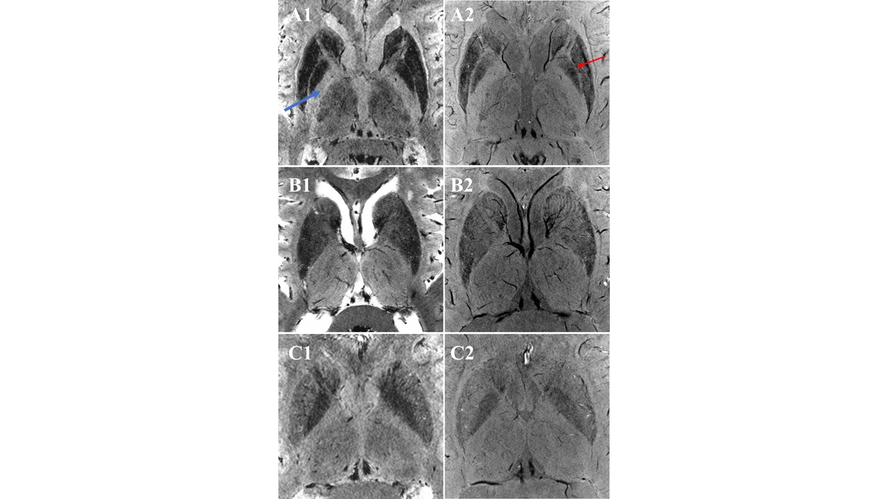
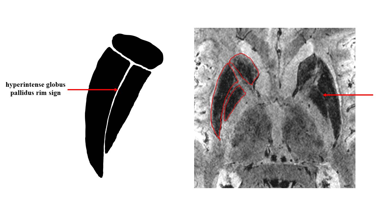
Compared with healthy controls, relatively low intensity in the putamen, globus pallidus and caudate could be observed in both WD and PD patients in SWI and T2*w sequence, which was related to iron concentration in neurodegenerative disorders. However, the patterns of iron deposition were different between WD and PD. The hypointensity in the thalamus was only observed in WD patients, and the internal capsule appears to be hyperintensity with a clear border (Figure 1). Moreover, bilateral linear signal intensity changes consisting of a lateral hyperintense rim at the lateral border of globus pallidus could only be observed in WD patients. We term the intensity change as the hyperintense globus pallidus rim sign, which was absent in PD and healthy controls in this study (Figure 1). A sketch map of hyperintense globus pallidus rim sign is shown in Figure 2.Discussion
To our best knowledge, this might be the first 7T MRI study focusing on the visible neuroimaging biomarker for the diagnosis of WD. In this preliminary case-control study, we found that the hyperintense globus pallidus rim sign could be a specific neuroimaging characteristic of WD. According to a post-mortem 7T MRI-histopathological study, T2/T2*w hypointensity observed in vivo in the basal ganglia of WD patients is related to the increased iron concentration rather than copper deposition1. Therefore, the hyperintense globus pallidus rim sign may be related to the unique iron deposition pattern in the striatum in WD patients, which is worth investigating in the histological study. Moreover, the relatively high intensity with a clear border of the internal capsule in WD can be associated with the increased iron deposition in globus pallidus, caudate and thalamus2. These findings have the potential to provide diagnostic biomarkers for WD. Future larger cohort study is needed to confirm the results.Conclusion
Using 7T MRI, our pilot case-control study suggested the hyperintense globus pallidus rim sign and relatively high intensity with a clear border of the internal capsule could be potential diagnostic biomarkers for WD.Acknowledgements
We thank the Siemens Healthineers for providing technical support. We also thank all participants for their cooperation.References
1. Dusek P, Bahn E, Litwin T, et al. Brain iron accumulation in Wilson disease: a post mortem 7 Tesla MRI - histopathological study. Neuropathol Appl Neurobiol. 2017;43(6):514-532. doi:10.1111/nan.12341
2. Yuan XZ, Li GY, Chen JL, Li JQ, Wang XP. Paramagnetic Metal Accumulation in the Deep Gray Matter Nuclei Is Associated With Neurodegeneration in Wilson's Disease. Front Neurosci. 2020;14:573633. doi:10.3389/fnins.2020.573633
Figures