3787
Pseudo-continuous arterial spin labeling reveal altered cerebral hemodynamics and blood-brain barrier dysfunction in CADASIL patients1State Key Laboratory of Brain and Cognitive Science, Institute of Biophysics, Chinese Academy of Sciences, Beijing, China, 2University of Chinese Academy of Sciences, Beijing, China, 3The Innovation Center of Excellence on Brain Science, Chinese Academy of Sciences, Beijing, China, 4Department of Neurology, Peking University First Hospital, Beijing, China, 5Beijing Key Laboratory of Neurovascular Disease Discovery, Peking University First Hospital, Beijing, China, 6Mark & Mary Stevens Neuroimaging and Informatics Institute, Keck School of Medicine, University of Southern California, Los Angeles, CA, United States, 7Siemens Shenzhen Magnetic Resonance Ltd., Shenzhen, China, 8Department of Radiology, Chaoyang Hospital, Capital Medical University, Beijing, China
Synopsis
Cerebral autosomal dominant arteriopathy with subcortical infarcts and leukoencephalopathy (CADASIL) is an inherited form of cerebral small vessel disease (cSVD). In this study, we investigated the changes of cerebral blood flow (CBF), arterial transit time (ATT) and water exchange rate (kw) measured by multi-delay pseudo-continuous arterial spin labeling (pCASL) and diffusion prepared pCASL. Increased ATT and decreased kw were found in CADASIL patients compared with healthy controls. The results indicated modified cerebral hemodynamics and dysfunction of blood-brain barrier in CADASIL patients. MD-pCASL and DP-pCASL are promising for evaluating hemodynamics and BBB function of cSVD.
Introduction
Cerebral autosomal dominant arteriopathy with subcortical infarcts and leukoencephalopathy (CADASIL) is a hereditary cerebral small vessel disease (cSVD) caused by NOTCH3 gene mutations which mainly express in vascular smooth muscle cells1. The pathology of CADASIL makes it an ideal model for the study of cSVD, and the imaging findings on CADASIL can be generalized to the diagnosis and the etiological study of cSVD. The dysfunction of blood-brain barrier (BBB) and abnormal hemodynamics were reported as critical factors in the pathogenesis of cSVD2. Multi-delay pseudo-continuous arterial spin labeling (MD-pCASL) has been widely accepted in the measurement of cerebral perfusion3. Diffusion prepared pCASL (DP-pCASL) is a newly proposed method to noninvasively measure the water exchange rate (kw) across BBB4. In this study, we used MD-pCASL and DP-pCASL to investigate the changes of cerebral hemodynamics and BBB in CADASIL patients.Methods
Twenty-seven genetically confirmed CADASIL patients and 28 age-matched healthy controls were included in the study. All participants signed informed contents approved by the local institutional review board. The data were acquired on a 3T Prisma system (Siemens Healthcare, Erlangen, Germany) with a 64-channel head coil. Structural images were obtained by T1-weighted magnetization-prepared rapid gradient echo (T1w-MPRAGE) with 1.0 mm isotropic resolution. In MD-pCASL, five post-labeling delays (PLDs) were used (0.5, 1.0, 1.5, 2.0 and 2.5 s with 2, 2, 2, 3, and 3 measurements respectively). The labeling duration was 1.5 s. Other imaging parameters were: resolution = 2.5x2.5x3.0 mm3, turbo factor = 12, EPI factor = 63. In DP-pCASL four sets of diffusion preparation modules and PLDs were repeatedly collected: b = 0/14 s/mm2 & PLD = 900ms for 15 times, b = 0/50 s/mm2 & PLD = 1800ms for 20 times. Other imaging parameters were: resolution = 3.5x3.5x8.0 mm3, turbo factor = 14, EPI factor = 63. The labeling duration was 1.5 s for both the pCASL sequences, with background suppression always applied. The degrees of disability/dependence of CADASIL patients were determined by the modified Rankin Scale (mRS).MD-pCASL data was post-processed by BASIL toolkit of FSL5. Cerebral blood flow (CBF, ml/100g/min) and arterial transit time (ATT, s) were calculated with a prior bolus arrival time of 1.3 s and a correction factor for the labeling efficiency of 0.73. The regions of interest (ROIs) were defined as whole brain, frontal lobe, parietal lobe, temporal lobe, occipital lobe, subcortical nuclei and white matter (WM). The kw map was reconstructed from DP-pCASL data by a custom Matlab toolbox which implemented a single-pass approximation model with total generalized variation regularization. All results were expressed as mean ± standard deviation. The differences between patients and controls were examined by independent samples t-test. Spearman’s rank correlation was computed between CBF, ATT, kw and mRS in patients. mRS higher than 1 were defined as abnormal mRS6 and CBF, ATT, kw were compared between the two groups. The statistics was performed in SPSS 26 software.
Results
Figure 1 shows representative results of CBF, ATT and kw maps from one patient and one control. The statistical results are presented in Table 1 and Figure 2. The ages of the patients matched well with the controls. The kw was significantly lower in CADASIL patients. While only CBF in WM was found lower in CADASIL patients, ATTs in all ROIs were significantly increased in patients. There was a positive correlation between ATT and mRS (r = 0.607, P < 0.001), and a negative correlation between kw and mRS (r = -0.627, P < 0.001). Figure 3 shows increased ATT and decreased kw in patients with abnormal mRS relative to normal mRS.Discussion
In this study, we quantified CBF, ATT and kw of CADASIL patients and healthy controls using MD-pCASL and DP-pCASL. The longer ATT indicates there may be collateral circulation which compensated impaired microcirculation to a certain extent, as comparable CBF was found in patients and controls. In patients, decreased CBF in WM may be attributed to the hypoperfusion in the watershed area7. The reduced kw in CADASIL patients indicates lower water exchange across BBB in this genetic form of cSVD. Reduced BBB water exchange has been hypothesized to impair glymphatic flow and brain waste clearance. Our finding is consistent with a recent study that reported positive associations of kw with CSF amyloid-beta42 and episodic memory score in a cohort of aged subjects with normal cognition8. The correlations between ATT, kw and mRS indicate the hemodynamic change and BBB dysfunction measured by pCASL may explain the pathology of neurologic disability in patients. Given CADASIL is a pure form of cSVD without confounding of amyloid-related dementia, our results suggest reduced BBB water exchange may be a biomarker of cSVD.Conclusion
By using MD-pCASL and DP-pCASL, increased ATT and decreased kw were found in CADASIL patients. These pCASL-based techniques are promising in the noninvasive evaluation of cerebral hemodynamics and BBB function of CADASIL and cSVD.Acknowledgements
This work was supported by the National Natural Science Foundation of China (81961128030, 82001804), the US NIH grant (R01NS114382), the Natural Science Foundation of Beijing Municipality (7191003), and the Strategic Priority Research Program of Chinese Academy of Science (XDB32010300).
References
1. Joutel, A. et al. Notch3 mutations in CADASIL, a hereditary adult-onset condition causing stroke and dementia. Nature 383, 707–710 (1996).
2. Bridges, L. R. et al. Blood-Brain Barrier Dysfunction and Cerebral Small Vessel Disease (Arteriolosclerosis) in Brains of Older People. J Neuropathol Exp Neurol 73, 1026–1033 (2014).
3. Alsop, D. C. et al. Recommended implementation of arterial spin-labeled perfusion MRI for clinical applications: A consensus of the ISMRM perfusion study group and the European consortium for ASL in dementia: Recommended Implementation of ASL for Clinical Applications. Magn. Reson. Med. 73, 102–116 (2015).
4. Shao, X. et al. Mapping water exchange across the blood-brain barrier using 3D diffusion-prepared arterial spin labeled perfusion MRI. Magn Reson Med 81, 3065–3079 (2019).
5. Chappell, M. A., Groves, A. R., Whitcher, B. & Woolrich, M. W. Variational Bayesian Inference for a Nonlinear Forward Model. IEEE Trans. Signal Process. 57, 223–236 (2009).
6. Ling, C. et al. Reduced Venous Oxygen Saturation Associates With Increased Dependence of Patients With Cerebral Autosomal Dominant Arteriopathy With Subcortical Infarcts and Leukoencephalopathy: A 7.0-T Magnetic Resonance Imaging Study. Stroke 50, 3128–3134 (2019).
7. Momjian-Mayor, I. & Baron, J.-C. The Pathophysiology of Watershed Infarction in Internal Carotid Artery Disease: Review of Cerebral Perfusion Studies. Stroke 36, 567–577 (2005).
8. Gold, B. T. et al. Water exchange rate across the blood‐brain barrier is associated with CSF amyloid‐β 42 in healthy older adults. Alzheimer's & Dementia alz.12357 (2021) doi:10.1002/alz.12357.
Figures
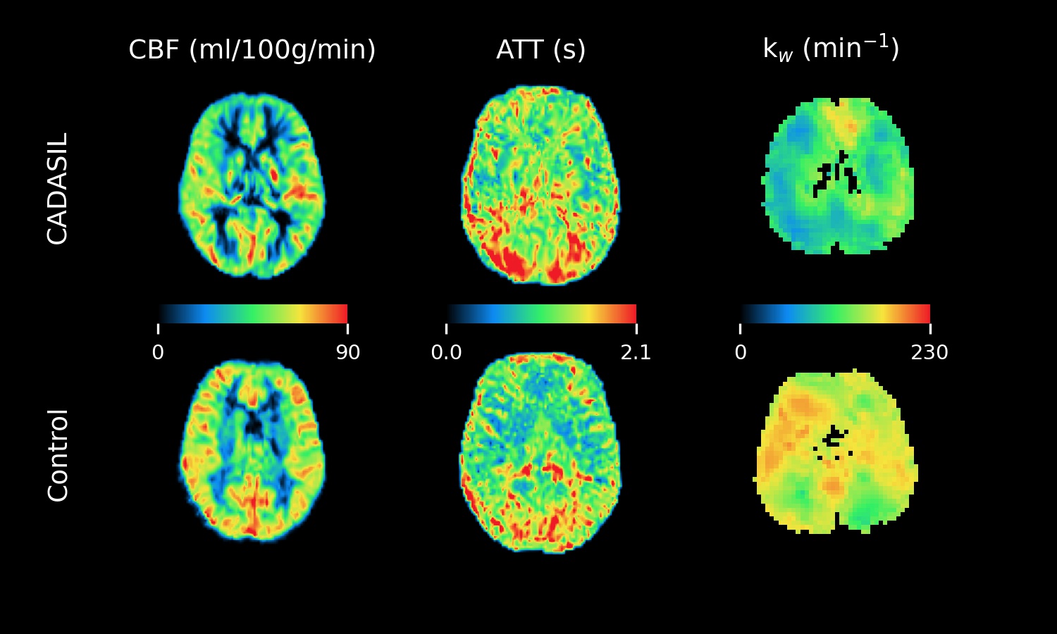
Figure 1. Example images of CBF, ATT, and kw from a CADASIL patient and a healthy control.
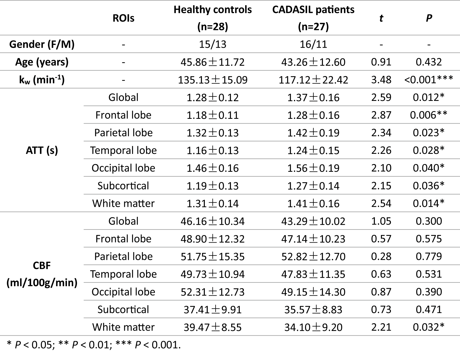
Table 1. Gender, age, water exchange rate (kw), cerebral blood flow (CBF) and arterial transit time (ATT) of CADASIL patients and healthy controls.
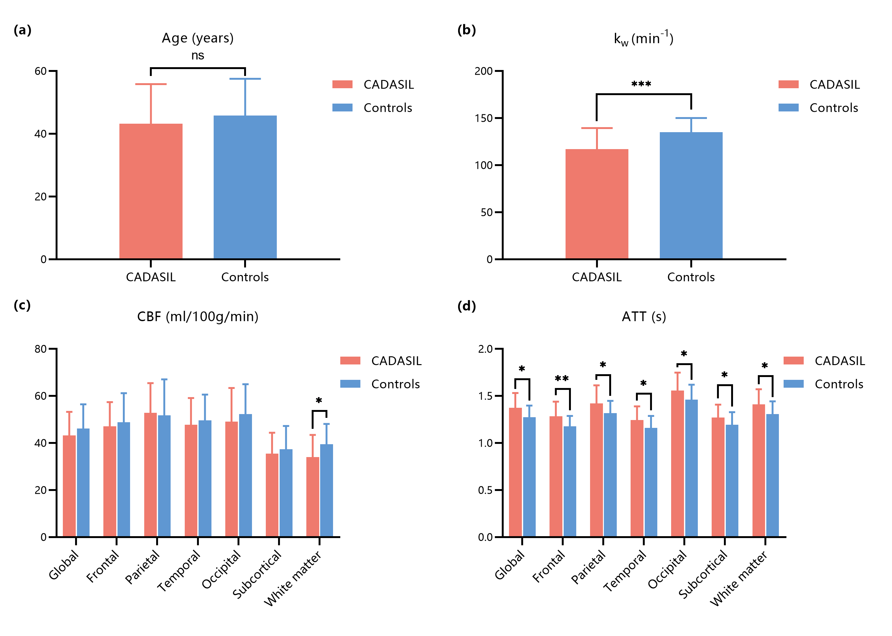
Figure 2. Comparison of age, water exchange rate (kw), cerebral blood flow (CBF) and arterial transit time (ATT) between CADASIL patients and healthy controls. Significant lower kw and longer ATT were found in the patients.
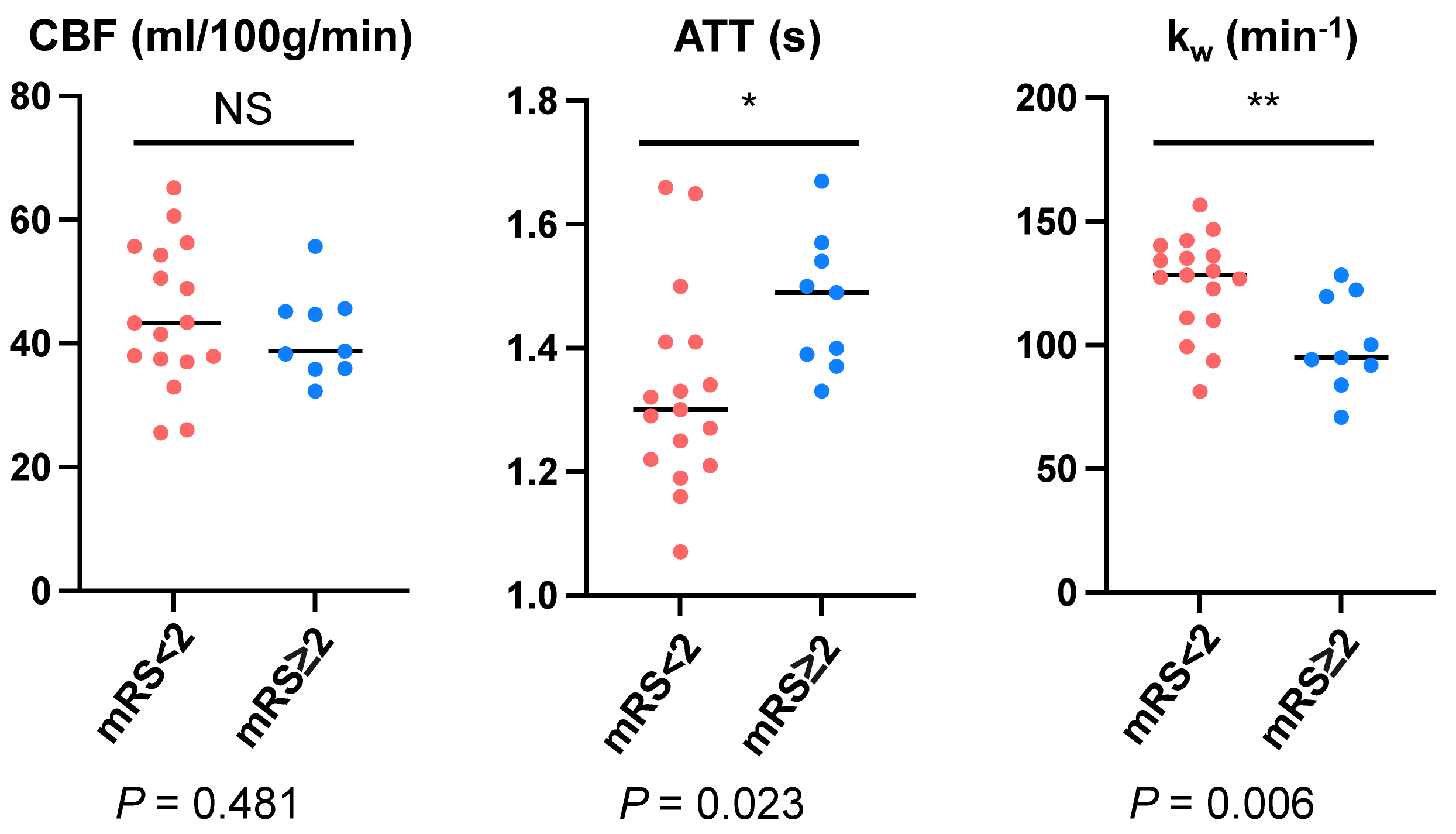
Figure 3. Comparison of CBF, ATT and kw between patients with abnormal and normal mRS.