3774
Value of Qualitative and Quantitative DCE-MRI Analysis based on GRASP Acquisition in Diagnosing of breast lesions1Department of Radiology, Chengdu Women's and Children's Central Hospital, School of Medicine, University of Electronic Science and Technology of China, Chengdu, China, 2MR Scientific Marketing, Siemens Healthineers, Shanghai, China
Synopsis
This study investigated the diagnostic capabilities of qualitative and quantitative parameters derived from high-resolution DCE-MRI with GRASP sequence. 31 patients (33 lesions, 13 benign and 20 malignant lesions) were prospectively included. The results showed that there were prominent discrepancies of quantitative and quantitative parameters between malignant and benign breast lesions. Ktrans, Kep, and iAUC(60) had excellent diagnostic efficiency for breast cancer.
Introduction
Golden-angle radial sparse parallel (GRASP) is a new acquisition technique which could achieve high temporal and spatial resolution. It combines compressed sensing, parallel imaging with radial sampling, which can speed up scanning and reduce motion artifacts1-2. Previous studies have confirmed that GRASP has good image quality in chest, body, and head/neck imaging3-6. Quantitative pharmacokinetic parameters derived from DCE-MRI can quantitatively evaluate the microvascular hemodynamics of tissue or tumor, which depends on high-temporal-resolution acquisition7. This study aimed to explore the diagnostic efficiency of quantitative analysis on high-resolution DCE-MRI of breast lesions by using GRASP sequence.Methods
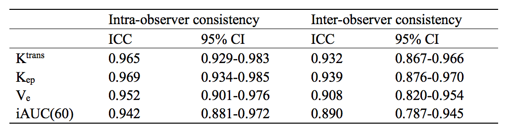
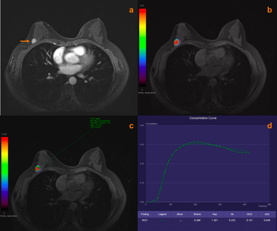
This IRB-approved and prospective study included 31 patients (33 lesions) with suspicious breast mass lesions and histopathologically verified results. All enrolled lesions were divided into benign group and malignant group according to the pathological results. All patients underwent breast MRI on a 3.0 Tesla clinical system (MAGNETOM Vida, Siemens Healthcare, Erlangen, Germany) with an 18-channel breast coil. Dynamic contrast-enhanced (DCE) MR data were acquired using GRASP sequence during free breathing, and reconstructed with a 14.8 sec/phase temporal resolution as well as B0 and B1 corrections. The detailed protocol were: TR/TE=4.09/1.86ms, FOV=350*350mm2, spatial resolution=1.1*1.1*2.0mm3, slice thickness=2.0mm, number of slice=72, total number of spokes=2671, total acquisition time=7min24sec. The quantitative perfusion analysis was performed based on a standard Tofts model (Tissue 4D Software, Siemens Healthcare, Erlangen, Germany) to generate the volume transfer constant (Ktrans, min-1), reflux rate constant (Kep, min-1), and interstitial volume fraction (Ve, ml/ml). iAUC(60) (the initial area under the curve when 60 seconds after enhancement) was considered as qualitative parameter in this analysis. Two radiologists evaluated MRI images and measured above-mentioned parameters of breast lesions independently. Mann–Whitney U-Test was used to compare the qualitative and quantitative parameters between benign and malignant groups. Intra-observer and inter-observer consistency were evaluated by intraclass correlation coefficient (ICC). ICC > 0.75 indicated high consistency.Results
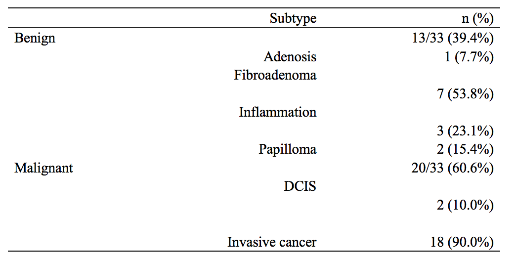
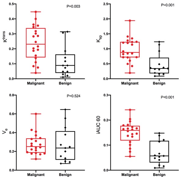
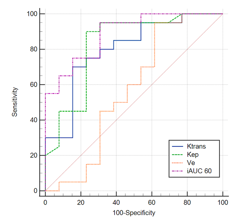
There were 13 (39.4%) benign and 20 (60.6%) malignant lesions according to the pathological results. The most common benign lesions were fibroadenoma (53.8%), while the most common malignant lesions were invasive cancer (90.0%). The values of Ktrans, Kep ,and iAUC(60) of malignant group were significant higher than those of benign group (0.23(0.14-0.34) vs 0.09(0.04-0.16), P=0.003; 0.88(0.71-1.23) vs 0.34(0.18-0.71), P=0.001; and 0.16(0.12-0.18) vs 0.06(0.03-0.12), P<0.001, respectively). However, there was no significant difference in Ve values between malignant and benign groups (0.25(0.19-0.34) vs 0.24(0.11-0.42), P=0.524). The measurement of Ktrans, Kep, Ve, and iAUC(60) all had high consistency between intra and inter-observers (all ICC>0.75). Area under the receiver operating characteristics curve (AUC) of Ktrans, Kep, Ve and iAUC(60) were 0.80, 0.83, 0.57, and 0.89.Discussion and Conclusions
We compared pharmacokinetic parameters of benign and malignant breast lesions on high-resolution DCE-MRI with GRASP sequence. Results showed that there were significant discrepancies of the value of Ktrans, Kep, and iAUC(60) between malignant and benign breast lesions. Furthermore, the measurement of these quantitative parameters had good repeatability. Ktrans, Kep, and iAUC(60) had high efficiency in differential diagnosis of breast lesions. This conclusion suggests that quantitative MRI analysis based on high-temporal-resolution MRI technique has a good application prospect in the differentiation of breast diseases.Acknowledgements
Not applicable.References
1.Feng L, Grimm R, Block KT, et al. Golden-angle radial sparse parallel MRI: combination of compressed sensing, parallel imaging, and golden-angle radial sampling for fast and flexible dynamic volumetric MRI. Magn Reson Med. 2014;72(3):707-717. doi:10.1002/mrm.24980
2. Chandarana H, Feng L, Block TK, et al. Free-breathing contrast-enhanced multiphase MRI of the liver using a combination of compressed sensing, parallel imaging, and golden-angle radial sampling. Invest Radiol. 2013;48(1):10-16. doi:10.1097/RLI.0b013e318271869c
3. Heacock L, Gao Y, Heller SL, et al. Comparison of conventional DCE-MRI and a novel golden-angle radial multicoil compressed sensing method for the evaluation of breast lesion conspicuity. J Magn Reson Imaging. 2017;45(6):1746-1752. doi:10.1002/jmri.25530
4. Chen L, Liu D, Zhang J, et al. Free-breathing dynamic contrast-enhanced MRI for assessment of pulmonary lesions using golden-angle radial sparse parallel imaging. J Magn Reson Imaging. 2018;48(2):459-468. doi:10.1002/jmri.25977
5. Yoon JH, Lee JM, Yu MH, et al. Evaluation of Transient Motion During Gadoxetic Acid-Enhanced Multiphasic Liver Magnetic Resonance Imaging Using Free-Breathing Golden-Angle Radial Sparse Parallel Magnetic Resonance Imaging. Invest Radiol. 2018;53(1):52-61.
6. Tomppert A, Wuest W, Wiesmueller M, et al. Achieving high spatial and temporal resolution with perfusion MRI in the head and neck region using golden-angle radial sampling. Eur Radiol. 2021;31(4):2263-2271. doi:10.1007/s00330-020-07263-0
7. Petralia G, Summers PE, Agostini A, et al. Dynamic contrast-enhanced MRI in oncology: how we do it. Radiol Med. 2020;125(12):1288-1300. doi:10.1007/s11547-020-01220-z
Figures