3773
Comparison of Computed DWI with actual scanned DWI for nasopharyngeal carcinoma detection using DWI TSE MVXD1Philips Healthcare, Beijing, China, 2Department of Radiology, Guangdong Provincial People’s Hospital, Guangdong Academy of Medical Sciences, Guangzhou, China
Synopsis
Diffusion MRI provides unique information on the structure, organization, and integrity of the nasopharyngeal carcinoma (NPC) non-invasively. Higher b-values diffusion-weighted imaging could improve the detection rate of malignancies. We use the combination of DWI TSE Multivane XD and computed DWI to generate the high b values diffusion for nasopharyngeal whithout distortion, with higher SNR and less scan time. A in vivo comparison between actual scanned DWI and cDWI study was also studied on both volunteer and patients with NPC. The image quality is comparable. It shows cDWI is a promising technique for the NPC diagnosis.
Purpose
The goal of this work is to do a preliminary study of the detection performance which combines the Computed DWI (cDWI) with DWI TSE Multivan XD (DWI TSE MVXD) to generate high b values diffusion imaging for nasopharyngeal carcinoma (NPC) without distortion and with less scan time.Introduction
Diffusion MRI provides unique information on the structure, organization, and integrity of the nasopharyngeal carcinoma (NPC) without the need for exogenous contrast agents1-2. Higher diffusion weighting (b-values) in diffusion-weighted imaging (DWI) is increasingly used to detect malignancies, it should be useful for improved the accuracy and efficiency of diffusion MRI in the NPC. Single-shot EPI is widely used for DWI, but it is sensitive to field inhomogeneity and suffers from distortion, high b-values (especially > 1000 s mm-2) can cause severe image distortions and the usage of longer echo times (TE) reduce the signal-to-noise ratio (SNR), especially in NPC due to the nasopharyngeal cavity will cause severe field inhomogeneity. It’s still a great technically challenge to do the DWI for NPC. Robust and distortion free DWI imaging is important for accurate diagnosis of NPC. DWI based on TSE Multivane XD (DWI TSE MVXD) was a distortion free methods3, which is very suitable for NPC. However, it uses multishot TSE and has low SNR, the scan time will be significantly increased for high b value imaging with large average. Computed DWI (cDWI) could be used to calculate the high b value images with high SNR4-5. Its performance has been widely studied for prostate diagnosis, it should be useful for NPC also6. It should be very useful to generated high b value images for NPC. We hereby do a preliminary study of cDWI using DWI TSE MVXD, and compared it with actual scanned DWI for NPC.Methods
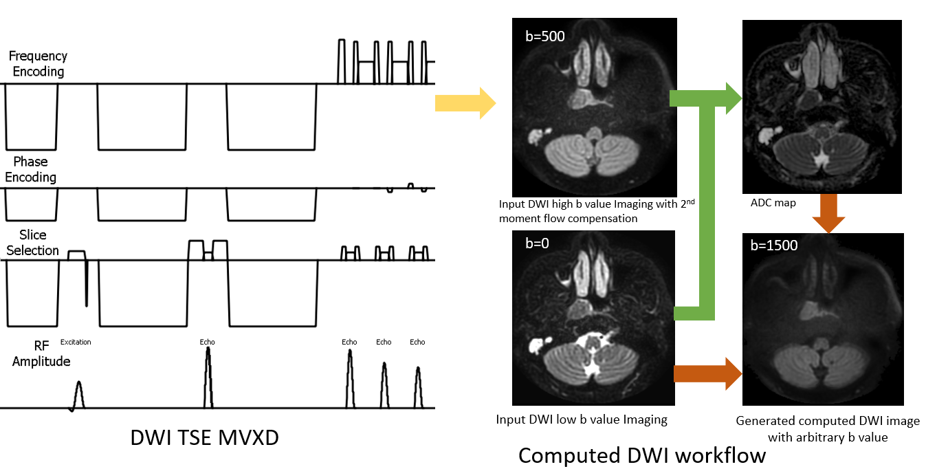
Fig 1 shows the pipeline for computed high b value for NPC diffusion. As the DWI based on EPI is unstable and has large distortion, it often has signal loss and fail, DWI TSE MVXD was used to get high resolution DWI images without any distortion. cDWI was implemented as reference 4. To get the high b values images with proper scan time for NPC, we use cDWI to generate high b value images. IMAge/engine was used for postprocessing to generate the cDWI images7.To evaluate the feasibility of cDWI for NPC, the computed DWI was compared with the actual scanned DWI, which is a golden standard for evaluating the computed high b value images. All scans were scanned on a Philips 3.0T Ingenia CX system (Philips Healthcare, Best, Netherland) with a 32-ch head and Neck coil. Two experienced observers used a five-point scoring method to subjectively evaluate the quality of images.
Results
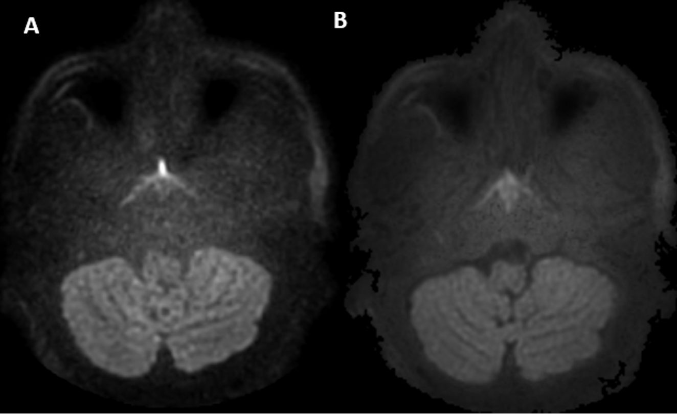
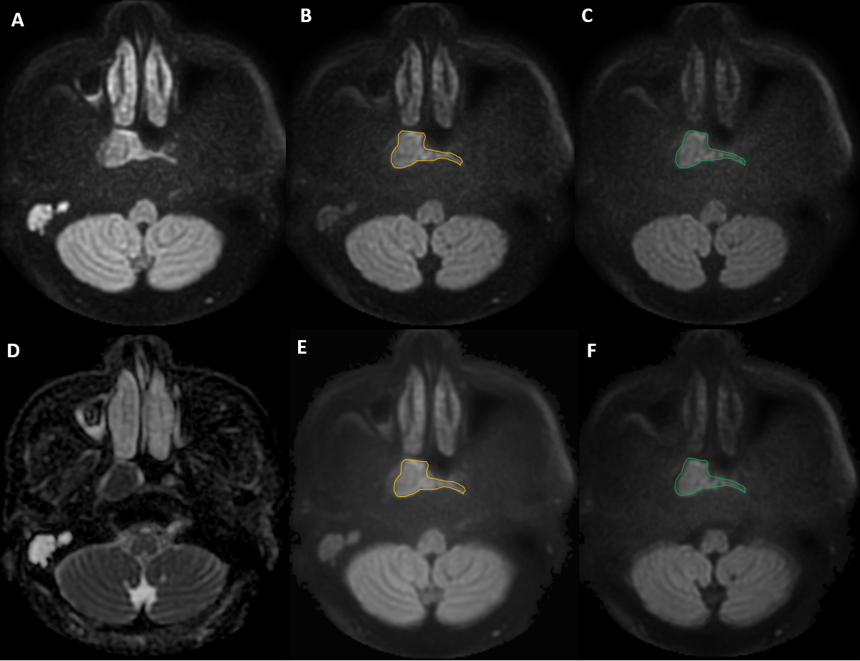
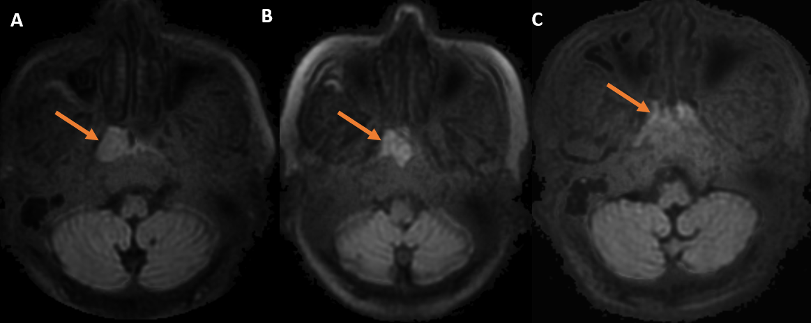
This research has been approved by the local IRB. The parameters for DWI TSE MVXD as following: FOV, 160x160 mm2; voxel size, 1.8 x1.8 mm2; slice thickness, 4mm; slice, 24; TR, 2500ms, TE, 120ms; acceleration factor, 2.0; MultiVane, 170%; for the b value = 500s/mm2, b value= 1000s/mm2 and b value = 1500s/mm2, the average is 2, 3 and 4 respectively. Fig 2 shows that the SNR of cDWI images is similar with actual scanned DWI even when the b value is 2000s/mm2. The cDWI images has similar sharpness also. Three patients with NPC which were confirmed by pathological biopsy, the data was collected, then high b value images were generated using IMAge/engine retrospectively.Compared with actual scanned diffusion images, Fig. 3 showed the computed DWI images (b = 1000 s/m2, b=1500 s/m2) have similar contrast and area of NPC, the area where was marked by brown freeform polygon for b = 1000s/m2, green freeform polygon for b = 1500s/mm2, the area of NPC was consistent with actual scanned images with same b values. Figure 4 shows higher b value images (b = 2000s/mm2) which was generated by cDWI, the actual DWI with same b value was not scanned. It shows the high b value images (b = 2000s/mm2) for all three patients have good SNR and image quality.
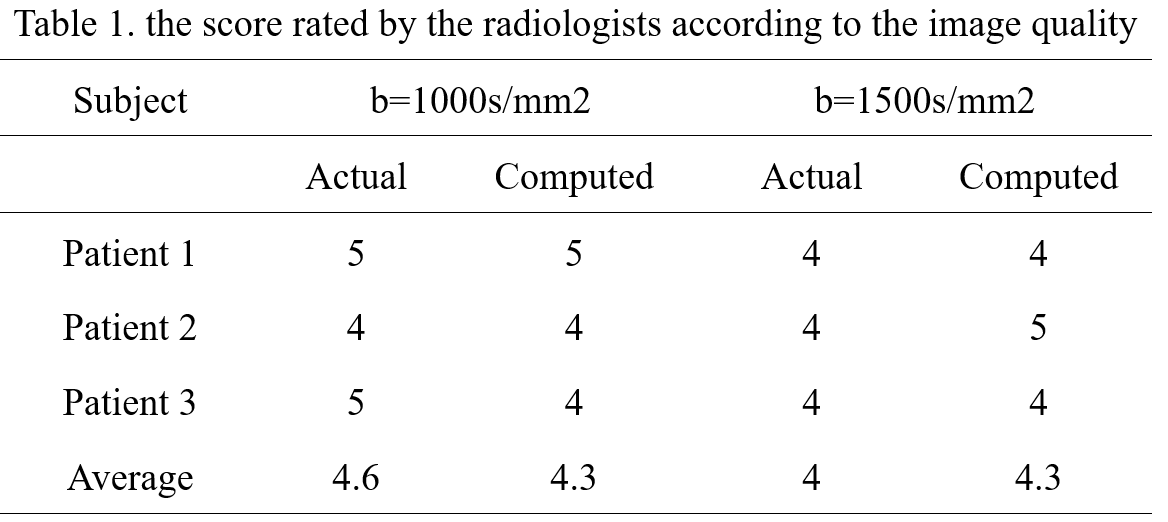
The scores by the two observers were in good agreement for computed DWI images and actual scanned DWI images both for b=1000s/mm2 and b=1500s/mm2. The results in Table 1 showed that the scores for images by cDWI is almost same with the scores by actual scanning. Due to the actual scanned low b value (b = 500s/mm2) scanning has much less average than the high b value scanning, it showed that the cDWI could generate high b value images with less scan time.
Discussion and conclusions
The preliminary study shows robust high b value NPC images with combination of DWI TSE MVXD and cDWI, the computed high b value could have similar image quality in contrast, sharpness, and resolution with actual scanned DWI, and it has less scan time than the actual scan but similar SNR. Considering the image quality of cDWI, the relatively short scan time, and the straightforward implementation, this technique holds the potential for wide clinical applications. This strategy could enhance the applicability of NPC diagnosis.Acknowledgements
No acknowledgement.References
1. Li H, Liu XW, 1,2, Geng ZJ, Wang DL and Xie CM, Diffusion-weighted imaging to differentiate metastatic from non-metastatic retropharyngeal lymph nodes in nasopharyngeal carcinoma, Dentomaxillofacial Radiology (2015) 44, 20140126.
2. Song CR,Cheng P, Cheng JL, et. al., Differential diagnosis of nasopharyngeal carcinoma and nasopharyngeal lymphoma based on DCE-MRI and RESOLVE-DWI, Eur Radiol, 2020;30(1):110-118.
3. Deng J, Omary RA, Larson AC, Multishot Diffusion-Weighted SPLICE PROPELLER MRI of the Abdomen, MRM 2008;59:947–953.
4. Matthew DB, Martin OL, David JC, Dow MK, Computed Diffusion-weighted MR Imaging May Improve Tumor Detection, Radiology 2011;261: 573-581.
5. Toru K, Yuko K, Fuminari K, et. al., Introduction to the Technical Aspects of Computed Diffusion weighted Imaging for Radiologists, RadioGraphics 2018; 38:1131–1144
6. Yoshiko RU, Tsutomu T,Satoru T, et. al., Computed Diffusion-Weighted Imaging in Prostate Cancer: Basics, Advantages, Cautions, and Future Prospects,KJR, 2018;19(5):832-837.
7. Ming Y, Yan YP, Wang H, IMAge-enGINE: a freely available software for rapid computation of high-dimensional quantification, Quant Imaging Med Surg 2018;1-9.
Figures