3772
Plain magnetic resonance imaging by black blood sequences to assess vascular invasion of complex retroperitoneal occupying1Southeast University, Nanjing, China
Synopsis
This study aimed to investigate the value of plain conventional and black-blood magnetic resonance imaging (MRI) for assessing the invasion of large and important vessels in patients with complex retroperitoneal tumors. The great advantages of vascular wall imaging are non-invasive and non-enhanced, as well as three-dementional, which provides a better presence and evaluation of wall structure, tumor contact of the vessel circumference, narrowing, and deformity. The researchers in the field of vascular imaging should be interested in this topic.
INTRODUCTION
Retroperitoneal space occupying lesion represent a heterogeneous group of rare with an overall incidence of 0.5–1/100 000, which mainly originated from retroperitoneal parenchymal organs, fat, muscle, nerve, lymphatic, and about 70%-80% are malignant tumors1. Since the location of the retroperitoneal is deep and concealed, the vascular invasion and organ metastasis have occurred at the initial diagnosis in 80% patients. In case of large artery invasion, surgical resection cannot be performed. The Da Vinci surgical robot assistance has been rapidly adopted by urological surgeons and has become particularly popular for oncological procedures involving the retroperitoneal space2. Although surgical resection can be performed without vascular invasion or venous and small artery invasion, the number and degree of vascular invasion will affect the operation method and difficulty. Therefore, precise evaluation severity of blood vessel invasion is an important reference standard for formulating treatment plans. Previous studies have shown that magnetic resonance black blood technology can display arterial wall lesions with high resolution, so it is expected to assess the extent of retroperitoneal vascular invasion3-4.METHODS
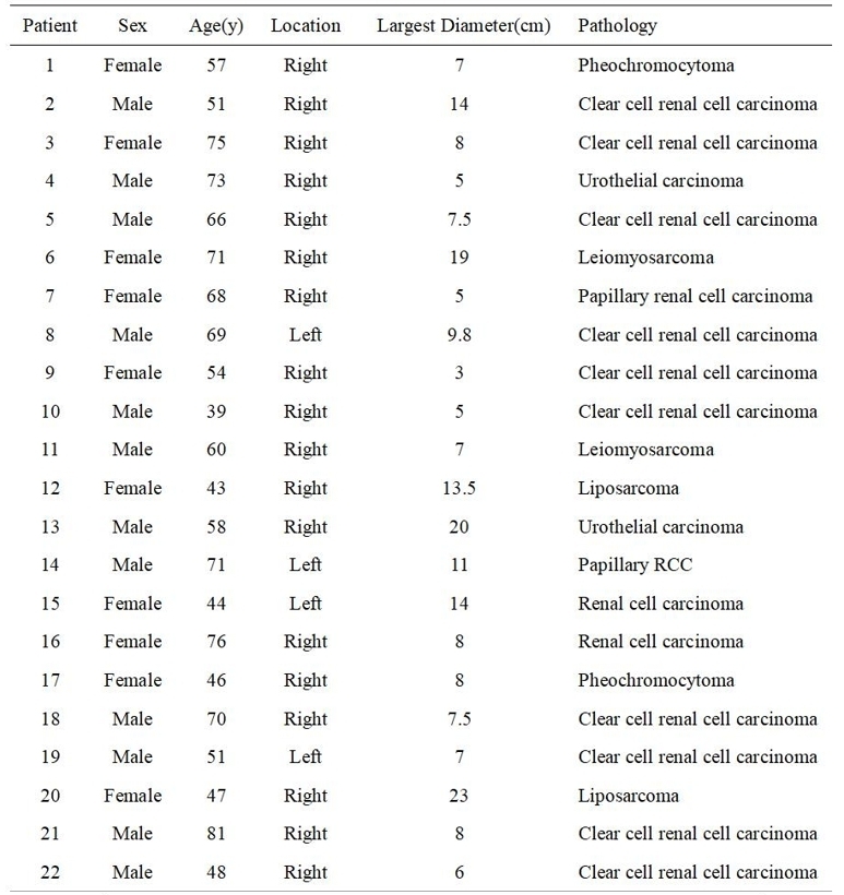
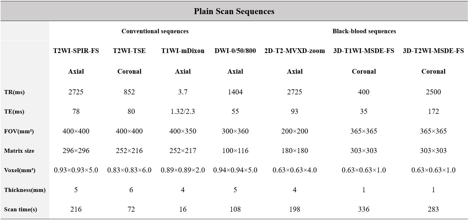
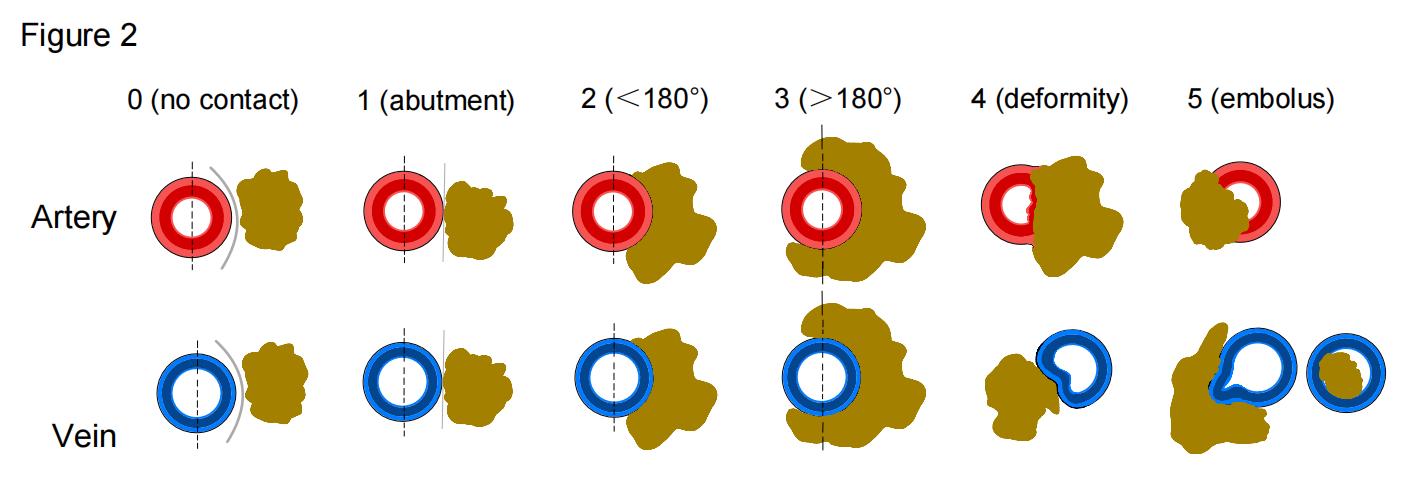
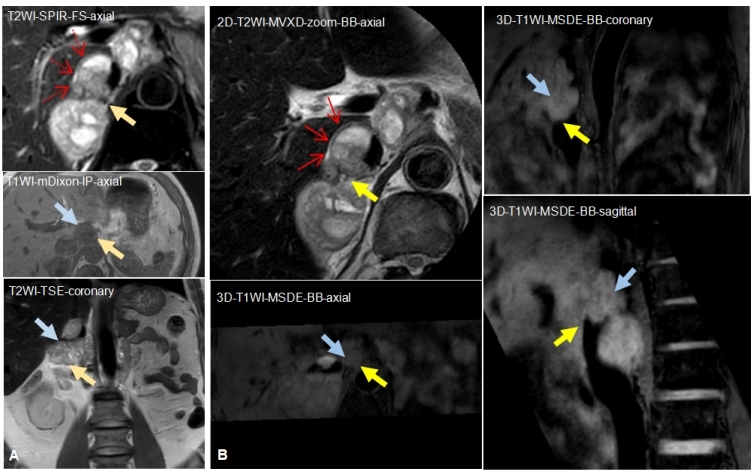
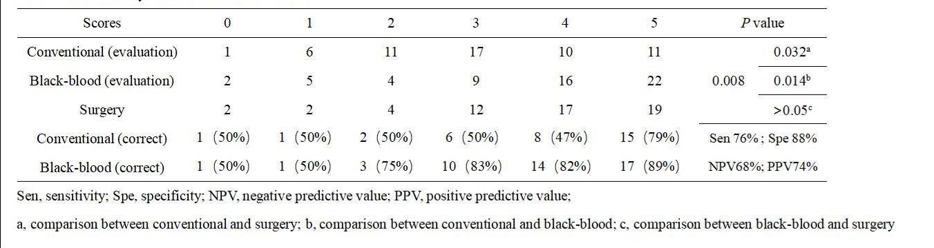
22 patients were prospectively collected who suspected of complicated retroperitoneal space occupying on computer tomography from May 2020 to November 2021 (Figure 1). All patients underwent 3.0 T magnetic resonance scan (Philips Ingenia II 3.0 T superconducting MR image, Netherlands), including conventional sequence (T2WI-SPIR-FS/ T2WI-TSE/ T1WI-mDixon/ DWI-0/50/800) and black blood sequence (2D-T2-MVXD-zoom/ 3D-T1WI-MSDE-FS/ 3D-T2WI-MSDE-FS)(Figure 2). Two experienced radiologists jointly evaluated the degree of vascular invasion in all images of abdominal aorta, abdominal trunk, superior mesenteric artery / vein, inferior vena cava, portal vein and renal artery and vein, respectively. As shown in the figure 3-4, according to the positional relationship and the degree of contact between the tumor and the surrounding arteries or veins, it is divided into no contact (0 points), edge contact (1 points), less than or equal to 180° tumor contact of the blood vessel circumference (2 points), more than 180° tumor contact of the vein circumference (3 points), vascular compression or deformation (4 points), intravascular tumor thrombus (5 points). All patients received surgical treatment after the MRI scan. Based on both operative notes and pathology records where relevant, the Kruskal-Wallis H test was used to compare the conventional scan sequence and black blood in SPSS for Windows (v.11.0, 2001; Chicago, IL). The accuracy of the sequence for the assessment of the degree of vascular invasion.RESULTS
The evaluation results of vessels through the three methods were different (H=10.793, P < 0.001), but there was no difference in evaluation consistency between the black-blood sequences and surgery findings(P > 0.05, sensitivity, 76%; specificity, 88%; NPV, 68%; PPV, 74%), compared with that between conventional and surgery (P = 0.026). In the analysis of all the 56 vessels which were recorded because of the closely association with masses and probably impact on the clinical treatment, the consistency of the two sequences was similar in evaluating the absence of contact and the presence of cancer embolism on the vascular wall, whether in the degree of vascular wall contact and invasion, the predicted results of black blood sequence were more consistent with the actual surgical situation (83%, 82%, 89%). About 33% of the vessels were over-evaluated in the conventional sequences, and some minor infiltrating vessels were ignored in both sequences (Figure 5).DISCUSSION
Our research shows that compared with the traditional scanning sequence, the black blood sequence has better performance in the grading diagnosis of vascular invasion. This is because magnetic resonance black blood sequence can realize multiplanar imaging of vascular wall and cavity, and clearly shows the relationship between the blood vessel and the surrounding adipose tissue4-5. Based on these, an operational evaluation method for converting qualitative data from vascular wall evaluation into quantitative data, which allows for a more precise and refined assessment of the relationship between vessels and tumors. Despite in terms of abdominal vessel wall imaging, there are still many challenges, such as the causes of imaging artifacts: breathing movement, gastrointestinal peristalsis, and blood vessel pulsation were difficult to avoid, there are still unexplored areas in vascular wall imaging, it shows a good prospect in indicating tumor resectability, guiding clinical treatment and further influencing prognosis6-7.CONCLUSION
Black-blood 3T-MRI had a good performance in evaluating the invasion of both arteries and veins in patients with retroperitoneal space occupying lesions. These techniques could be helpful for the elaboration of therapeutic protocols and surgical resection treatment plan.Acknowledgements
No acknowledge found.References
(1) Sassa N. Int J Urol. 2020; 27: 1058-1070.
(2) Bourgioti C., et al, Radiology. 2021; 298: 403-412.
(3) Al-Hawary MM., et al, Gastroenterology. 2014; 146: 291-304.
(4) Treitl KM., et al, Eur Radiol. 2016; 27: 1-10.
(5) Zhao D L., et al, J Clin Neurosci. 2015; 22: 700-704.
(6) Roes SD., et al, Magn Reson Med. 2009; 61: 35-44.
(7) Zhu C., et al, Magn Reson Imaging. 2016; 34: 18-25.
Figures