3770
Preoperatively Predict Microvascular Invasion in Hepatocellular Carcinoma - via Multi-region Radiomic Analysis in Multi-sequence MRI1The School of Medical Technology and Engineering, Fujian Medical University, Fuzhou, China, 2Department of Radiology, the First Affiliated Hospital of Fujian Medical University, Fuzhou, China
Synopsis
Microvascular invasion (MVI) affects postoperative prognosis in hepatocellular carcinoma (HCC) patients. Radiomics has shown great potential in providing valuable information for tumor pathophysiology. We aim to use multi-sequence MRI radiomics focused on not only intratumoral and peritumoral areas, but also the HCC-liver interface to predictive MVI status preoperatively. The radiomics and clinico-radiological indicator-based fusion model can well predict MVI status in HCC patients.
Introduction
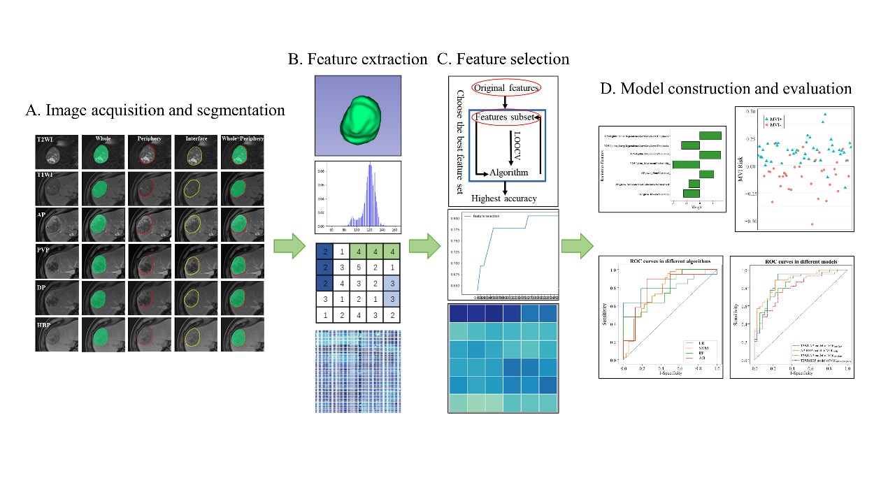
While hepatectomy and liver transplantation are potentially curative treatments for HCC, recurrence is still common. MVI affects postoperative prognosis in HCC patients, but there is a lack of reliable and effective tools for preoperative prediction. Radiomics refers to the automated quantification of the radiological phenotype using data-characterization algorithms. Radiomics strategies have shown great predictive potential by incorporating radiological features related to various diseases and clinical and/or pathological factors into a single fusion model.Methods
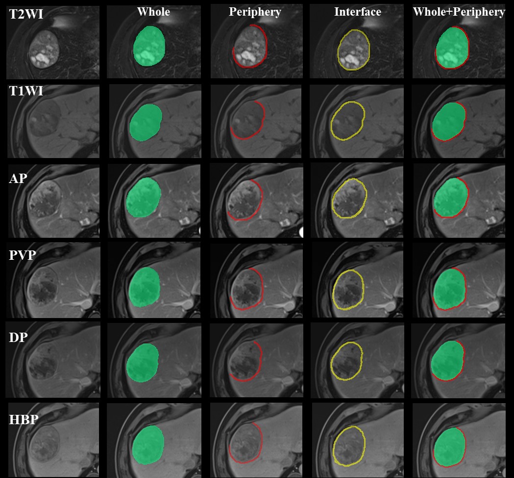
One hundred and fifteen patients (training set: n = 80; validation set: n = 35) with pathologically confirmed HCC who underwent preoperative magnetic resonance imaging (MRI) were retrospectively recruited. Radiomics features were extracted based on multi-sequence MRI and various regions (including intratumoral and/or peritumoral areas) using Pyradiomics and recursive feature elimination (RFE) method was performed for feature selection. Radiomics models were built using four classification algorithms (Logistic Regression, Support Vector machine, Random forest and AdaBoost) or combined with a clinico-radiological model to generate a fusion model by multivariable logistic regression. Model evaluation were performed with ROC curve and integrated discrimination improvement (IDI) to find out the most predictive model.Results
Among radiomics models, the model based on T2WI and arterial phase (T2WI-AP model) in the volume of the liver-HCC interface (VOIinterface) exhibited the best predictive power, with an area under the receiver operating characteristic curve (AUC) of 0.866 (95% CI: 0.783-0.947) in the training group and 0.855 (95%CI: 0.731-0.963) in the validation group. The fusion model that incorporated T2WI-AP radiomics model in VOIinterface and non-smooth tumor margin showed an excellent prediction efficacy in training group with AUC = 0.915 (95%CI: 0.853-0.976) and in validation group with AUC = 0.868 (95%CI: 0.749-0.988), outperforming clinico-radiological model and T2WI-AP radiomics model in the training and validation set (IDIs > 0).Discussion
The fusion model based on the combination of T2WI-AP radiomics signature in the VOIinterface and one radiological variable (tumor margin) demonstrated an excellent prediction efficacy. Despite AUCs not being significantly different from the T2WI-AP radiomics model in the VOIinterface, integrated discrimination improvement (IDI) showed significant improvement in the predictive value of the fusion model.Conclusion
The proposed multi-region multi-sequence Radiomics models are effective and non-invasive tools to preoperatively identify MVI status. The fusion model achieves desirable prediction of the individualized risk estimation of MVI in HCC patients, and may be beneficial to make precise decisions for personalized medicine.Acknowledgements
No acknowledgement found.References
1.Lee S, Kang TW, Song KD, et al. Effect of Microvascular Invasion Risk on Early Recurrence of Hepatocellular Carcinoma After Surgery and Radiofrequency Ablation. Ann Surg 2021;273(3):564-571.
2.Chan AWH, Zhong J, Berhane S, et al. Development of pre and post-operative models to predict early recurrence of hepatocellular carcinoma after surgical resection. J Hepatol 2018;69(6):1284-1293.
3.Tomaszewski MR, Gillies RJ. The Biological Meaning of Radiomic Features. Radiology 2021;298(3):505-516.
Figures