3752
Application of a modified tri-exponential DWI in detecting the change of glymphatic system: a preliminary exploration in gliomas1Department of Radiology, Department of Radiology, Renji Hospital, School of Medicine, Shanghai Jiao Tong University, Shangha, shanghai, China, 2Department of Radiology, Renji Hospital, School of Medicine, Shanghai Jiao Tong University, Shangha, shanghai, China, 3MR Collaboration, Central Research Institute, United Imaging Healthcare, Shanghai, China, shanghai, China
Synopsis
Spectral analysis using non-negative least squares in a modified tri-exponential muti-b-value diffusion-weighted imaging(DWI) enables estimation of an intermediate component in the diffusion spectrum between parenchymal diffusion and microvascular pseudo-diffusion. Several studies have suggested the intermediate component to be with the glymphatic alterations in different disease state. The aim of this this study aims to investigate whether the modified tri-exponential model are effective in evaluating the grading and IDH-1 gene type of gliomas.
Introduction
Intra voxel incoherent motion (IVIM) has the ability to separate parenchymal diffusion from microvascular pseudo-diffusion1. However, the bi-exponential model has been challenged for its lack of reproducibility and has been considered as oversimplified. Recently a spectral analysis method using the non-negative least squares (NNLS) as a modified tri-exponential muti-b-value DWI has been introduced to identify an intermediate component between the two classical components2-4. So far, some positive exploration of the source of the intermediate component have been made. This intermediate component is argued to represent interstitial fluid with the parenchyma and thus could investigate glymphatic alterations in small vessel disease5. Merel et al also get similar conclusions in Alzheimer’s disease2. Several previous studies have indicated the existence of water molecular pool with extremely low diffusion in tissues and may be related to expression of aquaporin-4 (AQP4) in the membrane of astrocytic6, 7.The glymphatic system has been recently recognized as a pathway for waste clearance and maintaining fluid balance in the brain parenchymal interstitium8. Cerebrospinal fluid (CSF) from the subarachnoid space flows into the brain parenchyma through periarterial spaces of the penetrating arteries and, under the influence of aquaporin-4 (AQP4) water channels, mixes with parenchymal interstitial fluid8. Recent studies have shown that the glymphatic system may play an essential role in gliomas5. The aim of this study was to investigate whether the modified tri-exponential model could provide more information about the malignant degree and progression of gliomas.
method
Subjects: A total of 60 patients were enrolled, including 19 grade II gliomas, 9 grade III gliomas and 32 grade IV gliomas. Totally 26 patients show IDH-1 mutation.MRI acquisition: All subjects underwent a 3.0-T MRI imager (LianYing uMR780). For DWI, we performed a single shot echo-planar sequence in the axial plane with the following parameters:
repetition time (TR) ms/echo time (TE) ms, 3000/106; section thickness, 5 mm; intersection gap, 1 mm; number of sections, 15; b-values, 0, 20, 50, 80, 150, 200, 300, 500, 800, 1000, 1500, 2000,2500, 3000, 3500, 4000, 4500 s/mm2;
Image analysis: For the model fitting, an iterative Levenberg Marquardt algorithm was employed for the non-linear least squares fitting using MATLAB 2021a (MathWorks, Natick, MA, USA). Then five parameters comprising F0, Df, Ds, Ff and Fs were calculated voxel-by-voxel with DWI of all the b values based on the predefined mathematical expression of the modified tri-exponential IVIM model.
$$S/S0=F0+Fs*exp(-ADCs*b)+Ff*exp(-ADCf*b), F0+Fs+Ff=1$$
The ROI was put in the enhancing region of the tumor on contrast-enhanced T1-weighted images. For the non-enhancing tumors, ROI was drawn at the center of the portion where the high T2-signal area and avoiding necrosis, cyst, hemorrhage areas. Mean value of each parameters (ADCfast, ADCslow, Fs, Ff and F0) were obtained and the differences in group characteristics were assessed using one-way analyses of variance. The diagnostic performance was evaluated by Receiver operating characteristic (ROC) curves.
result
The statistical result of the ADCfast, ADCslow, Fs, Ff and F0 in different groups of gliomas were summarized in Table 1. And the result of distinguish IDH-1 gene type (for all group) were showed in Table 2. Manifestations of LGG(low grade gliomas), HGG(high grade gliomas) in parametric maps generated were shown in Figure 1 and Figure 2. Figure 3 shows ROC curves and corresponding AUCs of each parameter for differentiation between the grade and IDH-1 gene type. All the five parameters showed significant in different among grade II, III and IV gliomas. The F0 and Fs showed significant difference between IDH-1 wide and mutation groups.discussion
In this study, we developed a modified tri-exponential muti-b-value DWI to detect the diffusion of transcellular, microvascular and interstitial fluid of gliomas. Five parameters (ADCfast, ADCslow, Fs, Ff and F0) were found to be effective in grading gliomas (all P < 0.05). The Ff and Fs showed significant difference between IDH-1 wide and mutation groups. The ADCfast were higher in high grade gliomas, and the ADCslow were lower in high grade gliomas. High-grade gliomas are often characterized by higher cellularity and thus lead to the restriction of water diffusion. And the exuberant vascularization often brought a high pseudo-diffusion. The Fs account for the largest proportion in grade IV gliomas and the F0 account for the largest proportion in grade II glioma, F0 account for the least in all grade and increase as the malignant degree increased, which may reflect the glymphatic alterations in higher gliomas.conclusion
The modified tri-exponential DWI and intermediate component may help to provide underling information about the malignant degree and progression of gliomas.Acknowledgements
This work was supported by grants from the National Natural Science Foundation of China (grants 81701642).References
1. Koh DM, Collins DJ. Diffusion-weighted MRI in the body: applications and challenges in oncology. AJR American journal of roentgenology 2007; 188(6): 1622-35.
2. van der Thiel MM, Freeze WM, Verheggen ICM, et al. Associations of increased interstitial fluid with vascular and neurodegenerative abnormalities in a memory clinic sample. Neurobiology of aging 2021; 106: 257-67.
3. Zeng Q, Shi F, Zhang J, Ling C, Dong F, Jiang B. A Modified Tri-Exponential Model for Multi-b-value Diffusion-Weighted Imaging: A Method to Detect the Strictly Diffusion-Limited Compartment in Brain. Frontiers in neuroscience 2018; 12: 102.
4. Keil VC, Madler B, Gielen GH, et al. Intravoxel incoherent motion MRI in the brain: Impact of the fitting model on perfusion fraction and lesion differentiability. Journal of magnetic resonance imaging : JMRI 2017; 46(4): 1187-99.
5. Toh CH, Siow TY. Factors Associated With Dysfunction of Glymphatic System in Patients With Glioma. Frontiers in oncology 2021; 11: 744318.
6. Xueying L, Zhongping Z, Zhoushe Z, et al. Investigation of Apparent Diffusion Coefficient from Ultra-high b-Values in Parkinson's Disease. Eur Radiol 2015; 25(9): 2593-600.
7. Yao X, Derugin N, Manley GT, Verkman AS. Reduced brain edema and infarct volume in aquaporin-4 deficient mice after transient focal cerebral ischemia. Neuroscience letters 2015; 584: 368-72.
8. Rasmussen MK, Mestre H, Nedergaard M. The glymphatic pathway in neurological disorders. The Lancet Neurology 2018; 17(11): 1016-24.
Figures
Table 1 :Mean values (mean ± SD) of F0, Fs, Ff, ADCslow, ADCfast in grade II-IV gliomas, results with P values less than 0.05 were considered to indicate a significant difference
Table 2: Mean values(mean ± SD) of F0, Fs, Ff, ADCslow, ADCfast in IDH-1 mutant and wild gliomas including grade II-IV grade, results with P values less than 0.05 were considered to indicate a significant difference
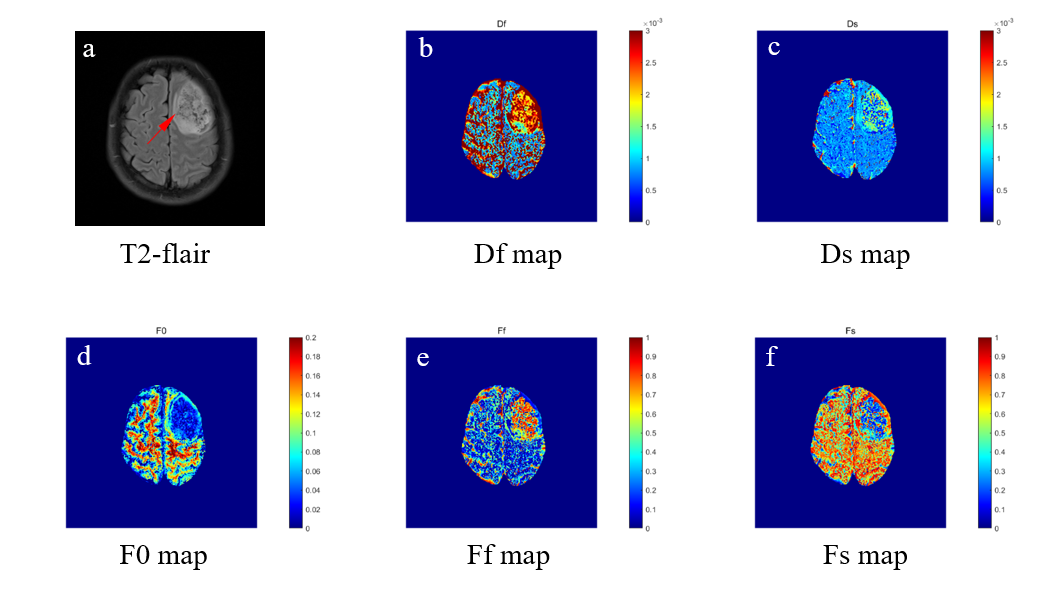
Figure 1
Illustrations of a 32-year-old female with astrocytoma (WHO grade II) The contrast-enhanced T2-flair maps, Df(ADCfast), Ds(ADCslow), F0, Ff and Fs maps are shown in first to sixth rows, respectively.
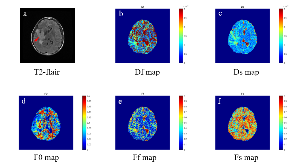
Figure 2
Illustrations of a 58-year-old male with glioblastoma (WHO grade IV) The contrast-enhanced T2-flair maps, Df(ADCfast), Ds(ADCslow), F0, Ff and Fs maps are shown in first to sixth rows, respectively.
Figure 3
Receiver operating characteristic curves of the mean F0, Fs, Ff, ADCslow, ADCfast values in distinguishing high-grade gliomas from low-grade gliomas (A), and distinguishing the IDH-1 gene type(B).