3750
Increased Cerebral Blood Flow in Recovered COVID-19 Patients Assessed by Arterial Spin Labeling MRI
Yan Bai1, Yaping Wu1, Wei Wei1, Xianchang Zhang2, and Meiyun Wang1
1Department of Medical Imaging, Henan Provincial People's Hospital, Zhengzhou, China, 2MR Collaboration, Siemens Healthineers Ltd., Beijing, China
1Department of Medical Imaging, Henan Provincial People's Hospital, Zhengzhou, China, 2MR Collaboration, Siemens Healthineers Ltd., Beijing, China
Synopsis
Increased global and regional cerebral blood flow were observed in recovered corona virus disease 2019 patients, which may reflect the brain’s compensatory responses following corona virus disease 2019.
INTRODUCTION
The severe acute respiratory syndrome coronavirus 2 has caused an ongoing global pandemic of corona virus disease 2019 (COVID-19). Although the COVID-19 has been shown to affect the brain (1), cerebral hemodynamics has not been characterized in patients with COVID-19. A multi-delay pseudo-continuous arterial spin labeling (pCASL) MRI provides measurements of cerebral blood flow (CBF) and arterial transit time (ATT) by using magnetically labeled arterial blood water as an endogenous tracer (2). The objective of the present study was to use a multi-delay pCASL MRI to investigate the cerebral hemodynamics of recovered COVID-19 patients.METHODS
Eight patients recovered from COVID-19 and 8 age and gender matched healthy controls underwent MRI on a 3-T Siemens Prisma scanner (Siemens, Erlangen, Germany) using a 64-channel head coil. A multi-delay pCASL pulse sequence with background suppressed 3D gradient and spin echo readout was applied with the following parameters: TR/TE/label time = 4100/36/1500ms; field of view = 24 cm; matrix size = 96×96, 40×3 mm slices, resolution = 2.5×2.5×3 mm, five post-labeling delay = 500/1000/1500/2000/2500 ms with 2/2/2/3/3 measurements respectively, label offset = 90 mm, two inversion pulses applied during the post-labeling delay for background suppression. Global mean CBF and ATT values were generated using a whole brain mask, and compared between the COVID-19 and control subjects by means of two-sample two-sided t-tests. In addition, the analysis was performed after deriving parameters from regions of interest defined by automated anatomical parcellation.RESULTS
CBF was elevated in the COVID-19 subject compared to the control subject, whereas ATT maps were comparable. The whole brain global mean CBF was significantly increased in COVID-19 patients than controls (p = 0.030, Figure 1), whereas the global mean ATT values were not significantly different between the two groups (p > 0.05). Accounting for global CBF differences, the relative CBF increases were mainly observed in the olfactory, sensorimotor and default mode networks including olfactory cortex, anterior and middle cingulate, bilateral insula, frontal gyri and posterior parietal cortex, as well as left superior temporal cortex and Rolandic operculum.DISCUSSION and CONCLUSION
PCASL MRI revealed increased global mean CBF and regional CBF. The increased CBF in olfactory cortex, insula and sensorimotor areas may be attributed to the recovery of olfactory and taste function in the patients. In addition, CBF was increased in the default mode network-related areas such as cingulum, frontal gyri, temporal and parietal cortices in the recovered COVID-19 patients. The recovered state from COVID-19 may contribute to increased CBF in default mode network. In summary, global mean and regional CBF were increased in recovered COVID-19 patients, which may reflect the brain’s compensatory responses following COVID-19.Acknowledgements
This work was supported by the Zhengzhou Collaborative Innovation Major Project (20XTZX05015).
References
1. Mao L, Jin H, Wang M, et al. Neurologic manifestations of hospitalized patients withcoronavirus disease 2019 in Wuhan, China. JAMA Neurol, 2020;77(6):683-690.
2. Wang R, Yu S, Alger JR, et al. Multi-delay arterial spin labeling perfusion MRI in moyamoya disease--comparison with CT perfusion imaging. Eur Radiol, 2014;24(5):1135-1144.
Figures
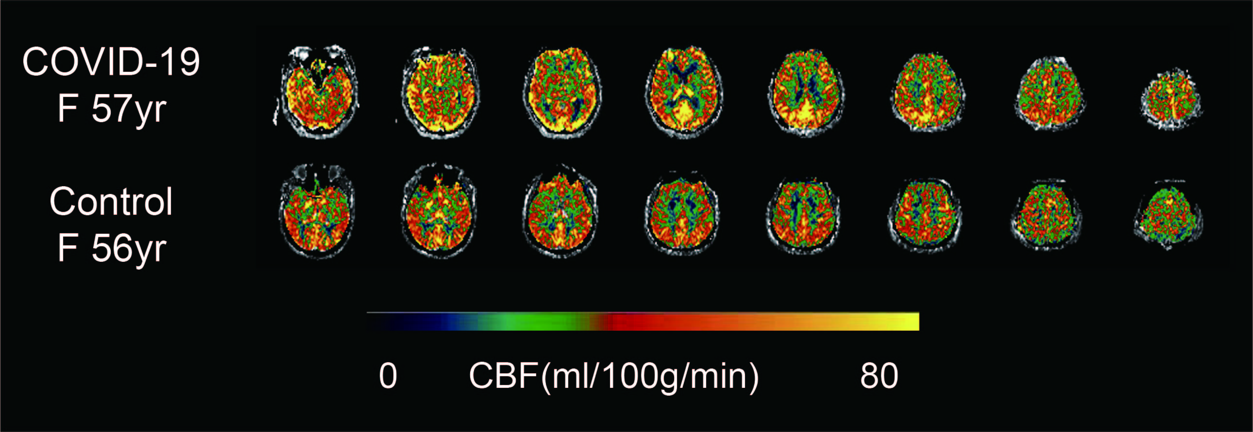
Figure 1. Cerebral blood flow maps of a representative corona virus disease 2019 (COVID-19) and a control subject.
DOI: https://doi.org/10.58530/2022/3750