3736
MRI-derived radiomics to guide initial prostate biopsy for initial biopsy patients1Department of radiology, West China Hospital, Chengdu, China, 2Philips Healthcare, Guangzhou, China
Synopsis
In this study, we applied the radiomics features of both suspected lesion and prostate gland on bi-parametric MRI (bpMRI) (T2W and ADC) to predict whether targeted biopsy alone is enough in detecting clinically significant prostate cancer (csPCa) in biopsy naïve men and compare their value with PI-RADS category.
Background
Multiparametric magnetic resonance imaging (mpMRI) with or without MRI-targeted biopsy has recently been a popularly alternative test to traditional transrectal ultrasonography-guided systematic biopsy in men with elevated prostatic specific antigen (PSA) levels and/or abnormal digital rectal examinations. It has been shown that MRI targeted biopsy performs better with greater PI-RADS score than systematic biopsy[1, 2]. Radiomics is a promising approach that permits high-throughput extraction of texture and image features from radiographic images to characterize the underlying tumor microarchitecture and heterogeneity which is subtle and not immediately visible on routine imaging[3]. In this study, we applied the radiomics features of both suspected lesion and prostate gland on bi-parametric MRI (bpMRI) (T2W and ADC) to predict whether targeted biopsy alone is enough in detecting clinically significant prostate cancer (csPCa) in biopsy naïve men and compare their value with PI-RADS category.Materials and Methods
This retrospective study included patients who performed combined systematic biopsy and cognitive MRI-targeted biopsy between May 2018 and February 2021 at department of radiology, West China Hospital . All patients underwent a pre-biopsy mpMRI for identifying the suspicious lesion and guiding targeted biopsy. There were finally 255 patients enrolled in this study, and they were randomly assigned to training set and testing set at a 8:2 ratio. All MRI examinations were acquired on 3.0-T MRIs (Magnetom Skyra, Siemens, and Achieva TX, Philips Healthcare) with phased-array body surface coil. Two radiologists, with 3 and 5 years of experience in abdominal imaging respectively, read all prostate mpMRI images separately, and suspicious lesions were assigned a Prostate Imaging Reporting and Data System Version 2.1 (PI-RADS v2.1) score of 1 to 5. The lesion with the highest PI-RADS score on mpMRI was defined as the index lesion. 3D segmentation was performed manually using the ITK- SNAP software. All patients underwent transperineal cognitive MRI-guided targeted biopsy and additional systematic biopsy. All specimens were individually labeled and analyzed by two pathologists. The pathological evaluation included the numbers of positive cores, Gleason score (GS) and grade group (GG) according to the 2014 International Society of Urologic Pathology (ISUP) criteria (19). A GG ≥2 (GS ≥ 3 + 4) was defined as csPCa. The radiomics features were extracted using Pyradiomics on Python (3.7.6). In total, 1316 radiomics features were extracted from each VOI. Redundant features were removed by One-way analysis of variance (ANOVA). Then, the least absolute shrinkage and selection operator (LASSO) regression method was applied to select the most distinguishable features. Finally, the logistic regression method would be used to build the prediction model.Results
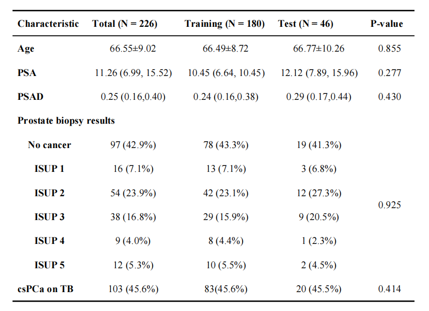
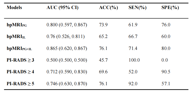
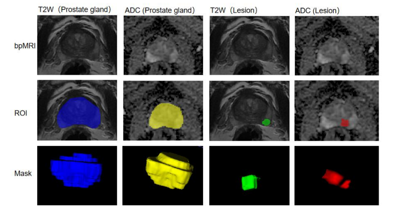
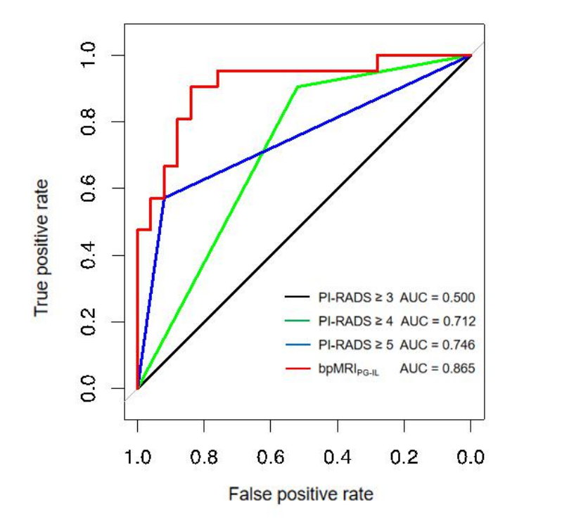
The clinical characteristics of the patients are summarized in Table 1. No significant difference was found between the training and test cohorts with respect to age, serum prostate specific antigen (PSA) level, PSA density (PSAD), grade group on prostate biopsy and csPCa detection on targeted biopsy (TB). Among the 226 patients underwent the combination of targeted and systematic biopsy, 113 (50%) were diagnosed as csPCa. Targeted biopsy alone detected 103 (45.6%) csPCa, with 83 (83/182, 45.6%) patients in the training cohort and 20 (20/44, 45.5%) patients in the test cohort. Targeted biopsy alone would miss 10 csPCa (8 with GS 3+4 and 2 with GS 4+3), with 6 patients in the training cohort and 4 patients in the test cohort. Figure 1 shows the prostate bpMRI and four different ROIs masking. In the training cohort, among the 1316 extracted radiomics features from the whole prostate gland (PG) and index lesion (IL) respectively, 166 and 178 stable features were selected for the subsequent analysis, according to the Kendall correlation coefficient assessment. In the LASSO regression analysis, 11 key PG features and 18 key IL features with nonzero coefficients were identified to most associated with csPCa risk and then used to construct the bpMRIPG and bpMRIIL models. Besides, 18 key PG features in combination with 6 key IL features were identified to construct bpMRIPG+IL model. The AUCs of bpMRIPG model, bpMRIIL model, and bpMRIPG+IL model were 0.800 (95% CI: 0.597 ~ 0.867) , 0.76 (95% CI: 0.526 ~ 0.811) , and 0.87 (95% CI: 0.620 ~ 0.867) respectively in the testing cohort (Table 2). The AUCs of PI-RADS ≥ 3, PI-RADS ≥ 4 and PI-RADS ≥ 5 model were 0.50 (95% CI: 0.500 ~ 0.500) , 0.71 (95% CI: 0.590 ~ 0.830) , and 0.76 (95% CI: 0.630 ~ 0.870) in the testing cohort (Table 2) . Figure 2 shows the ROC curves for bpMRIPG+IL radiomics model and PI-RADS assessment in the testing cohort.Conclusion
The bpMRIPG+IL radiomics model demonstrated superior predictive performance compared with PI-RADS scores in predicting whether targeted biopsy alone would detect csPCa. Radiomics demonstrated the potential to help reducing unnecessary systematic biopsy and making individualized prostate biopsy plan for biopsy naïve patients.Acknowledgements
No acknowledgement found.References
[1] Neale A, Stroman L, Kum F, et al. Targeted and systematic cognitive freehand-guided transperineal biopsy: is there still a role for systematic biopsy? BJU Int 2020;126(2):280-85. doi: 10.1111/bju.15092 [published Online First: 2020/04/23]
[2] Lee AYM, Yang XY, Lee HJ, et al. Multiparametric MRI-ultrasonography software fusion prostate biopsy: initial results using a stereotactic robotic-assisted transperineal prostate biopsy platform comparing systematic vs targeted biopsy. BJU Int 2020;126(5):568-76. doi: 10.1111/bju.15118 [published Online First: 2020/05/22]
[3] Lambin P, Leijenaar RTH, Deist TM, et al. Radiomics: the bridge between medical imaging and personalized medicine. Nat Rev Clin Oncol 2017;14(12):749-62. doi: 10.1038/nrclinonc.2017.141 [published Online First: 2017/10/05]
Figures