3723
The value of IVIM and mDIXON-Quant MR imaging in the identification of pediatric IgAN and HSPN1Radiology, Beijing Children's Hospital, Capital Medical University, National Center for Children's Health, Beijing, China, 2Beijing Children's Hospital, Capital Medical University, National Center for Children's Health, Beijing, China, 3Philips Healthcare, Beijing, China
Synopsis
It is difficult to identify IgA nephropathy (IgAN) and Henoch Schonlein purpura nephritis (HSPN) due to their similar clinical features. Functional magnetic resonance imaging (fMRI) can simultaneously obtain tissue structure and function imaging, and evaluate the functional state of kidney. In this study, intravoxel incoherent motion (IVIM) and mDIXON-Quant were performed in pediatric patients with nephropathy, and it was found that IgAN and HSPN were remarkably different in renal function. The values of medulla’s D and cortex’s R2* obtained by IVIM and mDIXON-Quant had the ability in differential diagnosis of IgAN and HSPN.
Introduction
Pediatric nephropathies are common chronic abdominal diseases in children, which are often repeated and prolonged. Improper treatment often progresses to chronic kidney diseases, and the prognoses are poor1-2. Pediatric nephropathy mainly includes IgA nephropathy (IgAN) and Henoch Schonlein purpura nephritis (HSPN). Although the early symptoms of the two are different, the clinical characteristics and laboratory results in the development of the disease are similar, and the diagnosis can only be confirmed by renal biopsy in clinic. However, the puncture biopsy is an invasive examination with poor repeatability, low patient compliance and unable to further assess the progress of patient’s condition3. Functional magnetic resonance imaging (fMRI)4-8, as a new MR imaging method, can simultaneously obtain tissue structure and function imaging, and its clinical application is valuable. Among them, intravoxel incoherent motion (IVIM) can evaluate renal perfusion and diffusion and mDIXON-Quant can evaluate renal oxygenation capacity. The purpose of this study is to investigate the feasibility of IVIM and mDIXON-Quant in differentiating IgAN from HSPN.Methods
Twenty-five pediatric patients with nephropathy (IgAN, 12 cases; HSPN, 13 cases) for kidney examinations were performed on a 3.0T MR scanner (Ingenia CX, Philips Healthcare, the Netherlands) using 32 channel abdominal coil. The scan protocol consisted of T1 weighted(T1WI), T2 weighted(T2WI), IVIM and mDIXON-Quant, the detailed information were shown in Table 1. MITK-Diffusion software (4.13.2, https://www.mitk.org/) was used to process IVIM images and outline ROIs of renal cortex and medulla. The IVIM measurement parameters include pure diffusion coefficient (D), the perfusion fraction (f) and the pseudo diffusion coefficient (D*). The values of R2* obtained from mDIXON-Quant images were processed by using Philips’s workstation (ISP, Philips Healthcare, the Netherlands) and ROIs of renal cortex and medulla were delineated respectively. Independent Student's T test or Mann-Whitney U test were used to analyze the difference of IVIM and mDIXON-Quant parameters between IgAN and HSPN. The ROC curve of the above parameters was used to analyze the diagnostic efficiency between IgAN and HSPN. P values < 0.05 were considered significant.Results
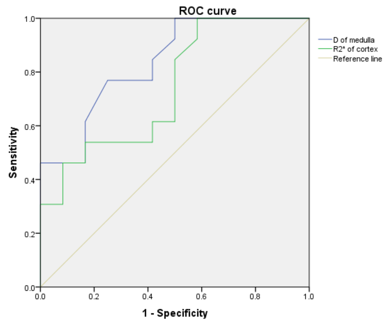
Corresponding parameter mapping of IVIM and mDIXON-Quant were showed in Figure 1. The D values of renal medulla in IgAN group were higher, but the R2* values were lower when compared with HSPN group (P=0.003 and 0.025 respectively) in Table 2 and 3. The area under the ROC cuvers (AUCs) of D of medulla and R2* of cortex were 83.7% and 74.0% for identify IgAN and HSPN, respectively (Table 4 and Figure 2). The sensitivity of the above parameters with feasible thresholds were 76.9% and 100%, the corresponding specificity were 75.0% and 41.7%.Discussion
Previous studies have shown that IVIM and mDIXON-Quant are associated with renal perfusion and diffusion4,7,8. This study further applied these techniques to evaluate pediatric nephropathy and to identify IgAN and HSPN. Although the pathogenesis of IgAN and HSPN is similar, their pathological changes are different. The IgAN has more serious renal lesions than HSPN, and the incidence of renal insufficiency is higher, which means that it is consistent with the clinical explanation9. In this study, although the renal medulla pure diffusion coefficient (D value) of IgAN patients was higher than that of HSPN, the diffusion capacity, perfusion capacity and perfusion fraction of cortex were lower than those of HSPN, which means that the overall renal lesions in IgAN patients were higher than those in HSPN patients. However, in this study, the degree of renal cortical hypoxia in patients with HSPN was higher than that in patients with IgAN, which is different from previous study9, and the results also means that the lesion degree of renal cortex in HSPN patients was higher than that in IgAN patients. We speculate that there may be a certain correlation between the number of cases and data measurement error.Conclusions
IVIM and mDIXON-Quant can significantly distinguish diagnosis of IgAN and HSPN. This result revealed that the value of D and R2* may evaluate the progression and prognosis of pediatric nephropathy in the further study.Acknowledgements
No.References
[1] Rodrigues JC, Haas M, Reich HN. IgA Nephropathy. Clin J Am Soc Nephrol. 2017;12(4):677-686.
[2] Zheng X, Chen Q, Chen L. Obesity is associated with Henoch-Schönlein Purpura Nephritis and development of end-stage renal disease in children. Ren Fail. 2019;41(1):1016-1020.
[3] Corapi KM, Chen JL, Balk EM, Gordon CE. Bleeding complications of native kidney biopsy: a systematic review and meta-analysis. Am J Kidney Dis. 2012;60(1):62-73.
[4] Zhang JL, Rusinek H, Chandarana H, et al. Functional MRI of the kidneys. J Magn Reson Imaging. 2013;37(2):282-293.
[5] Zhang JL, Lee VS. Renal perfusion imaging by MRI. J Magn Reson Imaging. 2020;52(2):369-379.
[6] Zhang JL, Morrell G, Rusinek H, et al. New magnetic resonance imaging methods in nephrology. Kidney Int. 2014;85(4):768-778.
[7] Mahmoud H, Buchanan C, Francis ST, Selby NM. Imaging the kidney using magnetic resonance techniques: structure to function. Curr Opin Nephrol Hypertens. 2016;25(6):487-493.
[8] Neugarten J, Golestaneh L. Blood oxygenation level-dependent MRI for assessment of renal oxygenation. Int J Nephrol Renovasc Dis. 2014; 7:421-435.
[9] Calvo-Río V, Loricera J, Martín L, et al. Henoch-Schönlein purpura nephritis and IgA nephropathy: a comparative clinical study. Clin Exp Rheumatol. 2013;31(1 Suppl 75): S45-S51.
Figures
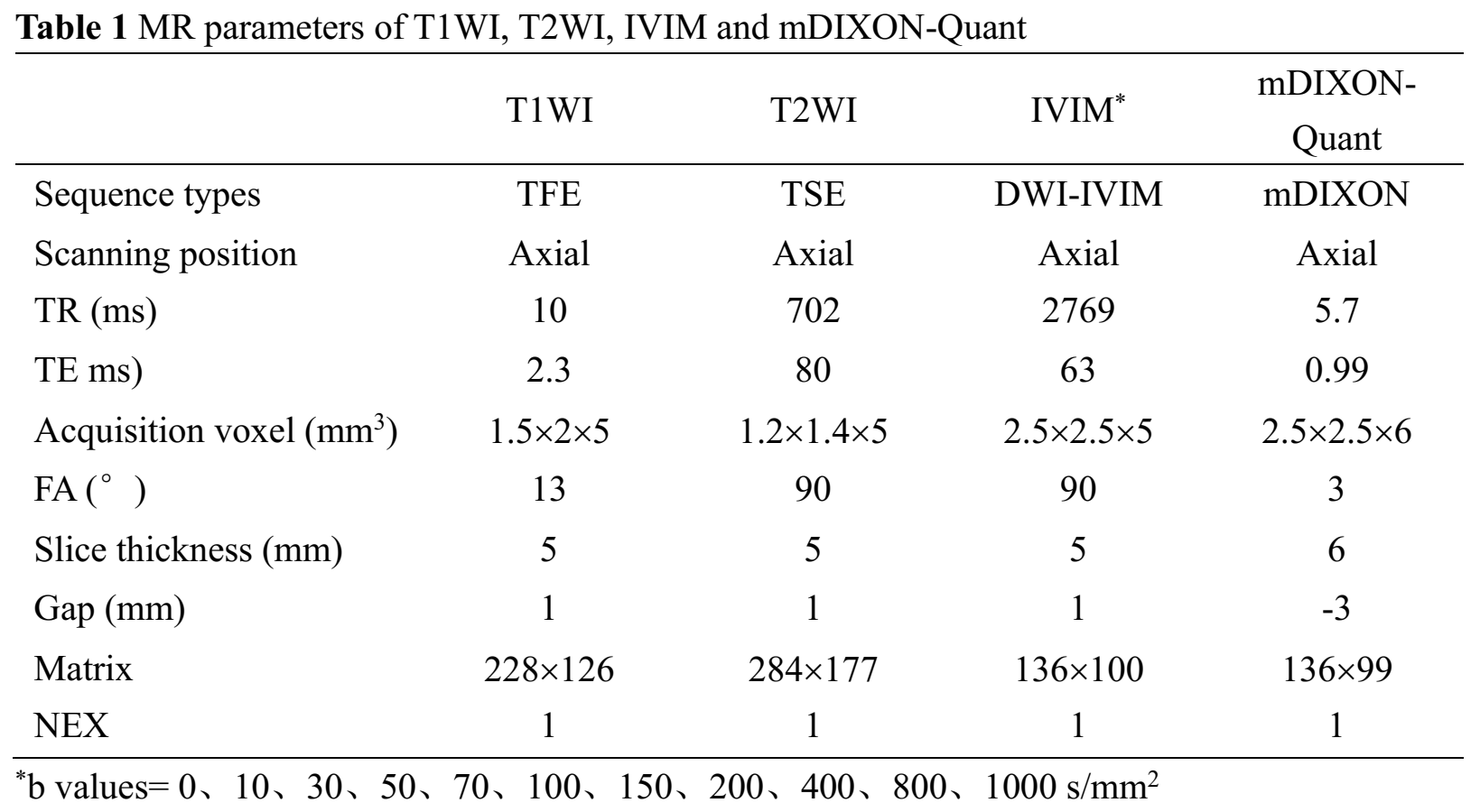
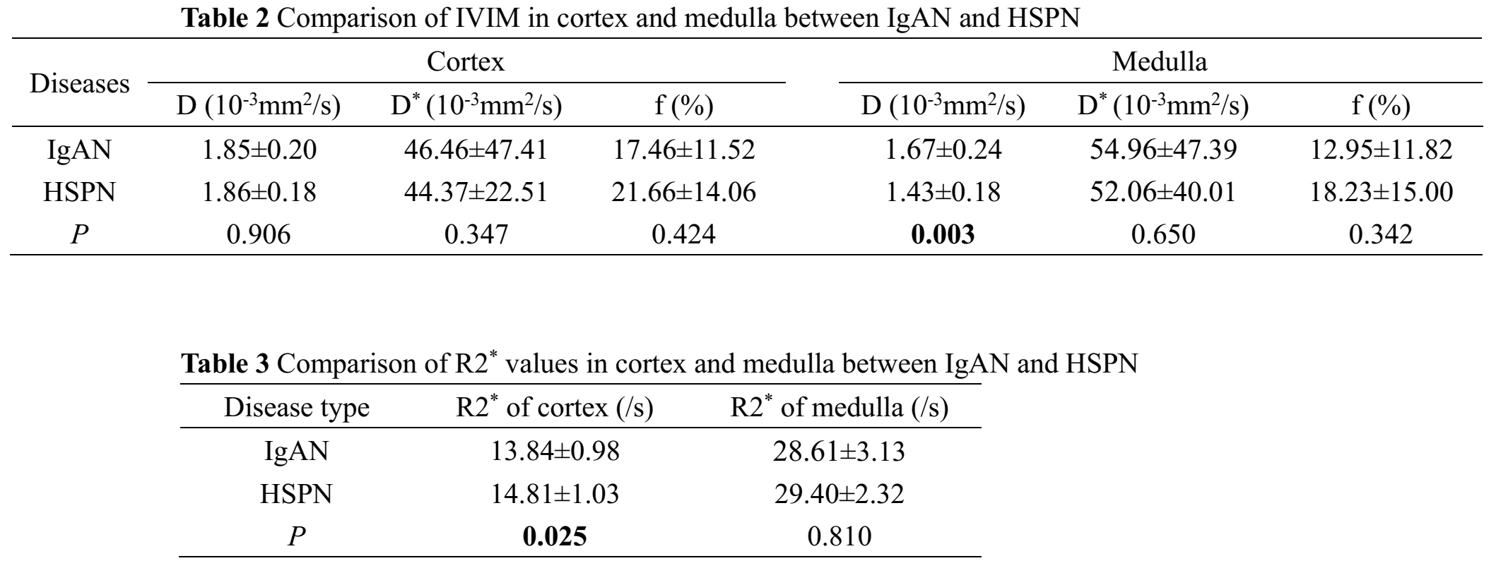
Table 2: Comparison of IVIM in cortex and medulla between IgAN and HSPN.
Table 3: Comparison of R2* values in cortex and medulla between IgAN and HSPN.
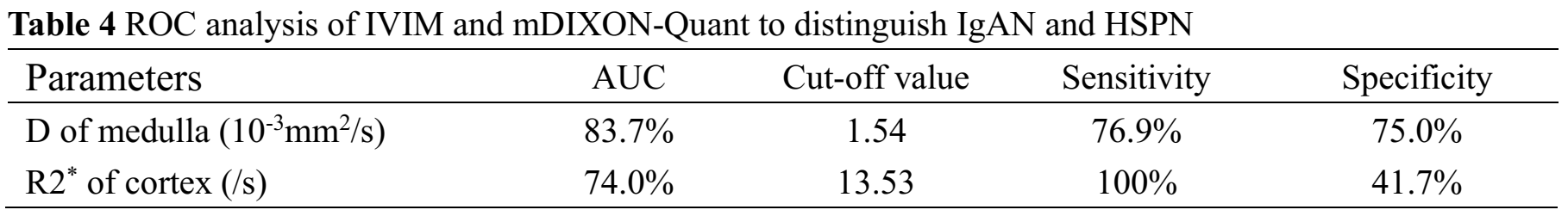
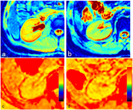
Figure 1. a. A 15.2 years old boy with IgAN. b. A 11.5 years old boy with HSPN. Although the renal medulla diffusion capacity of IgAN patients was higher than that of HSPN, the diffusion capacity, perfusion capacity and perfusion fraction of cortex were lower than those of HSPN.
c. A 12.7 years old girl with HSPN. d. A 13.9 years old boy with IgAN. Hypoxia in renal cortex of HSPN patient was more common than that of IgAN patient.