3722
Relationship between fetal brainstem growth and corresponding gestational weeks: an MRI study
jie LI1, xiao Ling1, songhong Yue1, haoyuan Li1, tao Wen1, jing Zhang1, and kai Ai2
1Lanzhou University Second Hospital, Lanzhou, China, 2Philips Healthcare, Xi'an, China
1Lanzhou University Second Hospital, Lanzhou, China, 2Philips Healthcare, Xi'an, China
Synopsis
We use midsagittal T2WI-based structural MRI images to investigate the relationship between fetal brainstem growth and corresponding gestational weeks. The changes of fetal brainstem substructure in of 153 pregnant women in the middle and late stages were quantitatively evaluated. We found that all measurement indices (height, anteroposterior diameter and cross-sectional area) of normal fetal brainstem were significantly correlated with gestational age. Our study demonstrates that MRI can clearly show the fetal brainstem structure, and the changes of brainstem substructure follow certain rules in fetus.
Introduction
Brainstem is an important structure and pathway of human brain, cerebellum and spinal cord. In the process of embryonic development, any risk factor may cause the development disorder of brainstem and its adjacent structures. Further, it can lead to the development disorder of central nervous system and increase the number of deformed children. Therefore, prenatal diagnosis of brainstem is particularly important and necessary for the development of fetus after birth1. The structure and development of embryonic brainstem has been the focus of recent researches in fetal neuroimaging2. However, the development of fetal brainstem was rarely investigated, and most of the previous literatures used ultrasound to evaluate the developmental process.Magnetic resonance imaging (MRI) technique can clearly display the fetal brainstem structures due to its high resolution and accurate soft tissue contrasts3. Besides, compared with ultrasound, MRI is not affected by gravida weight and fetal position. Therefore, our aim is to explore the correlation between MRI measurement indices (height, anteroposterior diameter and cross-sectional area) of normal fetal brainstem and gestational age, and to seek a quantitative evaluation method of MRI for normal fetal brainstem development.and to seek a quantitative evaluation method of MRI for normal fetal brainstem development.and to seek a quantitative evaluation method of MRI for normal fetal brainstem development.Methods
In this retrospective study, we collected the midsagittal MRI images of 153 normal fetuses between January 2016 and September 2021 at Magnetic Resonance Department, Lanzhou university second hospital. Examinations were performed using a 3.0T scanner (Ingenia, Philips Healthcare, The Netherlands) with 16 channel phase array body coil. In accordance with the 2015 ACR-SPR Fetal MRI Guidelines, gravidae with gestational age ranged from 22 to 39 weeks were finally chosen for research. Focus on T2WI sequences (single-shot turbo spin echo ,SSH): TR 15000ms, TE 115ms, thickness 4mm.For convenience, all images were transferred to PACS (model: V1.5.5+P23).All measurements of this study were analyzed and measured independently by two radiologists (both of them had at least 5 years' experience in fetal MRI diagnosis) who were blinded to the any clinical information. And the intraclass correlation coefficient was then calculated. All measurements were measured in the median sagittal position on T2WI. The indicators we measured include the height, anteroposterior diameter and cross-sectional area of the midbrain, pons,medulla oblongata, as well as the anteroposterior diameter of tegmentum. And all of the indicators were measured three times to get the average value. Figure 1 showed the example of measuring method for typical images. Correlation analysis was performed to assess relationship between gestational age and all measurements. All statistical analyses were performed by SPSS 22.0.Results
The measurement results of diameter and cross-sectional area of each component of brainstem was shown in Table 1, 2. ANOVA analysis and Welch test showed that there were significant differences between groups at different gestational weeks (P <0.05). The height, anteroposterior diameter and cross-sectional area of fetal brainstem components were positively correlated with gestational age. The correlation between pontine cross-sectional area and gestational age was the highest (r=0.963, P <0.05), and the correlation of anteroposterior diameter of tegmentum was the weakest (r=0.746, P <0.05). The correlation of measurement indices of pons was higher than midbrain and medulla oblongata (Table 3). The variation trend of diameter and cross-sectional area of brainstem components with gestational age was shown in Figure 2.Discussion
The fetal brainstem proportions and morphology significantly differs from the adult. The substructure of fetal brainstem follows a regular growth pattern4. Prenatal diagnosis of brainstem malformations is still a challenge because its requires the knowledge of normal brainstem anatomy, development process and imaging diagnosis model. This study demonstrated that all diameters and areas of normal fetal brainstem showed a significant and strong correlation with gestational age. The correlation of biological indices of pons was higher than midbrain and medulla oblongata, and the pontine structure had a large growth range in the second and third trimester of pregnancy, This can be interpreted as pons belongs to white matter fiber concentration area. In addition, we also found that the height of pons was always greater than the anterior posterior diameter of pons before 28 weeks of gestation. And after 28 weeks of gestation, the anterior posterior diameter of pons was always greater than the height. To our knowledge, this result has not been mentioned in previous studies. This would provide new information for clinicians to better evaluate the fetal brain development.Conclusion
The biological indices of fetal brainstem are positively correlated with gestational age, and can be used as an indicator to evaluate the growth and development of fetal brainstem.Acknowledgements
No acknowledgement found.References
- 1. Haratz KK, Lerman-Sagie T. Prenatal diagnosis of brain stem abnormalities. Eur J Paediatr Neurol. 2018;22(6):1016-1026.
- 2.Angeles Fernández-Gil M, Palacios-Bote R, Leo-Barahona M, et al Brain stem anatomy: staring at the stem of life. Semin ultrasound CT MR. June 2010; 31(3): 196-219.
- 3. Conte G, Milani S, Palumbo G, etc. Prenatal MR imaging of the brain: reference linear biometric percentiles between 20 and 24 gestational weeks. AJNR is J Neuroradiol. 2018;39(5):963-967.
- 4. Dovjak GO, Schmidbauer V, Brugger PC, etc. Normal human brainstem development in vivo: Quantitative fetal MRI study. Ultrasound Obstet Gynecol. 2021;58(2):254-263.
Figures
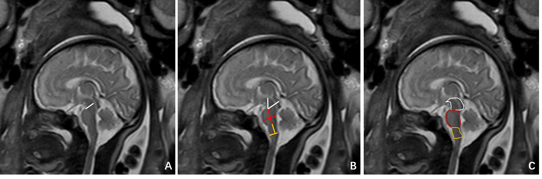
Fig.
1 Measurement of height, anterior posterior diameter and cross-sectional area. A:
the anterior posterior diameter of tegmentum. B: The anterior and posterior
diameter and height of midbrain (white lines). The anterior posterior diameter
and height of pons (red lines). The anterior and posterior diameter and height
of medulla oblongata ( yellow lines).C: midbrain cross-sectional area (white
line area); pons cross-sectional area (red line area); cross sectional area of
medulla oblongata (yellow line area).
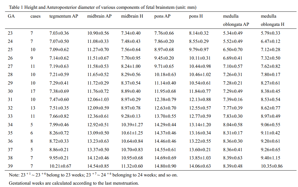
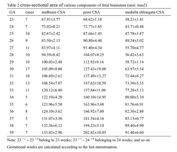
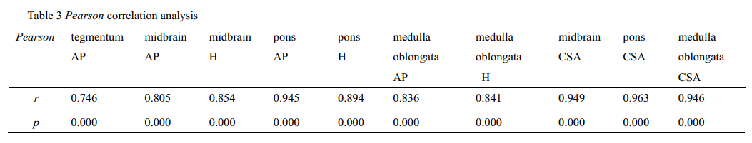
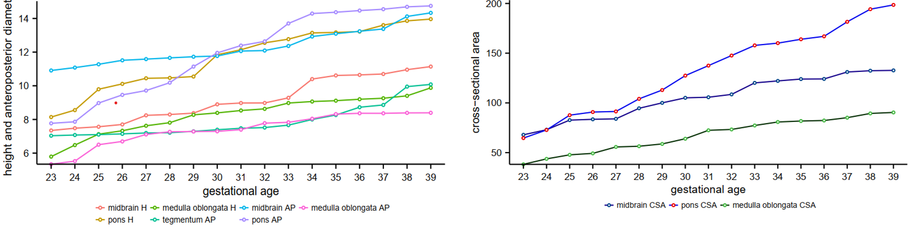
Fig.
2 Variation Trend of diameter and cross-sectional area of brainstem components
with gestational age from 23 to 39 weeks
DOI: https://doi.org/10.58530/2022/3722