3719
Fetal brain T1-weighted imaging with a deep learning constrained compressed SENSE reconstruction1Radiology, the First Hospital of Jilin University, Changchun, China, 2Philips Healthcare, Beijing, China
Synopsis
The quality of commonly used fetal T1-weighed inversion recovery(IR) images is relatively poor. Compressed SENSE(CS) technique allows shortening of scan time, but the overall image quality has not been significantly improved. In this study, Compressed-SENSE Artificial intelligence(CS-AI) framework was applied to reduce the scan time and increase the spatial resolution. This study aims at acquiring high-resolution fetal brain T1-weighted image with reduced scan time and compare the image quality among images reconstructed with CS-AI, CS and conventional SENSE.
Introduction
Magnetic resonance imaging(MRI) is an invaluable diagnostic tool for assessing brain development, and has been widely used in fetal imaging for diagnosis and research. Meanwhile, the T2-weighted single-shot-fast-spin-echo(ssFSE) sequence is the backbone of fetal brain MRI and is characterized by both high contrast and high quality. The T1-weighted sequence plays an important role in evaluating fetal intracranial hemorrhage which is related to neonatal outcome1. However, the quality of commonly used fetal T1-weighed inversion recovery(IR) images with high resolution is relatively poor2. Compressed SENSE(CS) technique allows shortening of scan time, but the overall image quality has not been significantly improved. Recently, deep learning AI algorithms have been combined with CS to improve the quality of the final reconstructed image3. In this study, Compressed-SENSE Artificial intelligence(CS-AI) framework was applied to reduce the scan time and increase the spatial resolution. This study aims at acquiring high-resolution fetal brain T1-weighted image with reduced scan time and compare the image quality among images reconstructed with CS-AI, CS and conventional SENSE.Materials and Methods
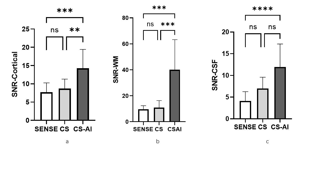
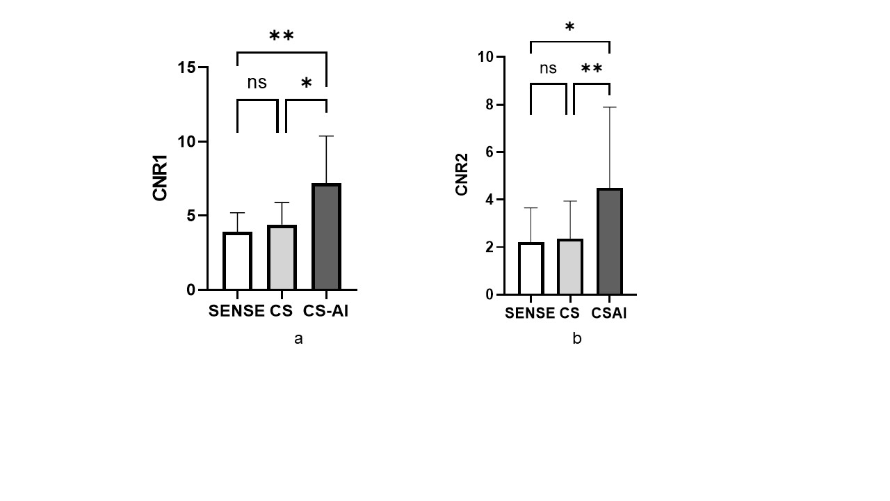
The study was approved by the local Institutional Review Board(IRB),and written consent was obtained from all subjects. Six pregnant women(median gestational age,30 weeks, gestational age range, 243/7-37 weeks) were examined on a 3.0T system(Elition, Philips Healthcare) with body coil. The inspection purposes were ventriculomegaly(2/6), blurry cavum septum pellucidum(2/6) , small head circumference(1/6), and megacisternmagna(1/6). The T1-weighted images were obtained by IR turbo-field-echo(TFE) and utilizing CS-SENSE or CS-AI technology to reconstruction. According to the institutional standard T1-weighted TSE sequence, SENSE technology with the acceleration of 2 was also obtained as a reference. Following parameters were applied to all examinations: FOV=375X306,Voxel=1.6X1.6, slice thickness(mm)/gap=4.0/0, matrix=236X193, TR(ms)=12, TE(ms)=2.3, CS acceleration factor=3, NSA=1, scan time=1’41’’(SENSE) or 1’10’’(CS and CS-AI). Reconstruction parameter denoising level was set to system default for all cases. Signal-to-noise ratio (SNR)(SNR=mean signal intensity(SI)/standard deviation(SD)of SI) and contrast-to-noise ratio (CNR) ($$CNR_{tissure1-tissrue2}=\frac{|SI_{ROI1}-SI_{ROI2}|}{\sqrt{SD_{ROI1}^{2}+SD_{ROI2}^{2}}}$$ )(CNR1, between cortical and cerebrospinal fluid(CSF), CNR2 between cortical and white matter ) of T1-SENSE were compared with those of CS and CS-AI. Image quality of three sequences was analyzed by two readers independently, who were blinded to any clinical information (8 years of experience in MR imaging and 13 years of experience in MR imaging). The statistical analysis was performed with Graphpad 9.0. The Friedman test and Dunn‘s post-hoc analysis was performed to test for an influence of the different sequences. P values< 0.05 were considered significant.Results
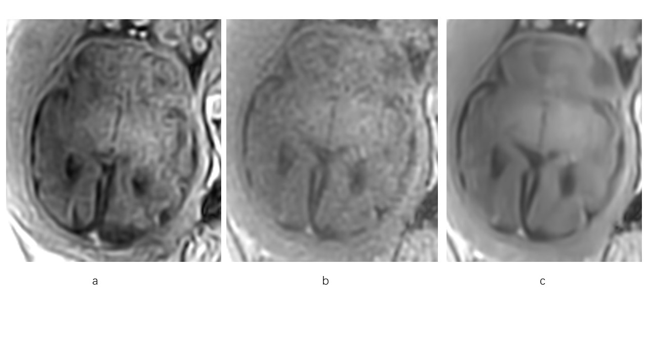
Representative high resolution T1W images using the SENSE, C-SENSE and CS-AI reconstructions are shown in Figure 1. As Figure 2 showed, compared to SENSE, the CS-AI produced higher SNR of CSF, cortical and white matter, and they showed significant difference (p<0.05). Compared to CS, the CS-AI images showed higher SNR too, while the SNR of cortical and white matter demonstrated significant difference (p<0.05), but the SNR of CSF did not show statistical difference(p=0.0742). Figure 3 demonstrates the CNR comparison of the SENSE, CS, and CS-AI for T1WI images. The CS-AI produced higher CNR of cortical/CSF, and cortical/white matter, compared to SENSE(p=0.0066 and 0.0429 respectively) and CS(p=0.0429 and 0.0066 respectively). The CS images quality is comparable to that of the SENSE(p>0.9999).Conclusions
Overall, in this study, we found that the CS-AI technology can reduces the scan time for the high resolution T1-IR sequence in fetal brain MR imaging about 31 seconds as well as the image quality is improved (measured by physical parameters such as SNR and CNR). This technology may be particularly useful in patients whom unable to remain stationary for a long time. T1-IR-CS-AI outperforms conventional SENSE sequence and CS sequence in fetal brain MR imaging which is a reliable alternative for clinical use in fetal brain MR imaging.In the future study, more patients will be enrolled in this study, and more comprehensive subjective assessments and objective measurements will be performed in different denose level for CS-AI.Acknowledgements
No acknowledgement found.References
1.Malamateniou C, McGuinness AK, Allsop JM, O'Regan DP, Rutherford MA, Hajnal JV. Snapshot inversion recovery: an optimized single-shot T1-weighted inversion-recovery sequence for improved fetal brain anatomic delineation. Radiology. 2011 Jan;258(1):229-35. doi: 10.1148/radiol.10100381. Epub 2010 Oct 27. PMID: 20980451.
2.Sun T, Jiang L, Zhang Z, Zhang C, Zhang H, Wang G, Qian Z. Feasibility of free-breathing T1-weighted 3D radial VIBE for fetal MRI in various anomalies. Magn Reson Imaging. 2020 Jun;69:57-64. doi: 10.1016/j.mri.2020.03.004. Epub 2020 Mar 20. PMID: 32171775.
3.Knoll F, Murrell T, Sriram A, Yakubova N, Zbontar J, Rabbat M, Defazio A, Muckley MJ, Sodickson DK, Zitnick CL, Recht MP. Advancing machine learning for MR image reconstruction with an open competition: Overview of the 2019 fastMRI challenge. Magn Reson Med. 2020 Dec;84(6):3054-3070. doi: 10.1002/mrm.28338. Epub 2020 Jun 7. PMID: 32506658; PMCID: PMC7719611.
Figures