3715
Evaluation of fetal spinal anatomy and vertebral malformation using three-dimensional T2-star FFE sequence at 3T: clinical experience1Chengdu Women's and Children's Central Hospital, School of Medicine, University of Electronic Science and Technology of China, Chengdu, China, 2Philips Healthcare, Chengdu, China
Synopsis
Magnetic resonance image (MRI) has been increasingly used to evaluate fetal malformations. Currently, the commonly used fetal spine scanning sequences include conventional 2D Single-Shot TSE (2D SSH TSE) and 2D balanced fast field echo sequence (2D BTFE), However, 2D MR sequences have disadvantages such as unclear display of tiny structures especially the fetal cervical vertebra, furthermore the image quality is strongly affected by fetal postures. In this study we compared 3D T2-star-weighted FFE sequence with conventional 2D SSH TSE sequence and 2D BTFE sequence. The 3D T2-star FFE sequence has high prenatal diagnosis value for fetal spine malformation, and can be used as an important supplementary sequence for fetal spine diagnosis in clinical practice.
Introduction
Fetal vertebral malformation is one of the common fetal malformations. Congenital vertebral malformation can lead to abnormal appearance of the spine after birth, and some patients may be combined with other system abnormalities, such as intestinal atresia, brain atrophy, hypospadias, etc. [1]. The routinely used spinal MRI protocol include two-dimensional (2D) turbo spin echo (TSE) sequence and balanced fast field echo sequence (2D BTFE). Compared to 2D MR sequence, three-dimensional (3D) sequence can improve anatomical delineation to allow the characterization of the topographic distribution of diseases, hence enhancing diagnostic confidence with better understanding of the spine pathophysiology. To the authors’ knowledge, there are rarely previous studies investigated the application of the 3D T2-star-weighted gradient echo pulse sequence of the cervical spine at 3T for anatomical delineation in normal participants only [3-5].The aim of this study was to compare 3D T2-star-weighted FFE sequence with conventional 2D Single-Shot TSE (2D SSH TSE) sequence and 2D BTFE for its clinical diagnostic performance of fetal spinal anatomy at 3T, especially to assess the detectability of the vertebral malformation.
Methods
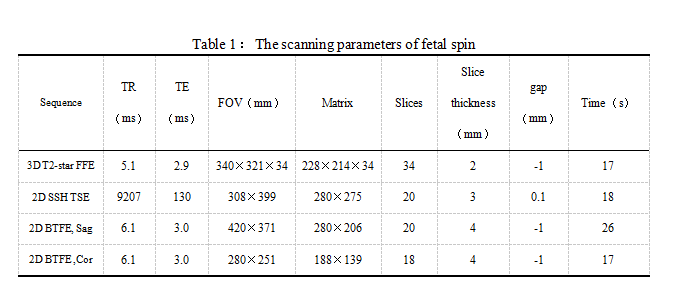
Experiment: This study was approved by the institutional ethics committee. 21 middle and late pregnant women, whose prenatal ultrasound showed abnormal fetal vertebral bodies or unclear spine parts, were consecutively enrolled. The gestational age was 25.0-37.4 weeks, with an average of (30.1±3.6) weeks. All fetuses underwent corresponding spinal MRI examination on a 3T MR with a 16-channel body matrix coil (Ingenia, Philips). The scanning MR sequences included three-dimensional T2 star fast field echo sequence (3D-T2 star FFE), two-dimensional single shot spin echo sequence (2D SSH TSE) and two-dimensional balanced fast field echo sequence (2D BTFE). Detailed scanning parameters were listed in Table 1.Image Analysis: The image quality of each segment of the spine of the three sequences was scored by two diagnosticians, and the difference in scores between the sequences was compared using the non-parametric Friedman test. For quantitative assessment ,region of interest (ROI) was carefully delineated for all cases to calculate the signal difference ratio *SI of the corresponding vertebral bodies and intervertebral discs in the cervical, thoracic and lumbar segments with the three sequences respectively to evaluate image contrast. The *SI of region of interest a & b is defined as:
$$SI=(SIb-SIa)/(SIb+SIa)$$
The signal difference ratio was compared using the one-way analysis of variance among the three sequence. A p value less than 0.05 was considered statistically significant.
Results
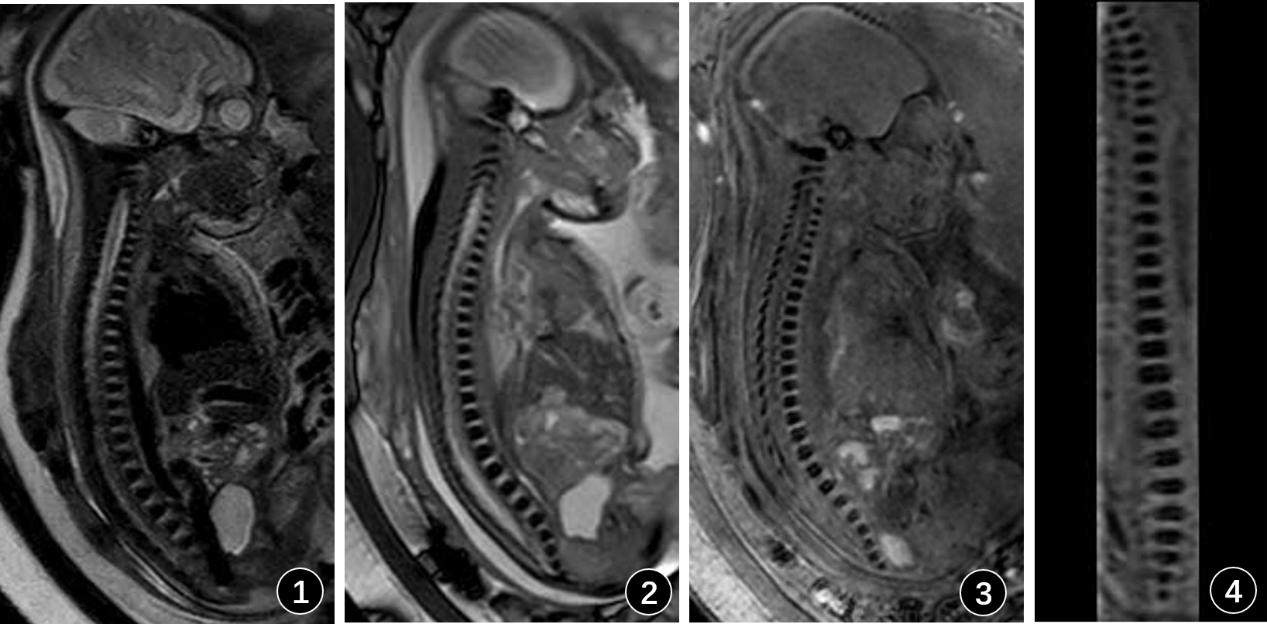
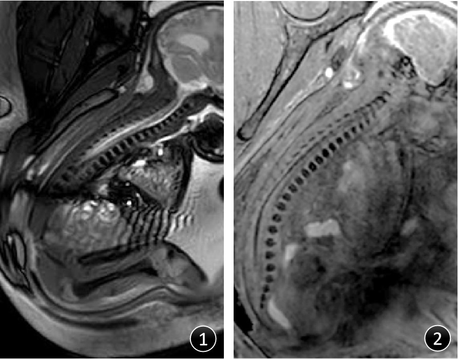
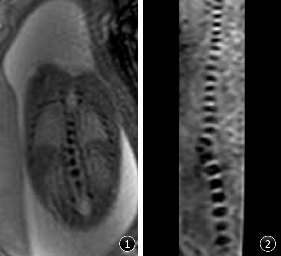
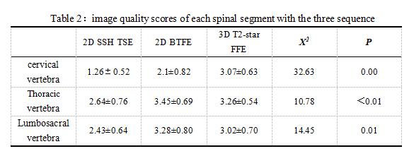
The examples are shown in Figure 1, Figure2 and Figure 3. Regarding visual analysis of artefacts, 3D T2-star FFE displayed significantly better image quality for anatomical delineation of spinal anatomy compared to 2D BTFE sequence (Figure 2). The differences of image quality score of each spinal segment among the three sequences were statistically significant (P < 0.05) (Table 2), which indicated that 3D-T2 star FFE can achieve better image quality of the cervical spine when compared to the 2D SSH TSE and 2D BTFE sequences. In addition, the image quality of thoracic and lumbosacral vertebra with BTFE sequence was also higher than that of SSH TSE sequence (Table 2).For the delineation of fetal cervical spine, the *SI values of 3D-T2 star FFE and 2D BTFE sequences were higher than that of 2D SSH TSE sequence, and the difference was statistically significant (P < 0.05). There was no statistical difference in * SI of fetal thoracic and lumbar spine.
Discussion and Conclusion
2D BTFE and 3D-T2 star FFE sequences performed well on fetal vertebral bodies imaging at 3T. However, the 3D-T2 star FFE is superior in delineation of anatomical details of the spine compared to 2D sequences, without being affected by fetal position, thus having high prenatal diagnosis value for fetal spine malformation. In addition, the scanning time of 3D-T2 star FFE is short ( 17s), which can reduce the influence of fetal movement on image quality and can be used as an important supplementary sequence for fetal spine diagnosis during in clinical practice. A large clinical study is underway to validate the diagnostic value of this technique.Acknowledgements
No acknowledgement found.References
[1] Passias P G, Poorman G W, Jalai C M, et al. Incidence of Congenital Spinal Abnormalities Among Pediatric Patients and Their Association With Scoliosis and Systemic Anomalies[J]. J Pediatr Orthop,2019,39(8):e608-e613.
[2] Goodall A F, Barrett A, Whitby E, et al. T2*-weighted MRI produces viable fetal "Black-Bone" contrast with significant benefits when compared to current sequences[J]. Br J Radiol,2021,94(1123):20200940.
[3] Matsubara Y, Higaki T, Tani C, et al. Demonstration of Human Fetal Bone Morphology with MR Imaging: A Preliminary Study[J]. Magn Reson Med Sci,2020,19(4):310-317.
[4] Xiao L, Siu C W, Yeung K, et al. MRI of the cervical spine with 3D gradient echo sequence at 3 T: initial experience[J]. Clin Radiol,2015,70(9):926-931.
[5] Lemire G T, Beauregard-Lacroix E, Campeau P M, et al. Retrospective analysis of fetal vertebral defects: Associated anomalies, etiologies, and outcome[J]. Am J Med Genet A,2020,182(4):664-672.
Figures