3707
Intravoxel Incoherent Motion (IVIM) DiffusionWeighted imaging in quantitatively evaluation of WT-1 expression in Ovarian Cancer1Department of Radiology, the First Affiliated Hospital of Dalian Medical University, Dalian, Dalian, China
Synopsis
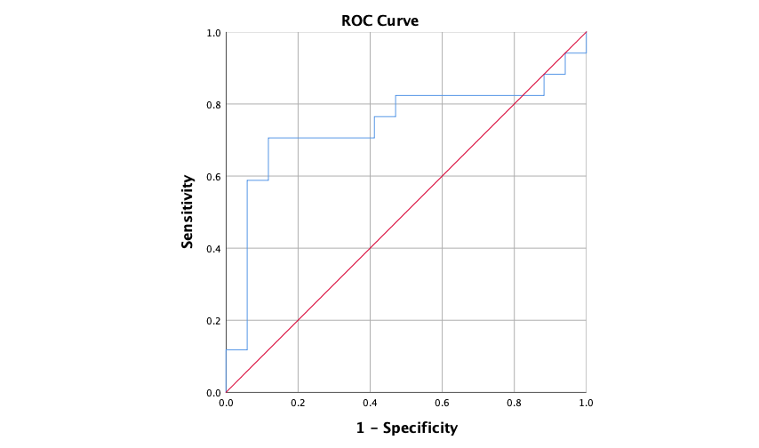
Ovarian cancer (OC) behaves more aggressively and has a worse prognosis than any other cancer involving the female genital tract. WT-1 was significantly upregulated in human OC tissues and closely associated with OC type, grade and FIGO stage. This worked aimed at exploring the value of Intravoxel incoherent motion (IVIM) quantitative parameters for diagnosis of WT-1 positive OC. The results showed that D value provided a promising performance (AUC = 0.740, sensitivity= 88.2%, specificity= 70.6 %) in quantitatively evaluating WT-1 positive OC.
Purpose
To explore the value of Intravoxel incoherent motion (IVIM) quantitative parameters for difference WT-1 positive and negative OC.Introduction
Ovarian cancer (OC) behaves more aggressively and has a worse prognosis than any other cancer involving the female genital tract. Research suggest that a single WT-1 immunohistochemistry can be used to assess both the tumor cells and micro-vascular density in ovarian tumors[1]. WT-1 is expressed in both tumor and endothelial cells, the development of therapeutic agents to target WT-1 may provide an effective treatment option for ovarian cancer. IVIM can be used to evaluate the pure molecular diffusivity (performed by the parameters apparent diffusion coefficient [ADC] and diffusion coefficient value [D]) and perfusion-related diffusivity (performed by parameters perfusion coefficient value [D∗] and perfusion fraction value [f]). The b-value range are (0 20 50 100 150 200 400 800 1200 2000). Therefore, the purpose of this study is to evaluate the efficacy of Intravoxel incoherent motion (IVIM) parameters in the preoperative evaluation of WT-1 expression in OC.Methods
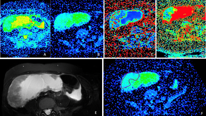
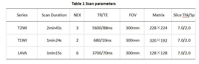
A total of 34 patients of ovarian cancer confirmed by surgery and pathology from 2015 to 2021 were retrospectively collected. Including 17 WT-1 positive OC (age (51.71±11.08) years old) and 17 WT-1 negative OC (age, (60.82±8.65) years old). All patients underwent abdominal MR examinations (Signa HDxt, GE Medical Systems, USA) included T2WI, IVIM (Scan parameters show in table 1). The original axial digital images from the IVIM sequence were transmitted to the GE SDC-ADW 4.6 workstation (Sun Microsystems, Santa Clara, Calif) and the post-processing was performed by Functool software. Referring to the anatomical location of lesion obtained on T2-weighted, The stand ADC, D, D* and f maps were automatically constructed, and were reviewed by two observers who were blinded to clinical information and histopathologic results with 10 and 15 years of experience in pelvic imaging, respectively. The manual regions of interest (ROIs) were drawn along the edge of tumors on the slice with maximal solid area (we choose the solid in tumors), according to fat suppression T2WI (Fig. 1). The measurement was repeated for three times, and the averages of three measurements were calculated. The stand ADC, D, D* and f were recorded. Difference between above values and the expression of WT-1 in OC was compared by Mann-Whitney U test. Receiver operating characteristic (ROC) analysis was performed to evaluate diagnostic performance.Results
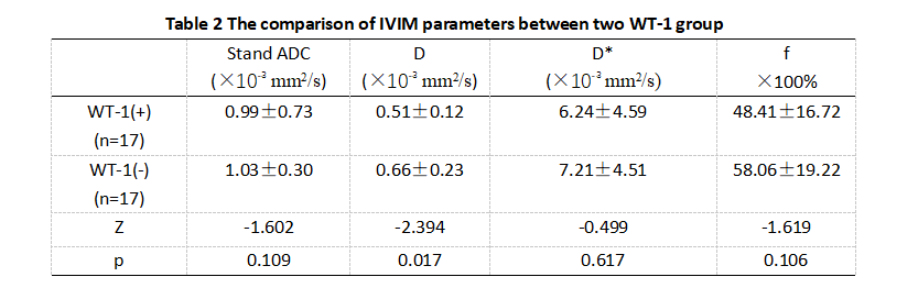
The D value of WT-1 positive group was higher than WT-1 negative group, and P value=0.017(table 2). The stand ADC, D* and f were not significantly different between the two groups (P values, 0.109, 0.617 and 0.105). The AUC of D value for discriminating WT-1 positive group and WT-1 positive group was 0.740, when the D value less than 0.596×10⁻³ mm2/s, the sensitivity was 88.2%, and the specificity was 70.6 % (Figure 2).Discussion
WT-1 immunohistochemistry can be used to assess both the tumor cells and micro-vascular density in ovarian tumors[1]. Our findings suggest that a single WT-1 immunohistochemistry can be used to assess both the tumor cells and micro-vascular density in ovarian tumor [1]. The D value is mainly affected by water molecule diffusion of the lesion tissue rather than by microcirculation perfusion. The shrunken extracellular space due to abnormal proliferation of cancer cells and following incremental local cell density in malignant tumor tissues generates more severe diffusion limitation in malignant lesions than in benign lesions[2]. In the present study, we proposed that D value could be a promising imaging biomarker in evaluation of WT-1 expression.Conclusion
D value of IVIM image could be a promising imaging biomarker in evaluation of WT-1 expression in ovarian cancer.Acknowledgements
NothingReferences
[1] Hsiao YH, Siddiqui S, Man YG. Dual use of a single Wilms' tumor 1 immunohistochemistry in evaluation of ovarian tumors: a preliminary study of 20 cases.J Cancer. 2010 Jul 13;1:93-7.
[2] Long L, Zhang H, He X, Zhou J, Guo D, Liu X. Quantitative evaluation of intravoxel incoherent motion diffusion-weighted imaging (IVIM) for differential diagnosis and grading prediction of benign and malignant breast lesions. Medicine (Baltimore). 2018 Jun;97(26):e11109.
Figures