3706
Texture analysis of multiparametric MRI in the diagnosis of the ovarian sex cord-stromal tumors (SCSTs)1Beijing Hospital, Beijing, China, 2Nanjing Meishan Hospital, Nanjing, China
Synopsis
The accurate diagnosis of malignant SCSTs is still a challenge. We retrospectively analyzed the MRI texture features of 69 cases, and used t-test, one-way ANOVA, the Mann-Whitney U-test and ROC curve to evaluate these parameters. The results showed that the derived texture parameters may be valuable tool in predicting malignant SCSTs.
Introduction and Purpose
Different types of SCSTs have different treatment options and clinical prognostic outcomes, so accurate preoperative diagnosis is important1. However, the appearance of different types of SCSTs on conventional images overlaps2,3, and this assessment is usually subjective, which may result in differences based on radiologists, experience and expertise. Therefore, quantitative index to accurate diagnosis and achieve consensus are of great importance. Texture analysis evaluates the spatial distribution of signal intensities, which may have the potential to give more comprehensive information and reflect tumor heterogeneity. It has been successfully applied to the lesions of the brain, thyroid, breasts, lungs, heart, liver, kidneys, and prostate (4-6). Hence, our study aims to explore the diagnostic value of texture features of multiparametric MRI in malignant SCSTs.Materials and Methods
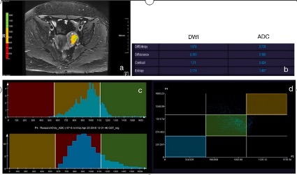
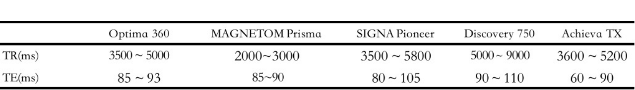
This retrospective study included 57 benign SCSTs in 54 patients and 12 malignant SCSTs in 12 patients. 19 patients with 19 benign SCSTs, and 5 patients with 5 malignant SCSTs underwent MRI examinations on 1.5T systems (Optima 360, GE Healthcare, Milwaukee, WI, USA), while 35 patients with 38 benign SCSTs, and 7 patients with 7 malignant SCSTs underwent MRI examinations on 3T systems (1. MAGNETOM Prisma, Siemens Healthcare, Erlangen, Germany; 2. SIGNA Pioneer, GE Healthcare, Milwaukee, WI, USA; 3. Discovery 750, GE Healthcare, Milwaukee, WI, USA; 4. Achieva TX, Philips HealthCare, Best, Netherlands). The MR protocols included T2 weighted images with fat saturation (The parameters are shown in Table 1.) and diffusion weighted imaging (DWI) with two b-values (0 and 1000s/mm2). The ADC parametric maps were generated inline after data acquisition.The texture analysis for the FS-T2WI, DWI images of b=1000 s/mm2, and ADC parametric maps were performed on a prototypic postprocessing software (MR Multiparametric Analysis, Siemens Healthineers, Erlangen, Germany). Four texture-based features (including entropy, and diff-entropy, diff-variance, contrast) were extracted based on the solid component (the region of interest avoided cystic degeneration or necrosis). Univariate analyses were performed by t-test or one-way ANOVA when normally distributed otherwise by the Mann-Whitney U test. In addition, logistic regression models were then run on the diagnostic features identified in the univariate analyses, and the receiver operating characteristic (ROC) curves were established to evaluate the value of texture features for the diagnosis of malignant SCSTs.
Results
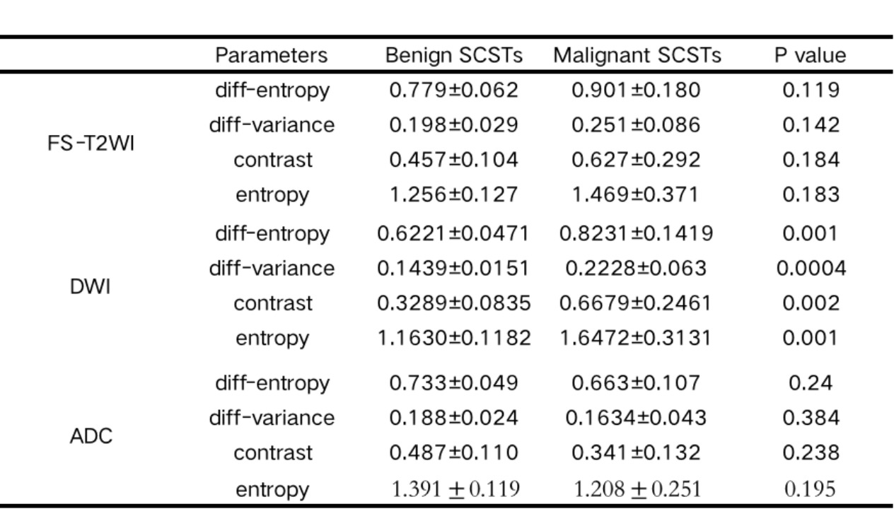
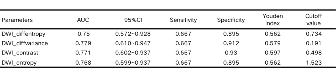
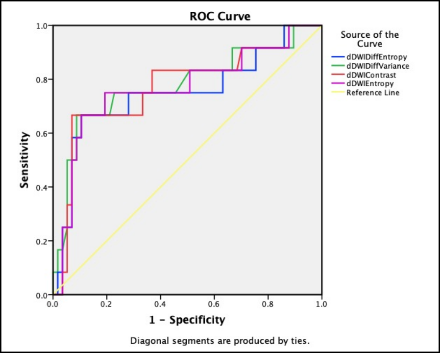
Comparison of texture features between the benign and malignant SCSTs are shown in Table 2. Four texture-based features of FS-T2WI and ADC (including entropy, and diff-entropy, diff-variance, contrast),were obtained,respectively,both of them no differences (P>0.05), Four texture-based features of DWI had significantly differences. The results of the ROC analyses for diagnostic performance of texture features for malignant SCSTs are shown in Table 3, Fig.2. The sensitivity of DWI_diff-variance and DWI_contrast for diagnosing malignant SCSTs were 66.7%, and the specificity of those were 91.2% and 93.0%, respectively. And combining all DWI texture features resulted in a higher diagnostic power than the individual parameters to predict malignant SCSTs (AUC = 0.792, sensitivity 75.0%, specificity 87.7%, 95% CI = 0.630~0.955) (Fig.3).Discussion and Conclusion
Our study found that Four texture-based features of DWI had the potential to predict malignant SCSTs, and the combination of all DWI texture parameters could achieve higher diagnostic power to predict malignant SCSTs. The DWI texture features may better reflect the heterogeneity of tumors,than the others.Acknowledgements
We sincerely thank the participants in this study.References
1.Shim SH, Kim DY, Lee SW, et al. Laparoscopic management of early-stage malignant nonepithelial ovarian tumors: surgical and survival out- comes. Int J Gynecol Cancer 2013; 23:249–255.
2.Prat J, editor. Pathology of the ovary. Philadelphia: Saunders; 2004.
3.Hanley KZ, Mosunjac MB. Practical Review of Ovarian Sex Cord-Stromal Tumors. Surg Pathol Clin. 2019;12(2):587-620.
4.LuoDan Qian, JiaLiang Ren. MR imaging of epithelial ovarian cancer: a combined model to predict histologic subtypes. European Radiology 2020; 30:5815–5825.
5.Castellano G, Bonilha L, Li LM, Cendes F. Texture analysis of medical images. Clin Radiol 2004;59(12):1061–9.
6.Shun Zhang, Gloria Chia-Yi Chiang. MRI based texture analysis to classify low grade gliomas into astrocytoma and 1p/19q codeleted oligodendroglioma.Magn Reson Imaging. 2019; 57: 254–258.
Figures