3700
Evaluation of isotropic high-resolution IRIS diffusion-weighted MRI in bladder cancer: clinical experience1radiology, westchina hospital, Chengdu, China, 2westchina hospital, Chengdu, China, 3Philips Healthcare, Chengdu, China
Synopsis
Conventional single-shot echo planar imaging is widely used in bladder DWI for evaluating the malignant degree of lesions, however, it usually suffers from magnetic susceptibility artifacts and image blurring, which have limitations to identify the small lesions or lesions presented as slightly thickened bladder wall. This study this study was to investigate the feasibility and effectiveness of isotropic high-resolution IRIS DWI for the evaluation bladder lesions at 3T. The results suggested that the IRIS DWI with isotropic high resolution can effectively improve image quality and diagnostic confidence for bladder lesions.
Introduction
Bladder cancer is the 11th most commonly diagnosed cancer worldwide, and the second common cancer of the urinary system. Diffusion-weighted imaging (DWI) has been increasingly applied in the management of bladder cancer, until VI-RADS became widely available in 2017 and the accuracy of diagnosis is improved effectively. However, conventional DWI is most commonly adopted on single-shot echo planar imaging (ss-EPI) which usually suffers from magnetic susceptibility artifacts and image blurring [1], which is limited to diagnose small lesions or lesions presented as slightly thickened bladder wall. In contrast, multi-shot EPI (MS-EPI) was proposed and demonstrated the feasibility of producing high-resolution DW images to identify the small lesions with improved resolution and reduced distortion. Nonetheless, MS-EPI suffers from strong ghost artifacts due to motion-induced inter-shot phase variations. Image-space sampling functions (IRIS) MS-EPI have been introduced and reported could solve the phase inconsistency issue [2]. The aim of this study was to investigate the feasibility and effectiveness of isotropic high-resolution IRIS DWI for the evaluation bladder lesions, especially for the small lesions or lesions presented as slightly thickened bladder wall, in comparison with ss-EPI DWI.Material and methods
The study was approved by the institutional ethics committee. Twenty patients diagnosed with bladder cancer were recruited and underwent MR examinations on 3T scanner (Elition, Philips Healthcare) using a 32-channel body-array coil. The scanning parameters for conventional ss-EPI include: FOV=60X160mm2, imaging matrix=64X63, flip angle= 90o, thickness= 3.5mm, b values include 50, 800 and 100s/mm². In comparison, the scanning parameters for isotropic high-resolution IRIS (ISO-IRIS) DWI include: FOV=250X250mm2, imaging matrix=124X126, flip angle 90o, thickness 2 mm, MS number=7, and b value includes 50, 800 and 100s/mm². All images scored by 2 radiologists (with 3 and 5 years’ experience respectively) using a 5-point scale (1 point indicated poor image quality and cannot diagnostic; 2point indicated below average, 3 point indicated average image quality and basically meet the diagnostic requirements,4 point indicated above average level and provide some diagnostic confidence 5point indicated the image quality is excellent and provides sufficient evidentiary detail for diagnosis). T-test was used to evaluate the statistical difference of the image quality scores. Statistical significance was defined as p < 0.05.Results
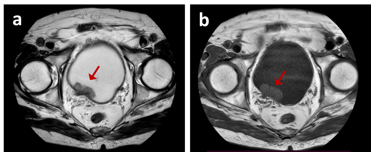
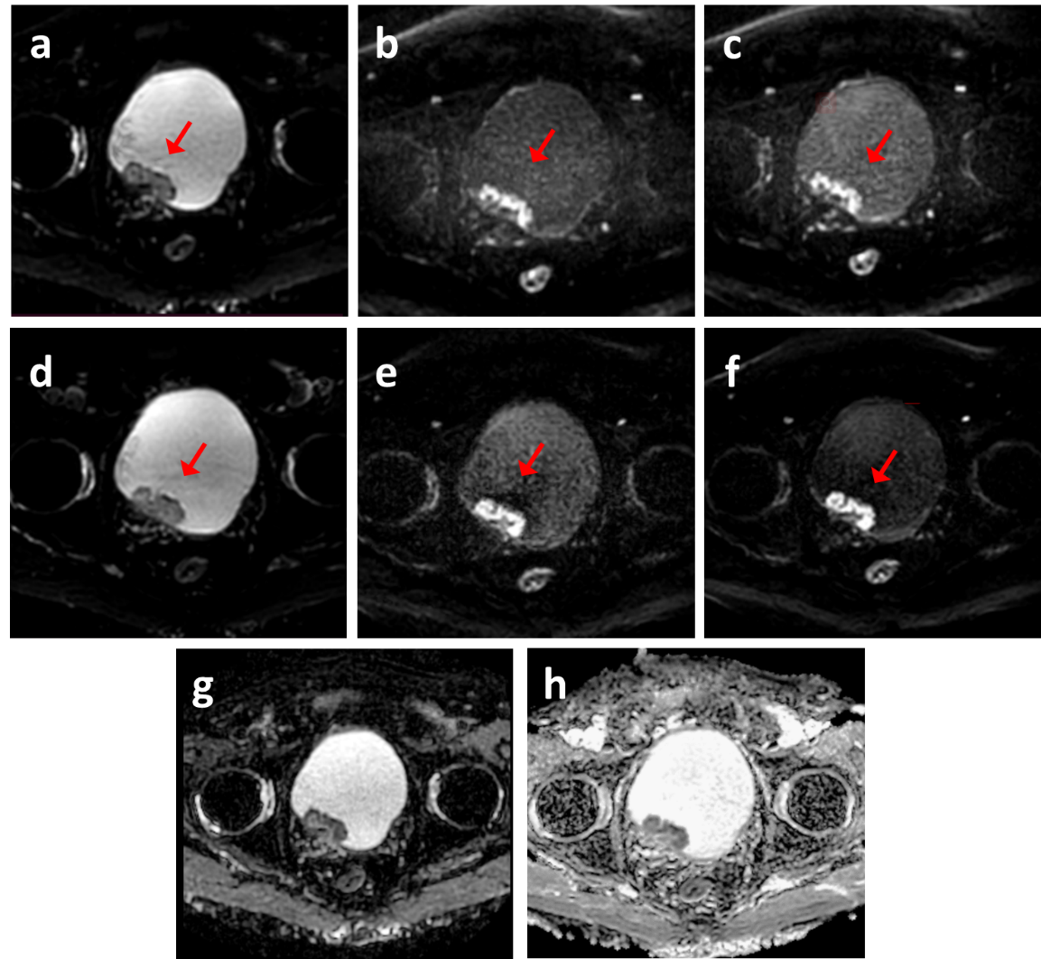
The examples were shown in Figure 1 and Figure 2. Compared with conventional ss-EPI DWI, the ISO-IRIS provides less tissue geometric deformation (4.3±0.7 vs 3.2±0.6,p<0.01), better tissue contrast, especially in the presence of lesions (4.1±0.5 vs 3.5±0.6, p<0.01), and less artifacts at bladder layer (3.8±0.3 ,3.3±0.5, p<0.01) (Figure 2).Conclusion
The study results suggested that the IRIS DWI with isotropic high resolution can effectively improve image quality and diagnostic confidence for bladder lesions. The relationship between muscle layer and lesion can be well demonstrated in the thin-walled organ of bladder cancer.Acknowledgements
noneReferences
1. Porter DA, Heidemann RM (2009) High resolution diffusionweighted imaging using readout-segmented echo-planar imaging, parallel imaging and a two-dimensional navigator-based reacquisition. Magn Reson Med 62:468–475
2. Li Guo, Feng Huang, Zhongbiao Xu, Yingjie Mei, Wenxing Fang, Xiaodong Ma, Erpeng Dai, Hua Guo, Qianjin Feng, Wufan Chen, Yanqiu Feng, eIRIS: Eigen-analysis approach for improved spine multi-shot diffusion MRI, Magnetic Resonance Imaging, Volume 50, 2018, Pages 134-140,
Figures