3695
The value of diffusion-weighted imaging and dynamic contrast-enhanced imaging in the differential diagnosis of male breast diseases1Department of Magnetic Resonance Imaging, The First Affiliated Hospital of Zhengzhou University, Zhengzhou, China
Synopsis
The male breast is often ignored because of its rudimentary and nonfunctional nature. However, the male breast can develop many benign and neoplastic diseases, including breast cancer.Much less is known regarding the appearance of benign and malignant male breast conditions. Therefore, in this retrospective study, we explored the value of diffusion-weighted imaging (DWI) and dynamic contrast-enhanced imaging (DCE-MRI) in the differential diagnosis of male breast diseases and confirmed the importance of DWI and DCE-MRI in the end.
Introduction
The male breast is often ignored because of its rudimentary and nonfunctional nature. However, the male breast can develop many benign and neoplastic diseases, including breast cancer1. Gynecomastia is the most common disease in men. Although breast cancer in men is a rare entity, it often presents at a more advanced stage than it does in women due to a delay in diagnosis2. Most studies in the literature have reported on the features of primary breast carcinoma in men. However, very little information has been reported on the appearance of benign and malignant male breast conditions3-6.Much less is known regarding the characteristics of MRI in men than in women. Therefore, in this retrospective study, we describe the features of male breast diseases and the role of MRI in the evaluation of male breast diseases.Objective
To explore the value of diffusion-weighted imaging (DWI) and dynamic contrast-enhanced imaging (DCE-MRI) in the differential diagnosis of male breast diseases.Materials and Methods
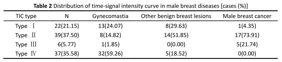
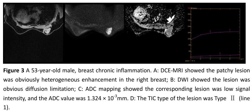
Conventional MRI, DWI and DCE-MRI images of 104 male breast diseases confirmed by surgical pathology from June 2012 to June 2021 were retrospectively analyzed. The original images were transmitted to GE ADW 4.5 workstation, and the DWI and DCE-MRI images are post processed by functool toolkit. The apparent diffusion coefficient (ADC) images generated by DWI were imported into ADC software, and the region of interest (ROI) was manually placed to avoid the part of hemorrhage and necrotic cystic changes. The ADC value was measured for three times to calculate the average. The time-signal intensity curve (TIC) was obtained by the dynamic contrast-enhanced imaging (DCE-MRI) into Functional. According to its shape, it was divided into four types: ① type Ⅰ (persistent type): the signal intensity of the lesion gradually enhanced during the dynamic time; ② Type Ⅱ (plateau type): the signal intensity of the lesion gradually enhanced in the early stage, maintained after reaching the peak, and formed a platform in the middle and late stage; ③ Type Ⅲ (washout type): the signal intensity of the lesion enhanced rapidly in the early stage, and gradually decreased after reaching the peak; ④ Type Ⅳ (non-enhancement type): the signal intensity of the lesion remained at a low level without obvious enhancement. The ADC value and time-signal intensity curve (TIC) of the lesions were statistically analyzed.Results
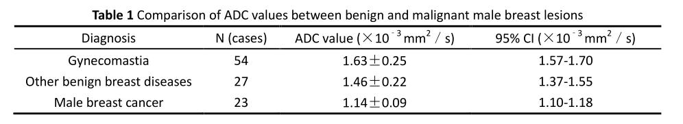
The average ADC values of gynaecomastia, other benign breast lesions and male breast cancer were (1.63 ±0.25) ×10﹣3 mm2, (1.46 ±0.22) × 10﹣3 mm2 and (0.99 ±0.09) × 10﹣3 mm2, respectively. Taking the ADC value of 1.297 × 10﹣3 mm2 as the optimal critical value, the area under the curve (AUC) for differential diagnosis of benign and malignant male breast lesions was 0.938, and the sensitivity, specificity and accuracy of diagnosis were 100.000% (23/23), 85.19% (69/81) and 88.46% (92/104), respectively. There were statistically significant differences in the distribution of TIC curve of gynaecomastia, other benign breast lesions and male breast cancer (value =53.190, P<0.05), and further pairwise comparison showed statistically significant differences. The TIC curves of gynaecomastia were mainly type Ⅳ and type Ⅰ, type Ⅱ and type Ⅰ were dominant in other benign lesions of male breast, and type Ⅱ and type Ⅲ were mainly type Ⅱ and type Ⅲ in male breast cancer.Conclusions
Diffusion weighted imaging and dynamic contrast-enhanced imaging is of great significance in the diagnosis and evaluation of benign and malignant male breast lesions.Acknowledgements
We thank the National Natural Science Foundation of China and the Henan Medical Science and Technology Research Program.References
1. Iuanow E,Kettler M,Slanetz PJ. Spectrum of disease in the male breast [J]. AJR American journal of roentgenology, 2011,196(3):W247-259.
2. Nguyen C,Kettler MD,Swirsky ME,et al. Male breast disease: pictorial review with radiologic-pathologic correlation [J]. Radiographics : a review publication of the Radiological Society of North America, Inc, 2013,33(3):763-779.
3. Doyle S,Steel J,Porter G. Imaging male breast cancer [J]. Clinical radiology, 2011,66(11):1079-1085.
4. Mathew J,Perkins GH,Stephens T,et al. Primary breast cancer in men: clinical, imaging, and pathologic findings in 57 patients [J]. AJR American journal of roentgenology, 2008,191(6):1631-1639.
5. Muñoz Carrasco R,Alvarez Benito M,Muñoz Gomariz E,et al. Mammography and ultrasound in the evaluation of male breast disease [J]. European radiology, 2010,20(12):2797-2805.
6. Günhan-Bilgen I,Bozkaya H,Ustün E,et al. Male breast disease: clinical, mammographic, and ultrasonographic features [J]. European journal of radiology, 2002,43(3):246-255.
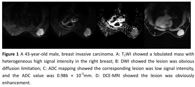
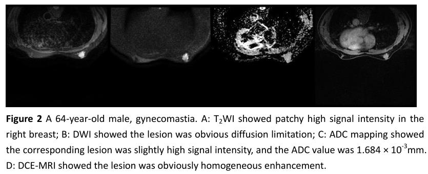
Figures