3694
3D Amide Proton Transfer-weighted Imaging In Evaluation Of Breast Lesions: Comparison With Diffusion-weighted Imaging1Department of Magnetic Resonance Imaging, The First Affiliated Hospital of Zhengzhou University, Zhengzhou, China, 2Philips Healthcare, Beijing, China
Synopsis
This purpose of this study is to investigate the value of 3D APTWI and evaluate the diagnostic efficacy of its combination with DWI for discriminating breast benign from malignant lesions. Results showed the MTRasym(3.5 ppm) value calculated by APTWI for malignant breast lesions were significantly lower than those of benign lesions, and APTWI combined with DWI obtained a better diagnostic performance than the single method. Therefore, APTWI is a novel technique, which can be used to differentiate breast benign from malignant lesions noninvasively.
Introduction
Diffusion-weighted imaging is a traditional technique which can obtain the tissue’s diffusion information of water molecules. And it is a useful tool for tumor detection and characterization1, which allows measurement of water molecular movement using apparent diffusion coefficient (ADC) values2. The importance of breast DWI has been confirmed in the multiparametric breast MRI protocol to differentiate between breast cancer and benign lesions3-4, distinguish in situ from invasive lesions, and predict the responses to neoaduvant therapy over time5-7.Amide proton transfer-weighted imaging (APTWI) is a noninvasive molecular imaging technique that can measure the concentration of free proteins and polypeptides in tissues without the use of exogenous contrast agents based on a chemical exchange between amide protons and water protons8, and it may reflect the microscopic information of diseased tissues and has the potential to evaluate many biological factors for breast cancer9-10.
Therefore, both DWI and APTWI can reflect the microscopic information of diseased tissues theoretically. However, few studies discussed the differences in APTWI and DWI between benign and malignant lesions. So the purpose of this study is to investigate the value of 3D amide proton transfer-weighted imaging (APTWI) and its combination with diffusion-weighted imaging (DWI) for discriminating breast benign from malignant lesions for the first time.
Methods and Materials
Two hundred and twenty-six patients were enrolled in the study prospectively with suspected breast lesions clinically. 3.0T MRI scan was performed in all patients, including T1WI, T2WI, DWI, APTWI and dynamic contrast-enhanced magnetic resonance imaging. The ADC and MTRasym(3.5 ppm) values were obtained from DWI and APTWI. The pathology results were confirmed by surgery or biopsy (totally 226 lesions, with 102 benign lesions and 124 malignant lesions). Receiver operating characteristic (ROC) curve was used to evaluate the diagnostic efficacy of APTWI and DWI for distinguishing breast benign from malignant lesions.Results
The ADC and MTRasym(3.5 ppm) values were significantly lower in breast malignant than in benign lesions (P<0.05, respectively). The areas under ROC curves of ADC and MTRasym(3.5 ppm) values for differentiating breast benign from malignant lesions were 0.817 and 0.752, respectively. Taking the maximum Youden's index of ADC value as the ROC optimal cut-off, the diagnostic sensitivity, specificity and accuracy for distinguishing breast benign from malignant lesions were 94.4% (117/124), 62.7% (64/102) and 80.1% (181/226), respectively. Taking the maximum Youden's index of MTRasym(3.5 ppm) value as the ROC optimal cut-off, the diagnostic sensitivity, specificity and accuracy for distinguishing breast benign from malignant lesions were 73.4% (91/124),64.7% (66/102),69.5% (157/226), respectively. Combing the parameters of both DWI and APTWI, the diagnostic performance was superior to either single parameter (P<0.05, respectively), which the area under the ROC curve was 0.868, and the diagnostic sensitivity, specificity and accuracy for distinguishing breast benign from malignant lesions were 82.3% (102/124),79.4% (81/102) and 81.0% (183/226).Conclusions
APTWI can be used to distinguish breast benign from malignant lesions, and the combination of APTWI and DWI parameters could obtain a better diagnostic performance than the single method.Acknowledgements
We thank the National Natural Science Foundation of China and the Henan Medical Science and Technology Research Program.References
1. Kul S,Cansu A,Alhan E,et al. Contribution of diffusion-weighted imaging to dynamic contrast-enhanced MRI in the characterization of breast tumors [J]. AJR American journal of roentgenology, 2011,196(1):210-217.
2. Vidić I,Egnell L,Jerome NP,et al. Support vector machine for breast cancer classification using diffusion-weighted MRI histogram features: Preliminary study [J]. Journal of magnetic resonance imaging : JMRI, 2018,47(5):1205-1216.
3. Ei Khouli RH,Jacobs MA,Mezban SD,et al. Diffusion-weighted imaging improves the diagnostic accuracy of conventional 3.0-T breast MR imaging [J]. Radiology, 2010,256(1):64-73.
4. Hirano M,Satake H,Ishigaki S,et al. Diffusion-weighted imaging of breast masses: comparison of diagnostic performance using various apparent diffusion coefficient parameters [J]. AJR American journal of roentgenology, 2012,198(3):717-722.
5. Wang Q,Mao N,Liu M,et al. Radiomic analysis on magnetic resonance diffusion weighted image in distinguishing triple-negative breast cancer from other subtypes: a feasibility study [J]. Clin Imaging, 2020,72:136-141.
6. Suo S,Zhang D,Cheng F,et al. Added value of mean and entropy of apparent diffusion coefficient values for evaluating histologic phenotypes of invasive ductal breast cancer with MR imaging [J]. European radiology, 2019,29(3):1425-1434.
7. Shin JK,Kim JY. Dynamic contrast-enhanced and diffusion-weighted MRI of estrogen receptor-positive invasive breast cancers: Associations between quantitative MR parameters and Ki-67 proliferation status [J]. Journal of magnetic resonance imaging : JMRI, 2017,45(1):94-102.
8. Ohno Y,Yui M,Koyama H,et al. Chemical Exchange Saturation Transfer MR Imaging: Preliminary Results for Differentiation of Malignant and Benign Thoracic Lesions [J]. Radiology, 2016,279(2):578-589.
9. Zhou J,Heo HY,Knutsson L,et al. APT-weighted MRI: Techniques, current neuro applications, and challenging issues [J]. Journal of magnetic resonance imaging : JMRI, 2019,50(2):347-364.
10. Meng N,Wang XJ,Sun J,et al. Comparative Study of Amide Proton Transfer-Weighted Imaging and Intravoxel Incoherent Motion Imaging in Breast Cancer Diagnosis and Evaluation [J]. Journal of magnetic resonance imaging : JMRI, 2020,52(4):1175-1186.
Figures
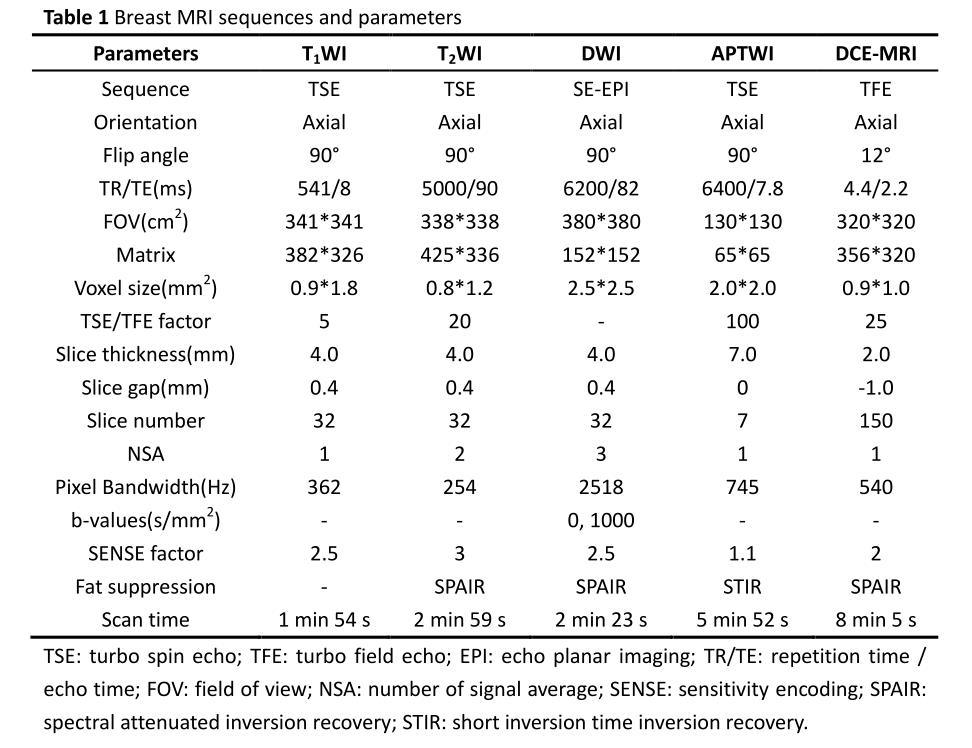
Figure 1 Box plots of DWI and APTWI parameters with breast benign and malignant lesions. The top and bottom lines of the box represent the 25th to 75th percentile values, the line in the box represents the median value, the lines outside the boxes represent maximum and minimum values.
Table 2 Comparison of Different Parameters between Benign and Malignant Lesions

Table 3 ROC Analysis of the Diagnostic Performance for Different Parameters or Methods for Distinguishing Breast Benign From Malignant Lesions
Table 4 Comparison of Diagnostic Efficacy of Different Methods for Distinguishing Breast Benign From Malignant Lesions


Figure 3 (a–d) Female, 65 years, with an invasive ductal carcinoma in the right breast (Grade 2, ER-positive; PR-positive; HER-2-negative, Ki-67-positive, 30%), ADC=0.93×10⁻³mm2/s, MTRasym (3.5 ppm)=1.4%.
(e–h) Female, 35 years, with phyllodes tumor in the right breast, ADC=1.62×10⁻³mm2/s, MTRasym (3.5 ppm)=5.4%.
In these images, a/e are DCE-MRI, b/f are DWI images (b = 1000s/mm2), c/g are ADC pseudocolored maps, d/h are MTRasym (3.5 ppm) pseudocolored maps.