3692
Differentiation of benign and malignant breast lesions using diffusion-weighted imaging with a fractional order calculus model1Department of Radiology, Xinyang Central Hospital, Xinyang, China, 2East China Normal University, Shanghai, China, 3Central Research Institute, United Imaging Healthcare, Shanghai, China
Synopsis
Diffusion-weighted imaging (DWI) is a powerful tool for cancer imaging. Many advanced DWI techniques have been developed and applied during clinical practice. Since fractional order calculus (FROC) model was proposed, it has shown huge clinical potential for cancer diagnosis, prognostic prediction and so on. However, scarcely has the FROC-DWI been utilized for breast imaging. The purpose of this study is to demonstrate that a new set of parameters (D, β and μ) from a fractional order calculus (FROC) diffusion model can be used to improve the accuracy of differentiating among benign and malignant breast lesions. Results suggested it’s feasible to use FROC diffusion model to improve the differentiation between benign and malignant breast lesions.
Introduction:
Breast cancer is widely regarded as a huge threat to women’s health (1). Early detection, diagnosis and treatment of breast cancer have essential roles in reducing mortality and improving the prognosis of patients. Magnetic resonance imaging (MRI) is able to offer high diagnostic sensitivity and specificity (2).In particular, due to the capacity of providing various biological information via characterizing the diffused water molecules, the Diffusion-Weighted MRI has shown great clinical potential for evaluating the breast cancer. Previous studies concluded that characterizing the tissue heterogeneity of breast cancer is of high importance for guiding the following clinical management. The non-invasive imaging technique is thus urgently needed for unveiling the micro-structural information. In order to explore the microstructure characteristics of breast cancer more accurately, a large number of more advanced and complex diffusion-weighted models have been developed. The fractional calculus (FROC) model (3), for example, yields a new set of parameters to describe the anomalous diffusion process in complex biologic tissues containing diffusion coefficient D, fractional-order derivative in space β (dimensionless), and a spatial parameter μ (in micrometers). Several previous studies demonstrated the huge potential of FROC-DWI for brain tumor. However, the FROC-DWI has hardly been performed for breast imaging. Therefore, the purpose of this study is to evaluate the efficacy of fractional order calculus (FROC) model in differentiating the malignant breast cancer from benign breast lesions.Methods:
A total number of 32 patients were recruited into this prospective study. MR measurements were performed on a 3-T MR scanner (uMR790, United Imaging Healthcare, Shanghai, China). Detailed MR protocols include T2WI, contrast-enhanced T1WI and DWI. The detailed parameters of the DWI were as the followings: TR/TE/FA:4036ms/75.3ms/90°; Matrix/FOV: 100×240/160×320 mm2; In total, 12 b values were applied including 0,50,100,200,400,600,800,1200,1600, 2000,2500 and 3000 s/mm2. Then the FROC-DWI derived metrics were calculated according to the fitting formula previously reported. Next, the metrics were statistically compared between the two subgroups with the Mann-Whitney U test. The ROC (receiver operating characteristic curve) analysis was performed to assess the performance of individual parameters for diagnosing malignant breast lesion.Results:
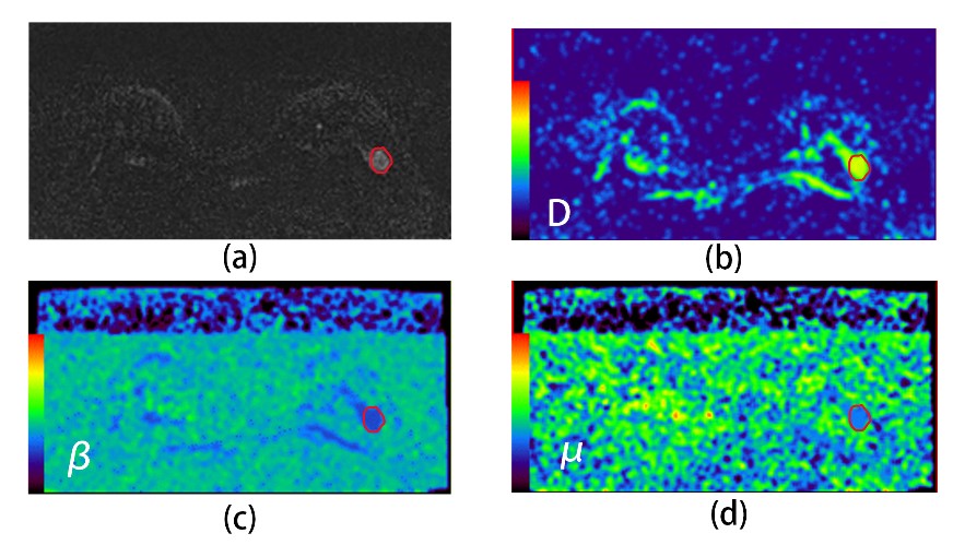
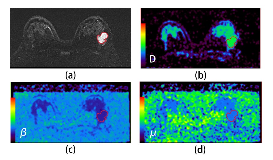
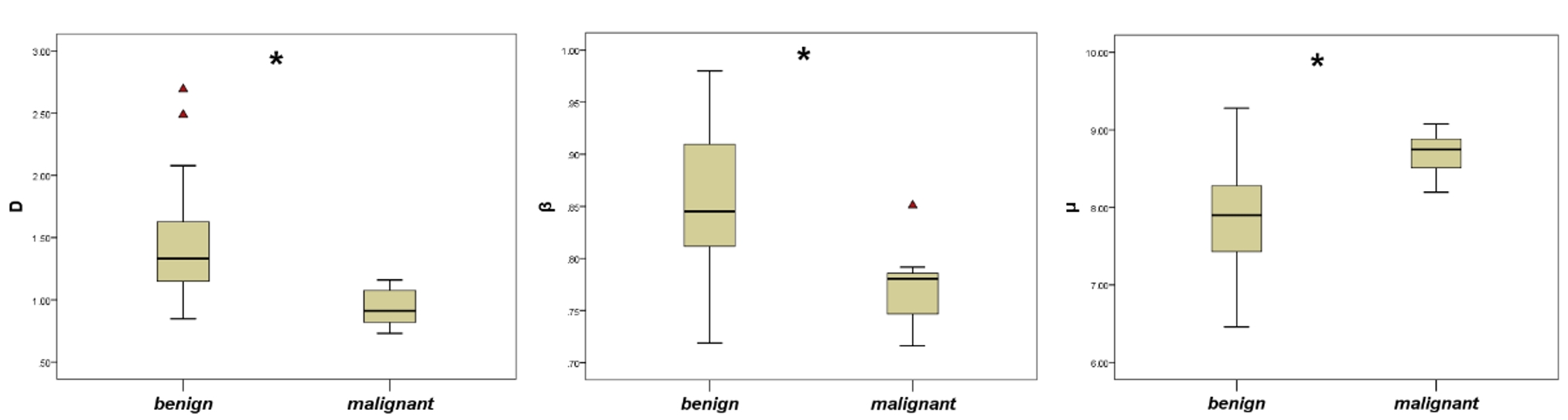
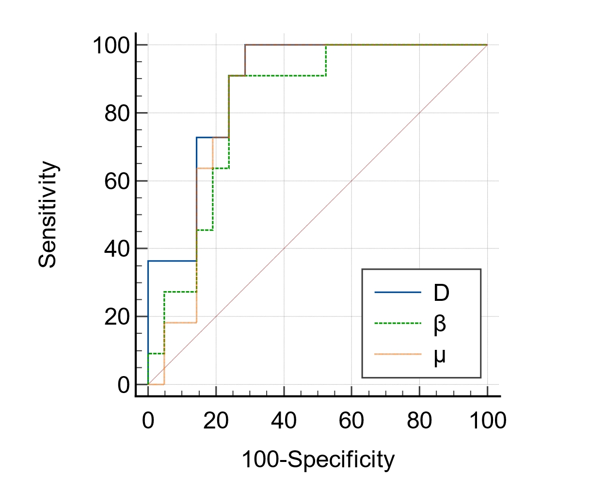
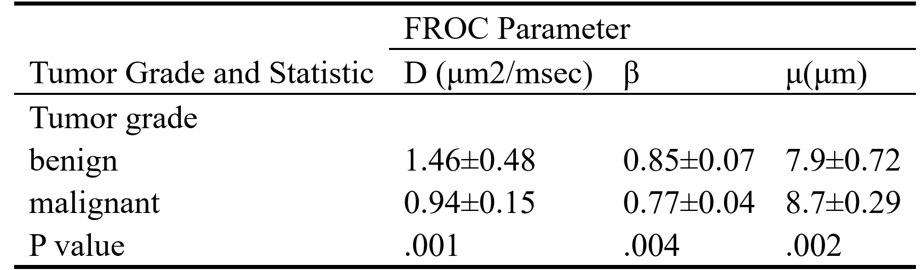
Comparison among Representative Patients in Each Group: Figure 1 shows a set of axial images of a patient with a benign breast lesion including diffusion-weighted imaging (DWI, b=800s/mm2) (Fig 1a), and the FROC maps (color images in Fig 1b-d). Figure 2 shows a set of axial images from a representative patient with the malignant breast lesion, including diffusion-weighted imaging (DWI, b=800s/mm2) (Fig 2a), and the FROC maps (color images in Fig 2b-d). Group Comparison Based on the FROC Parameters: The detailed statistical results of each FORC-derived metrics were shown in Table 1. The D and β values were significantly lower in the malignant lesions than those in the benign lesions (P=0.001, and P=0.004, respectively), whereas the μ values were higher in the malignant lesions than those in the benign lesions (P=0.002). ROC Analysis: The results of the diagnostic evaluation are shown in Fig 4. The AUC of D, β and μ for diagnosing the malignant lesion were respectively 0.88 (95% CI: 0.72-0.97), 0.82(95% CI: 0.64-0.93) and 0.84 (95% CI: 0.67-0.95). Besides, the results demonstrated that the AUC of D was significantly higher than those of β and μ.(Fig 4)Discussion:
Here we demonstrate the feasibility of using a set of FROC-derived diffusion parameters to differentiate malignant breast lesion from the benign lesions and found that D, β, and μ exhibited significant differences between the two subgroups. In this study, we found that the D values and β values were lower in malignant breast lesions than in benign lesions, which indicates that the diffusion rate of water molecules in malignant breast lesions is slower and that the internal tissue structure is more uneven, which is consistent with previous studies focusing on brain tumors(4, 5). However, the μ values in malignant lesions are higher than those in benign lesions. According to the previous studies, μ was found to be negatively correlated with the mean dispersion length of water molecules. The diffusion of water molecules in malignant lesions was limited, the average diffusion length was shorter, and the μ value was higher than that in benign lesions. Comparing the different parameters, D resulted in a higher diagnostic accuracy in terms of AUC for predicting malignancy.Conclusion:
This study demonstrated the feasibility of using the FROC diffusion model to improve the accuracy of identifying malignant breast lesions.Acknowledgements
NoneReferences
1. Sung H, Ferlay J, Siegel RL, Laversanne M, Soerjomataram I, Jemal A, et al. Global Cancer Statistics 2020: GLOBOCAN Estimates of Incidence and Mortality Worldwide for 36 Cancers in 185 Countries. CA: a cancer journal for clinicians. 2021;71(3):209-49.
2. Heller SL, Moy L. MRI breast screening revisited. Journal of magnetic resonance imaging : JMRI. 2019;49(5):1212-21.
3. Magin RL, Abdullah O, Baleanu D, Zhou XJ. Anomalous diffusion expressed through fractional order differential operators in the Bloch-Torrey equation. Journal of magnetic resonance (San Diego, Calif : 1997). 2008;190(2):255-70.
4. Sui Y, Wang H, Liu G, Damen FW, Wanamaker C, Li Y, et al. Differentiation of Low- and High-Grade Pediatric Brain Tumors with High b-Value Diffusion-weighted MR Imaging and a Fractional Order Calculus Model. Radiology. 2015;277(2):489-96.
5. Sui Y, Xiong Y, Jiang J, Karaman MM, Xie KL, Zhu W, et al. Differentiation of Low- and High-Grade Gliomas Using High b-Value Diffusion Imaging with a Non-Gaussian Diffusion Model. AJNR American journal of neuroradiology. 2016;37(9):1643-9.
Figures