3681
Added value of deep learning-accelerated T2-weighted imaging of the bladder on image quality and lesion evaluation1Peking Union Medical College Hospital, Beijing, China, 2Siemens Healthineers Ltd., Beijing, China, 3MR Application Predevelopment, Siemens Healthcare GmbH, Erlangen, Germany
Synopsis
This study evaluated deep learning accelerated T2w imaging (T2DL) of the bladder in terms of acquisition time (TA), overall image quality, presence of artifacts, diagnostic confidence, sharpness of lesions, and VI-RADS T2 score compared to a standard T2w (T2S) sequence in twenty-five patients. Two radiologists evaluated the images independently. TA of T2DL was reduced by nearly 50% compared to T2S. Overall image quality and sharpness of lesions were superior in T2DL, while artifacts, diagnostic confidence and T2 score were similar between T2S and T2DL. T2DL has the potential to replace T2S in bladder MR.
Introduction and purpose
Bladder cancer (BCa) is a common and sometimes potentially life-threatening disease.1 Multiparametric MRI (mpMRI) shows its great potential in BCa diagnosis and staging.2 But the long examination time of mpMRI is problematic especially in elderly patients. Recent novel deep learning (DL) reconstruction methods have been shown to be promising in reducing acquisition time.3 Thus, the purpose of this study was to investigate the impact of DL on T2-weighted imaging (T2DL) of the bladder regarding acquisition time (TA), image quality, and diagnostic confidence compared to standard T2-weighted imaging (T2S).Method
This prospective study consecutively enrolled 25 patients (19 female, 66 ± 14 years [range: 33~85 years]). Three patients had pathologically confirmed BCa and received neoadjuvant treatment before the examination, while the remaining 22 patients were clinically suspected of BCa. Following informed consent, all the patients underwent bladder mpMRI on a 3T MRI system (MAGNETOM Vida, Siemens Healthcare, Erlangen, Germany). Patients were asked to empty their bladders and then drink about 500 ml of water 30-60 minutes prior to the examination. Patients were scanned in supine position with a combined 18-channel body coil and 32-channel spine coil. The protocol of bladder mpMRI consisted of T2w imaging, diffusion weighted imaging, T1w imaging, and optional dynamic contrast enhanced imaging. T2w imaging was acquired in axial, coronal, and sagittal orientations and the prototypic T2DL turbo spin echo (TSE) sequence was acquired directly after T2S TSE. Acquisition parameters for T2S and T2DL TSE sequences were as follows: in-plane FOV 230 ´ 230 mm2, slice thickness 3 mm, 34 slices, voxel size reconstructed to 0.3 ´ 0.3 ´ 3.0 mm3, parallel imaging factor 2, turbo factor 20, echo trains per slice 12, TR = 7110 ms for T2S and 6920 ms for T2DL, TE = 106 ms for T2S and 108 ms for T2DL. Two averages were performed in T2S resulting in a TA of 2:58 min, whereas only one average was performed in T2DL resulting in a TA of 1:30 min in axial orientation and 1:40 min in coronal and sagittal orientations. All T2S and T2DL images were organized in a random order and evaluated by two radiologists (7 and 15 years of experience, blinded to sequence information) independently. A four-point Likert scale was used to rate the overall image quality (4=very good to 1=very poor), artifacts (4=no artifacts to 1=many artifacts), diagnostic confidence (4=very good to 1=non-diagnostic) and sharpness of lesions (4=no blurring to 1=very blurred) respectively. T2 score of the most malignant lesion was also evaluated using VI-RADS.4 The Wilcoxon signed-rank test was used for comparison. Cohen’s kappa was used to analyze inter-reader agreement. A two-sided P<0.05 was regarded as significant.Results
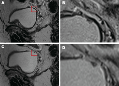
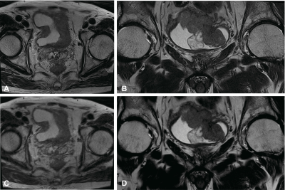
Of the 25 patients, 3 patients had diffuse thickening of bladder wall, 18 patients had focal bladder lesions, 2 patients had lesions in the left ureter and 2 patients had no visible lesions. The average size of focal bladder lesions was 24.0 ± 19.7 mm (range: 3.5~75.9 mm). Example sagittal comparison of lesion images for T2DL and T2S are shown in Figure 1; axial and coronal comparisons are shown in Figure 2. The results of both readers for image evaluation are summarized in Table 1. Inter-reader agreement for image quality parameters was good (kappa of 0.888 for overall image quality, 0.730 for artifacts, 0.865 for diagnostic confidence, 0.845 for sharpness). For both readers, on axial, coronal, and sagittal images, overall image quality and sharpness of lesions were rated significantly superior in T2DL, but there was no significant difference in diagnostic confidence between T2S and T2DL. For Reader 2, the severity and impact of artifacts on axial, coronal, and sagittal images was rated similar between T2S and T2DL . For Reader 1, the presence and impact of artifacts on axial images was rated superior in T2DL, but it was rated similar on coronal and sagittal images between T2S and T2DL. Inter-reader agreement for T2 score was very good for T2S (kappa of 0.885) T2DL (kappa of 0.833). No significant difference was found in T2 score of lesions between T2S and T2DL on axial, coronal, and sagittal images.Discussion and conclusion
This study investigated a novel deep learning T2w TSE sequence of the bladder and its influence on examination time, image quality, and lesion evaluation. Compared to T2S, acquisition time of T2w TSE imaging in T2DL could be reduced by 49.4% in axial orientation and 43.8% in coronal and sagittal orientations. Although the presence and impact of artifacts were observed similar in T2S and T2DL, bladder lesions in T2DL demonstrated superior sharpness compared to lesions in T2S, resulting in better overall image quality in T2DL. T2 score according to VI-RADS was also rated similar in T2S and T2DL. Our results indicate that T2DL might be able to replace conventional T2S with a significant reduction of acquisition time and preserved diagnostic confidence or superior lesion evaluation. But this study had a small patient population, and most bladder lesions were relatively large. Further investigation is needed to validate our results. In conclusion, a T2DL TSE sequence (with DL reconstruction) of the bladder could reduce acquisition time while maintaining image quality and lesion evaluation compared to T2S.Acknowledgements
NoneReferences
1. Antoni S, Ferlay J, Soerjomataram I, et al. Bladder Cancer Incidence and Mortality: A Global Overview and Recent Trends. Eur Urol. 2017 Jan;71(1):96-108.
2. Panebianco V, Pecoraro M, Del Giudice F, et al. VI-RADS for Bladder Cancer: Current Applications and Future Developments. J Magn Reson Imaging. 2020 Sep 17. doi: 10.1002/jmri.27361. Epub ahead of print. PMID: 32939939.
3. Hammernik K, Klatzer T, Kobler E, et al. Learning a variational network for reconstruction of accelerated MRI data. Magn Reson Med. 2018 Jun;79(6):3055-3071.
4. Panebianco V, Narumi Y, Altun E, et al. Multiparametric Magnetic Resonance Imaging for Bladder Cancer: Development of VI-RADS (Vesical Imaging-Reporting And Data System). Eur Urol. 2018 Sep;74(3):294-306.
Figures

