3679
Prostate cancer Diagnosis and risk stratification with combination of DISCO and MUSE: a feasibility study.1The Department of Radiology, The General Hospital of Ningxia Medical University, Yinchuan, China, 2GE Healthcare,MR Research, Beijing, China
Synopsis
In this study, we aim to investigate the value of DISCO quantitative parameters combined with MUSE in distinguishing benign and malignant prostate lesions. It was concluded that Ktrans, Kep, Ve and ADC can be used as imaging biomarkers to distinguish benign and malignant prostate lesions; Ktrans and ADC are helpful to predict aggressiveness of the tumor,and can be used as independent predictors of prostate cancer.
Summary of Main Findings
DISCO quantitative parameters and ADC can be used as imaging biomarkers to distinguish benign and malignant prostate lesions. There were significant differences in Ktrans, Kep, Ve and ADC between benign and malignant prostate lesions.Introduction
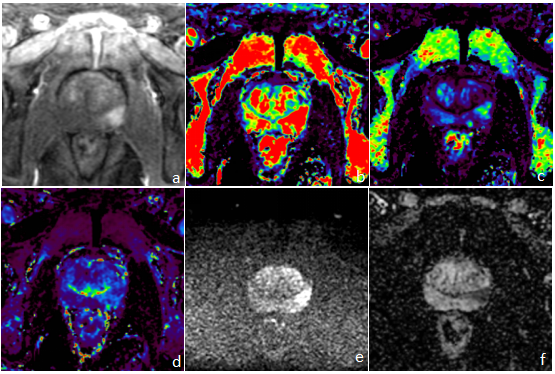
Dynamic contrast-enhanced (DCE) MRI and Diffusion-weighted imaging (DWI) both provide important basis for clinical diagnosis of prostate cancer (PCa). Traditionally, DCE MR imaging was performed with gradient echo sequence using cartesian k-space filling strategy. Under this circumstance, the high temporal resolution and high spatial resolution of DCE are often competing factors that cannot be achieved at same time. Low spatial resolution will lead to unclear display of small lesions, jeopardizing the differentiation of benign and malignant prostate lesions. Differential sub-sampling with cartesian ordering (DISCO) is novel DCE MR technique. Taking advantage of parallel imaging and a k-space view sharing reconstruction, it can improve temporal resolution while maintaining high spatial resolution[1-2]. As to diffusion imaging, to date the most widely used DWI is based on single shot echo planar imaging acquisition, which inherently has the limitation of low spatial resolution and spatial distortion. Multiplexed Sensitivity Encoding(MUSE) is multiple shot diffusion imaging technique, and has been reported to have better image quality in terms of spatial resolution and distortion [3-7]. At present, DISCO and MUSE have been successfully applied in liver, breast, pancreas and small intestine lesions etc [1-3,5-8], but their applications are rarely reported in prostate lesions. Therefore, the purpose of this study was to explore the value of DISCO combined with MUSE in differentiation of benign and malignant prostate lesions.Material and Methods
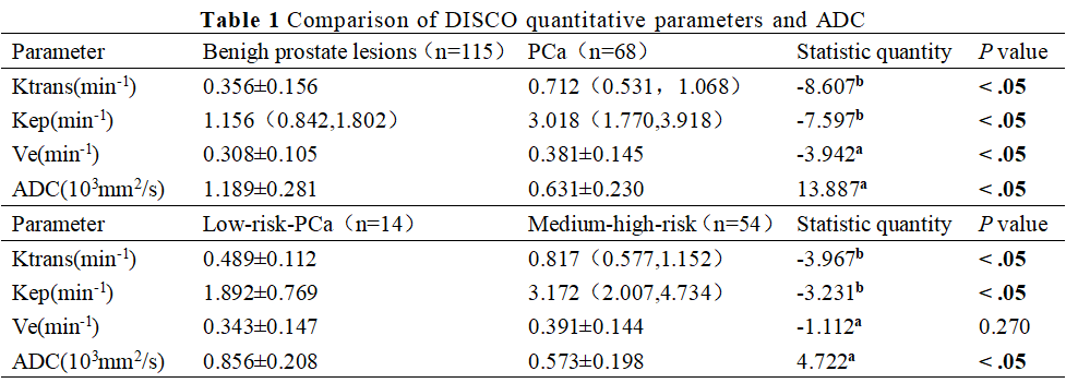
Our Institutional Review Board approved the protocol and written informed consent was obtained from each patient. From Jul 2020 to Aug 2021, the clinical and imaging data of 183 patients were included in our study, in which 68 cases were prostate cancer(Gleason score ≤ 6 of 14 cases,Gleason score ≥ 7 of 54 cases)and 115 cases were benign prostatic lesions (benign hyperplasia and / or inflammation ). All lesions were confirmed by biopsy or radical operation within one month of MR examination. Low-risk and Medium-high-risk PCa were defined as Gleason score ≤ 6 and Gleason score ≥ 7 respectively. All patients underwent 3.0T MR scanner (SIGNATM Architect, GE Healthcare, Milwaukee, USA ) with a 32-channel cardiac phased-array coil. The scan sequences included routine axial T1-weighted and T2-weighted imaging, sagittal T2-weighted and coronal T2-weighted imaging . MUSE and DISCO imaging parameters were as follows. MUSE: TR/TE=4800/76ms, FOV=26cm,slice thickness/spacing=3/0mm, NEX=1, number of shot = 2, b value =0,1000s/mm2 , matrix =180x108 ; DISCO: TR/TE=3.3/1ms, FOV=26cm, slice thickness/spacing=4/0mm, matrix = 320x256. A total 64 phases (5min12s) were scanned. All DCE and Diffusion post processing tasks were done on vendor provided advanced workstation. Ktrans、Kep、Ve、ADC of PCa group and benign prostate lesions group, low-risk PCa group and middle-high-risk PCa group were compared using Independent Student’s t-test or Mann-Whitney U test. The diagnostic performance was evaluated using ROC analysis. The correlation between the parameters and Gleason score was analyzed using Spearman correlation analysis. The parameters with statistically significant difference between PCa and benign prostate lesions group were analyzed using bivariate logistic regression analysis to determine the independent predictors of PCa.Results
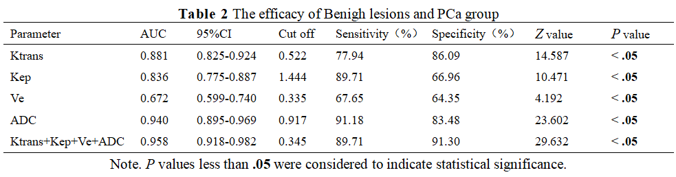
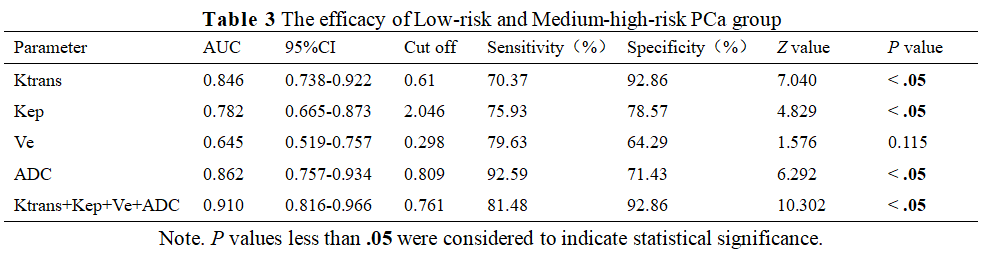
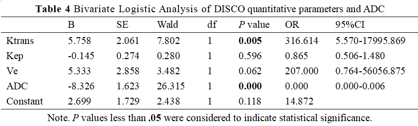
Among these parameters ,There were statistically significant differences in Ktrans、Kep、Ve and ADC between benign prostate lesions group and PCa group. There were no statistically significant differences in Ve between low-risk PCa group and medium-high-risk PCa group, while Ktrans, Kep and ADC were statistically significant different (Table 1, figure 1). The area under the curve (AUC) of Ktrans、Kep、Ve and ADC for differentiating benign prostate lesions from PCa、 low-risk PCa and medium-high-risk PCa is shown in Table 2. Gleason score has moderate negative correlation with ADC (r=-0.589, P<0.05), moderate positive correlation with Ktrans(r =-0.503, P<0.05), low positive correlation with Kep (r=0.375, P=0.02), and no significant correlation with Ve (r=0.144, P=0.242). Bivariate logistic regression analysis of Ktrans, Kep, Ve and ADC in benign prostate lesion group and PCa group was shown in Table 3.Discussion and Conclusion
Our study demonstrated DISCO quantitative parameters and ADC can be used as imaging biomarkers to distinguish benign and malignant prostate lesions. Ktrans, Kep and ADC could reflect the malignant degree of tumor to some extent. The approach combining all four parameters differentiated has the highest differential efficacy diagnosing benign prostate lesions from PCa,low-risk-PCa and medium-high-risk PCa. which is in accordance with the previous research results[9-10]. AUC of our study was higher than some previous literatures, which may be related to the higher spatial and temporal resolution of DISCO and MUSE. In addition, our study evaluated the correlation between Gleason score and parameters. One of the major limitations in our study is that the number of samples is too small. Therefore, future study with bigger cohort of subjects is needed. To conclude, Ktrans, Kep, Ve and ADC can be used to distinguish benign and malignant prostate lesions;Ktrans and ADC are helpful to predict aggressiveness of the tumor,and can be used as independent predictors of prostate cancer.Acknowledgements
We thank wiley for all physicians,assistance of The Department of Radiology, The General Hospital of Ningxia Medical University during the preparation of this manuscript. We thank all of the participants of this study. We also acknowledge the support provided by Dr.Xiaocheng Wei from GE Healthcare. Beijing,China.References
1.Guo Yafei, Lu Lin, Zhao Xin, et al. Guo Yafei, Lu Lin, Zhao Xin, et al. The value of disco imaging in the diagnosis of benign and malignant breast lesions [J]. Journal of Zhengzhou University (Medical Edition), 2021 556 (3): 397-400.
2.Donati F, Boraschi P, Cervelli R, et al. 3 T MR perfusion of solid pancreatic lesions using dynamic contrast-enhanced DISCO sequence: Usefulness of qualitative and quantitative analyses in a pilot study. Magn Reson Imaging. 2019;59:105-113.
3.Ichikawa S, Motosugi U, Oishi N, et al. Ring-Like Enhancement of Hepatocellular Carcinoma in Gadoxetic Acid-Enhanced Multiphasic Hepatic Arterial Phase Imaging With Differential Subsampling With Cartesian Ordering. Invest Radiol. 2018;53(4):191-199.
4.Baxter GC, Patterson AJ, Woitek R, Allajbeu I, Graves MJ, Gilbert F. Improving the image quality of DWI in breast cancer: comparison of multi-shot DWI using multiplexed sensitivity encoding to conventional single-shot echo-planar imaging DWI. Br J Radiol. 2021;94(1119):20200427.
5.Hu Y, Ikeda DM, Pittman SM, et al. Multishot Diffusion-Weighted MRI of the Breast With Multiplexed Sensitivity Encoding (MUSE) and Shot Locally Low-Rank (Shot-LLR) Reconstructions. J Magn Reson Imaging. 2021;53(3):807-817.
6.Daimiel Naranjo I, Lo Gullo R, Morris EA, et al. High-Spatial-Resolution Multishot Multiplexed Sensitivity-encoding Diffusion-weighted Imaging for Improved Quality of Breast Images and Differentiation of Breast Lesions: A Feasibility Study. Radiol Imaging Cancer. 2020;2(3):e190076.
7.Chang HC, Chen G, Chung HW, et al. Multi-shot Diffusion-Weighted MRI With Multiplexed Sensitivity Encoding (MUSE) in the Assessment of Active Inflammation in Crohn's Disease [published online ahead of print, 2021 Jun 25].J Magn Reson Imaging.2021;10.1002/jmri.27801.
8.Kim YY, Kim MJ, Gho SM, Seo N. Comparison of multiplexed sensitivity encoding and single-shot echo-planar imaging for diffusion-weighted imaging of the liver. Eur J Radiol. 2020;132:109292.
9.Zhu G, Luo J, Ouyang Z, et al. The Assessment of Prostate Cancer Aggressiveness Using a Combination of Quantitative Diffusion-Weighted Imaging and Dynamic Contrast-Enhanced Magnetic Resonance Imaging. Cancer Manag Res. 2021;13:5287-5295.
10.Ma XZ, Lv K, Sheng JL, et al. Application evaluation of DCE-MRI combined with quantitative analysis of DWI for the diagnosis of prostate cancer. Oncol Lett. 2019;17(3):3077-3084.
Figures