3667
Compressed Sensing and Deep Learning Reconstruction: Efficacy for Women’s Pelvic MRI at 1.5 T MR System in Routine Clinical Practice1Radiology, Fujita Health University School of Medicine, Toyoake, Japan, 2Canon Medical Systems Corporation, Otawara, Japan, 3Radiology, Fujita Health University Bantane Hospital, Nagoya, Japan, 4Joint Research Laboratory of Advanced Medical Imaging, Fujita Health University School of Medicine, Toyoake, Japan
Synopsis
We hypothesized that compressed sensing (CS) with deep learning reconstruction (DLR) can improve image quality and shorten examination time on not only T2-weighted imaging (T2WI), but also T1-weighted imaging (T1WI) on women’s pelvic MRI, when compared with PI at 1.5T MR system. The purpose of this study was to compare the utility of CS and DLR for shortening examination time and improving image quality on MRI at 1.5T system in patients with various female pelvic diseases.
Introduction
Compressed sensing has been introduced and tested by various MR sequences in the last decade1, 2. In addition, deep learning reconstruction (DLR) has been rapidly and clinically applied not on CT, but also MRI in the last a few years3-5. In 2020, the utility of CS and DLR was compared with routinely applied parallel imaging (PI) and demonstrated their utility on only T2WI at 3T MR system on women’s pelvic MRI6. Although T2WI is considered as one of the main sequences of the women’s pelvic MRI, it would be better to test these techniques with other MR sequences for women’s pelvic MRI. In addition, DLR can be applied at not only 3T, but also 1.5T MR systems in routine clinical practice. However, no major reports were assessed the utility of CS and DLR on women’s pelvic MRI as compared with PI at 1.5T MR system. We hypothesized that CS with DLR can improve image quality and shorten examination time on women’s pelvic MRI at 1.5T systems, when compared with conventional PI. The purpose of this study was to directly compare the capability of CS and DLR for shortening examination time and improving image quality with conventional PI at 1.5T system in patients with various female pelvic diseases.Materials and Methods
52 consecutive female patients (mean age 44 years, range 22–85 years) with various pelvic diseases underwent T1WI and T2WI by CS and PI. Then, each CS data was reconstructed with and without DLR, and examination times of CS and PI were recorded in all patients. For quantitative assessment, SNR of muscle and CNR between fat tissue and iliac muscle on T1WI, and myometrium and straight muscle on T2WI were determined by ROI measurement. For qualitative assessment, two board certified radiologists assessed overall image quality and diagnostic confidence level by 5-point scales, and each final score was determined as consensus of two readers. To compare the capability for examination time reduction, mean examination times of both sequences were compared among all data sets by Tukey’s HSD test. To determine quantitative image quality improvement on each CS data by DLR, SNRs and CNRs were compared among all methods by Tukey’s HSD test. On qualitative image quality evaluations, inter-observer agreement on each data set was assessed by κ statistics followed by χ2 test. Finally, both indexes on T1WI and T2WI were compared among all methods by Wilcoxon signed–rank test. A p value less than 0.05 was considered as significant in this study.Results
Representative cases are shown in Figure 1 and 2. Compared results of mean examination time, SNR and CNR are shown in Figure 3. Mean examination times of T1WI and T2WI obtained by CS with and without DLR were significantly shorter than those by conventional PI (p<0.05). SNRs of T1WI and T2WI obtained by CS with DLR were significantly higher than those of CS without DLR (p<0.05). SNRs of T1WI and T2WI obtained by CS with DLR were significantly higher than those of conventional PI (p<0.05). CNRs of T1WI and T2WI obtained by CS with DLR were significantly higher than those of CS without DLR (p<0.05). CNRs of T1WI and T2WI obtained by CS with DLR were significantly higher than those of conventional PI (p<0.05). Interobserver agreements for overall image quality and diagnostic confidence level and compared results of both qualitative indexes are shown in Figure 4 and 5. Inter-observer agreements were determined as ‘substantial’ or ‘almost perfect’ (0.71<κ<0.99, p<0.05). Overall image qualities of T1WI and T2WI obtained by CS with DLR were significantly higher than those by CS without DLR (p<0.05) and conventional PI (p<0.05). Diagnostic confidence level of T2WI obtained byCS with DLR was significantly higher than that by conventional PI (p<0.05) and CS without DLR (p<0.05).Conclusion
CS with DLR is considered as useful for image quality improvement with reducing examination time on women’s pelvic MRI at 1.5 T system, when compared with PI.Acknowledgements
This work was technically and financially supported by Canon Medical Systems Corporation.References
- Tsao J, Kozerke S. MRI temporal acceleration techniques. J Magn Reson Imaging. 2012 Sep;36(3):543-60.
- Feng L, Benkert T, Block KT, et al. Compressed sensing for body MRI. J Magn Reson Imaging. 2017 Apr;45(4):966-987.
- Higaki T, Nakamura Y, Tatsugami F, et al. Improvement of image quality at CT and MRI using deep learning. Jpn J Radiol. 2019 Jan;37(1):73-80.
- Singh R, Wu W, Wang G, et al. Artificial intelligence in image reconstruction: The change is here. Phys Med. 2020 Nov;79:113-125.
- Lin DJ, Johnson PM, Knoll F, et al. Artificial Intelligence for MR Image Reconstruction: An Overview for Clinicians. Magn Reson Imaging. 2021 Apr;53(4):1015-1028.
- Ueda T, Ohno Y, Kaori Y, et al. Compressed sensing and deep learning reconstruction for women's pelvic MRI denoising: Utility for improving image quality and examination time in routine clinical practice. Eur J Radiol. 2021 Jan;134:109430.
Figures
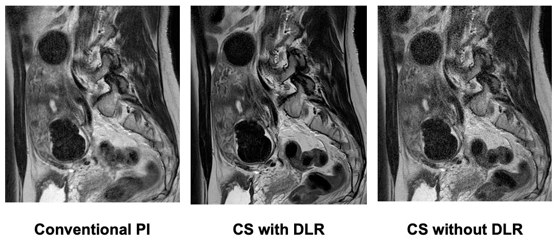
Figure 1. 50-year-old female patient with uterine myoma on T2WI.
(L to R: Conventional PI, CS with DLR, CS without DLR). MR examination times for all methods are as follows: Conventional PI, 285 s; CS with DLR, 107 s; CS without DLR, 103 s. CS with and without DLR reduces examination times as compared with conventional PI. CS with DLR reduces image noise and demonstrate uterine myoma as well as normal structure more clearly as compared with conventional PI and CS without DLR.
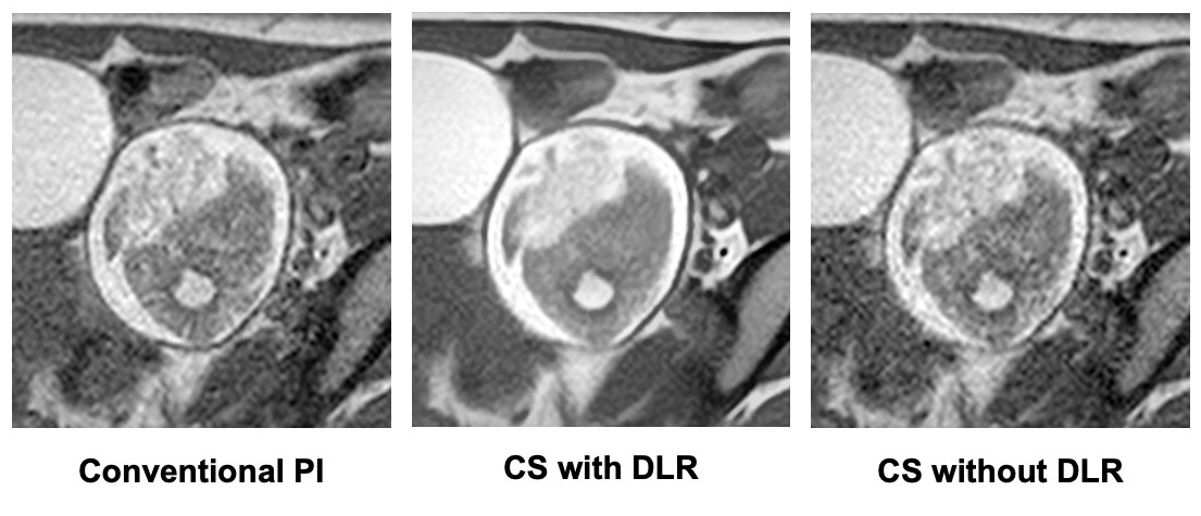
Figure 2. 32-year-old female patient with ovarian mature teratoma on T1WI.
(L to R: Conventional PI, CS with DLR, CS without DLR). All images clearly demonstrate ovarian mature teratoma. MR examination times for all methods are as follows: Conventional PI, 243 s; CS with DLR, 62 s; CS without DLR, 58 s. CS with and without DLR reduces examination times as compared with conventional PI. CS with DLR reduces image noise and demonstrate ovarian mature teratoma more clearly as conventional PI and CS without DLR.
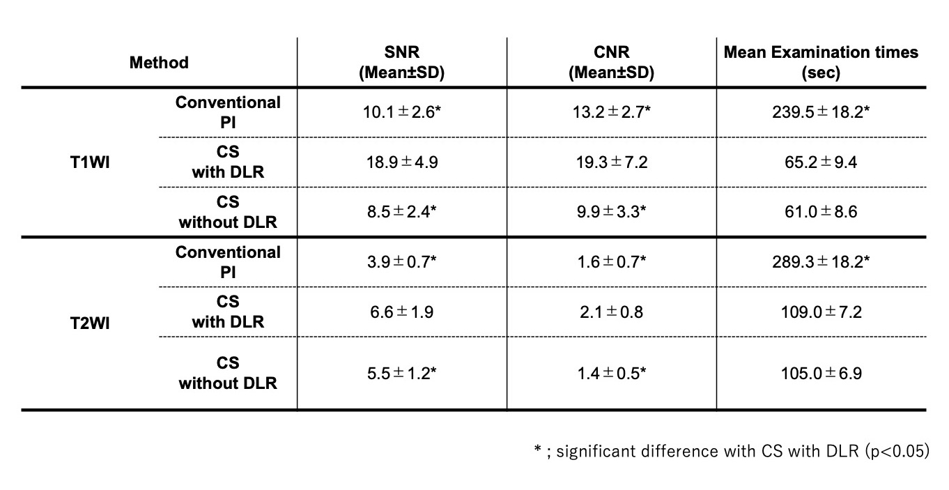
Figure 3. Compared results for mean examination time and each quantitative image quality on T1WI and T2WI among three methods.
Mean examination times of CS with and without DLR on T1WI and T2WI were significantly shorter than those of conventional PI (p<0.05). SNRs and CNRs of T1WI and T2WI obtained by CS with DLR were significantly higher than those of CS without DLR and conventional PI (p<0.05).
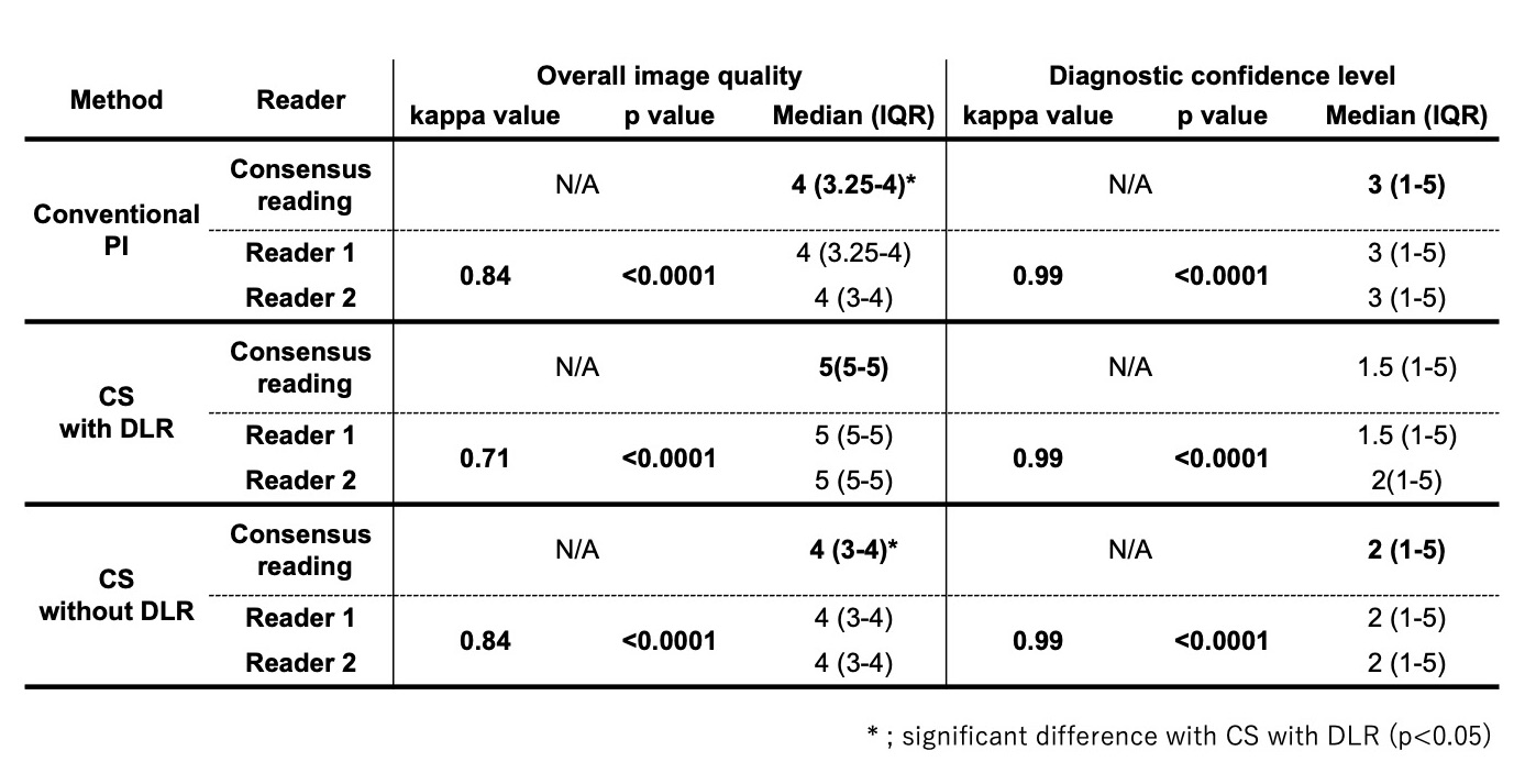
Figure 4. Overall image quality and diagnostic confidence level on three methods on T1WI.
Overall image qualities of T1WI (Median 5, IQR 5-5) by CS with DLR were significantly higher than that for conventional PI (Median 4, IQR 3.25-4, p<0.05) and CS without DLR (T1WI: Median 4, IQR 3-4, p<0.05). There were no significant differences in diagnostic confidence levels among any of the three methods on T1WI (PI: Median 3, IQR 1-5, CS with DLR: Median 1.5, IQR 1-5, CS without DLR: Median 2, IQR 1-5, p>0.05).
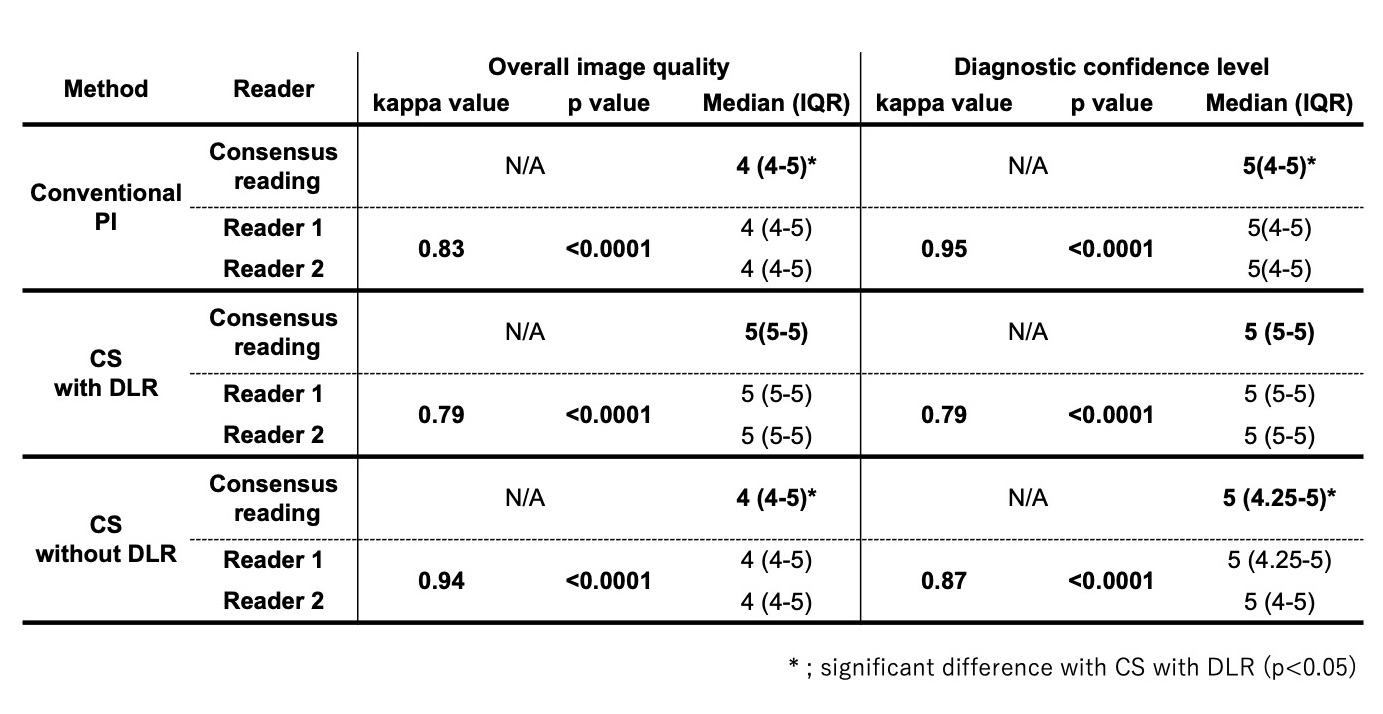
Figure 5. Overall image quality and diagnostic confidence level on three methods on T2WI.
Overall image qualities of T2WI (Median 5, IQR 5-5) by CS with DLR were significantly higher than that for conventional PI (Median 4, IQR 4-5, p<0.05) and CS without DLR (Median 4, IQR 3-4, p<0.05). Diagnostic confidence levels of T2WI (Median 5, IQR 5-5) by for CS with DLR were significantly higher than that for conventional PI (Median 5, IQR 4-5, p<0.05) and CS without DLR (Median 5, IQR 4.25-5, p<0.05).