3663
Differentiating Benign from Malignant Renal Cystic Masses Using Continuous-time Random-walk diffusion model1Department of Radiology, Sun Yat-Sen Memorial Hospital, Sun Yat-Sen University, Guangzhou, China, 22MR Scientific Marketing, Siemens Healthineers, Guangzhou, China, 3Shanghai Key Laboratory of Magnetic Resonance, School of Physics and Electronic Science, East China Normal University, Shanghai, China
Synopsis
This study evaluated whether continuous-time random-walk (CTRW) diffusion model could be used to distinguish benign from malignant renal tumors, as well as compared the diagnostic effectiveness between the diffusion parameters of CTRW and apparent dispersion coefficient (ADC). The results demonstrated that CTRW diffusion model as a new noninvasive MR technique, was able to differentiate benign from malignant renal masses in vivo, and further improve the diagnostic efficiency of renal malignant masses compared with conventional diffusion imaging.
Introduction
Solid renal masses are the most common tumors of the urinary system, with increasing detection rate in recent years [1]. Contrast-enhanced CT and conventional MRI are routinely used in the characterization of renal lesions. However, approximately 11%-16% of radiologic suspected malignant renal masses had benign pathologic findings on surgical resection [2, 3]. Accurate preoperative differentiation between benign renal mass and malignant tumor is thus important for appropriate treatment strategies. Recent studies implied that apparent diffusion coefficient (ADC) values derived from conventional mono-exponential model could be used for the characterization of solid renal masses, which is sensitive to tissue cellularity changes [4]. The continuous-time random-walk (CTRW) model using a more sophisticated algorithm has been reported to describe anomalous diffusion in biological tissues more specifically, which is characterized by diffusion wait time and jump length to reflect the temporal and spatial heterogeneity of diffusion in voxels, respectively [5]. At present, the value of CTRW model in renal research is unclear. Thus, the aim of this study was to explore the feasibility of CTRW model in distinguishing benign from malignant renal masses, and to compare with the diagnostic effectiveness of ADC.Methods
A total of 21 patients with renal tumors were prospectively recruited, including 11 malignant tumors and 10 benign masses according to pathologic findings. All patients underwent renal MRI on a 3T MR scanner (MAGNETOM Skyra, Siemens Healthcare, Erlangen, Germany) with a phased array body coil. Magnetic resonance (MR) examination, including conventional T1-, T2-weighted imaging, dynamic contrast-enhanced (DCE)-MRI and diffusion-weight imaging (DWI) sequence, was performed. The DWI data was acquired by using a single-shot echo planar imaging (EPI) sequence with 15 b values of 0, 10, 20, 30, 50, 70, 100, 200, 400, 600, 800, 1000, 1500, 2000, 2500, 3000 s/mm2. The imaging parameters were as follows: TE=69 ms, TR=6700 ms, field of view = 294 × 429 mm2, acquisition matrix = 96 × 140, slice thickness = 5 mm. The parameterric maps of CTRW (extract anomalous diffusion coefficient, Dm, and temporal and spatial heterogeneity parameters, α and β) and mono-exponential model (ADC) were calculated using a unified in-house developed software called BoDiLab, which is based on Python 3.7. The average of all diffusion parameters of each patient's renal mass was recorded for statistical analysis. Differences in Dm, α, β and ADC values were evaluated between benign and malignant renal masses by using the independent t-test. Diagnostic performances were assessed by receiver operating characteristic (ROC) analysis. P < 0.05 was considered statistically significant.Results
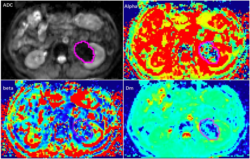
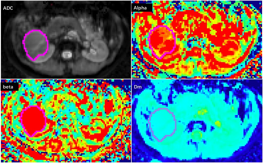
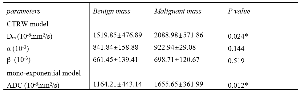
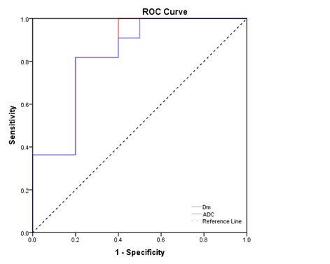
The typical cases of benign and malignant renal tumors are shown in Figure 1 and 2. The mean values of Dm and ADC of malignant renal masses was significantly increased compared with that of benign masses (P=0.024, 0.012). However, α and β showed no differences between benign and malignant renal tumors (Table 1). According to the ROC curve analysis, the AUCs of Dm and ADC were 0.836 and 0.827, the sensitivity was 0.818 and 0.818, and specificity was 0.8 and 0.8, respectively (Figure 3).Discussion & Conclusion
In our study, Dm derived from CTRW can be used in the differentiation of benign and malignant renal tumors, and it showed better diagnostic performance than ADC. Dm, as a novel indicator describing anomalous diffusion in tissue [6], was significantly higher in malignant renal tumors compared with benign masses, which may suggest that there is more anomalous diffusion in malignant renal tumors. Therefore, CTRW was more sensitive than conventional DWI in identifying benign and malignant renal tumors, and may contribute to better understanding of pathophysiology of renal tumors.Acknowledgements
No acknowledgement found.References
[1] Karaosmanoğlu AD, Onur MR, Shirkhoda A, Ozmen M, Hahn PF. Unusual benign solid neoplasms of the kidney: cross-sectional imaging findings. Diagn Interv Radiol. 2015 Sep-Oct;21(5):376-81. doi: 10.5152/dir.2015.14545.
[2] Kutikov A, Fossett LK, Ramchandani P, Tomaszewski JE, Siegelman ES, Banner MP, Van Arsdalen KN, Wein AJ, Malkowicz SB. Incidence of benign pathologic findings at partial nephrectomy for solitary renal mass presumed to be renal cell carcinoma on preoperative imaging. Urology. 2006 Oct;68(4):737-40. doi: 10.1016/j.urology.2006.04.011.
[3] Fujii Y, Komai Y, Saito K, Iimura Y, Yonese J, Kawakami S, Ishikawa Y, Kumagai J, Kihara K, Fukui I. Incidence of benign pathologic lesions at partial nephrectomy for presumed RCC renal masses: Japanese dual-center experience with 176 consecutive patients. Urology. 2008 Sep;72(3):598-602. doi: 10.1016/j.urology.2008.04.054. Epub 2008 Jul 23.
[4] Ludwig, D. R., Ballard, D. H., Shetty, A. S., Siegel, C. L., & Yano, M. (2020). Apparent Diffusion Coefficient Distinguishes Malignancy in T1-Hyperintense Small Renal Masses. AJR Am J Roentgenol, 214(1), 114-121. doi: 10.2214/AJR.19.21907
[5] Ingo, C., Magin, R. L., Colon-Perez, L., Triplett, W., & Mareci, T. H. (2014). On random walks and entropy in diffusion-weighted magnetic resonance imaging studies of neural tissue. Magn Reson Med, 71(2), 617-627. doi: 10.1002/mrm.24706
[6] Karaman, M. M., Wang, H., Sui, Y., Engelhard, H. H., Li, Y., & Zhou, X. J. (2016). A fractional motion diffusion model for grading pediatric brain tumors. Neuroimage Clin, 12, 707-714. doi: 10.1016/j.nicl.2016.10.003
Figures