3651
Improved Motion Correction in Dynamic Contrast Enhancement MRI Using Low Rank with Soft Weighting1SPMIC, The University of Nottingham Ningbo China, Ningbo, China, 2COMSATS University Islamabad, Islamabad, Pakistan, 3SPMIC, The University of Nottingham, Nottingham, United Kingdom
Synopsis
This work presents a robust motion corrected free breathing Dynamic Contrast Enhanced MRI (DCE-MRI) reconstruction method called Low rank plus sparse (L+S) with soft weighting which compressed motion blurring by employing a soft weighting matrix. The motion correction performance was quantified in this work. A computer simulation framework based on a modified shepp-logan model was developed with the ground truth to quantify the reconstruction errors. The proposed method achieved better motion correction and high reconstruction efficiency simultaneously.
Introduction
Golden Angle Radial Sparse Parallel (GRASP) MRI1 and L+S decomposition2 are proposed for free breathing DCE-MRI with prior temporal and spatial resolution by exploring the temporal sparsity among subdivided time frames. Stack-of-stars sampling pattern offers high motion robustness. However, periodic respiratory motion still results in blurring artefacts, degrading reconstruction quality. To alleviate respiratory motion blurring, extra dimension (XD) GRASP3 and motion-weighted GRASP (RACER-GRASP) MRI4,5 are updated with a further motion states subdivision within each time frame. XD-GRASP explores the additional motion sparsity among motion states. RACER-GRASP combines motion states with different weighting factors to lock the desired motion phase. However, additional motion dimension results in excessive computation burden. Grappa operator gridding (GROG) has been introduced to RACER-GRASP to accelerate the reconstruction4. L+S decomposition subdivides the image series into low rank background components (L) and sparse dynamic components (S), enabling efficient reconstruction of highly under-sampled DCE-MRI datasets. Rapid convergence is achieved by using Iterative Soft Thresholding Algorithm (ISTA). Here, we propose a soft weighting matrix to replace motion states subdivision into L+S decomposition model, compressing motion blurring and maintaining reconstruction efficiency simultaneously. Meanwhile, it is difficult to quantify motion correction performance in clinic scan without reference. A simulated phantom dataset with motion ground truth is created, enabling quantification of motion errors in different reconstruction schemes.Method
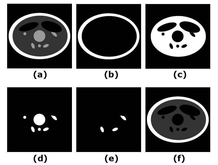
A simulated phantom dataset and a clinic free breathing liver DCE-MRI dataset offered by Li et al3 were implemented to evaluate the performance of the proposed method. A modified Shepp-logan computer model with 512*512 voxels was created as shown in Figure 1. Phantom contained six dynamic regions where the signal intensity was varied with designed dynamic curve. Three of dynamic regions were rotated periodically at a maximum angle of 12 degrees. A total of 1100 spokes with 768 readout points in each spoke and 8 virtual channels were acquired by the golden angle radial sampling pattern during the dynamic and motion variation period. The ground truth of phantom series was recorded as the reference. The liver dataset contains 8 channels, 1100 spokes with 512 readout points in each.The proposed reconstruction scheme is mathematically formulated as: $$ argmin_{L,S}=\frac{1}{2}\left \|W\{E(L+S)-d\}\right\|_{2}^{2}+\lambda_{L}\left \| L \right \|_{*}+\lambda_{T}\left \| TS \right \|_{1}\qquad \qquad [1]$$ where $$$ E $$$ is the multi-coil encoding operator and $$$ d $$$ is the acquired data. $$$ W $$$ is the soft weighting matrix for acquired spokes, produced by a modified sigmoid function6. $$$ T $$$ is the temporal TV sparsity transform. Parameters $$$ \lambda_{L} $$$ and $$$ \lambda_{T} $$$ trade off data consistency versus complexity of solution given by the nuclear norm $$$ \left \| \cdot \right \|_{*} $$$ and $$$ L_{1} $$$ norm $$$ \left \| \cdot \right \|_{1} $$$.
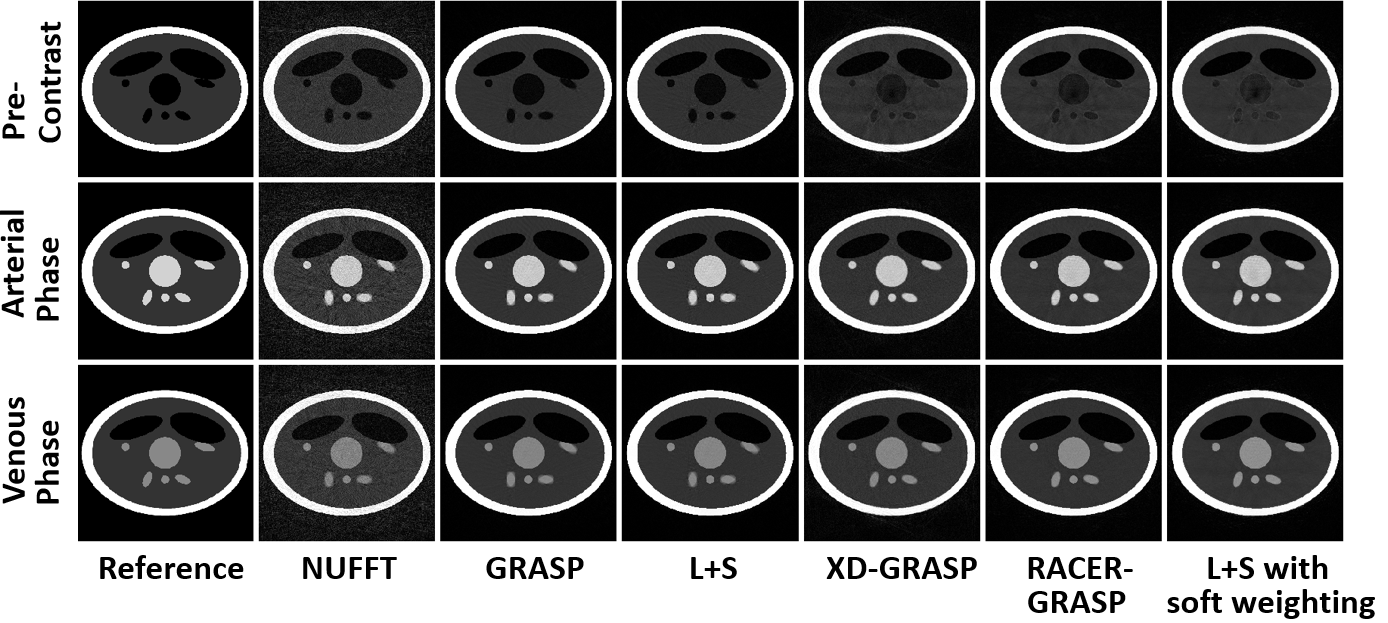
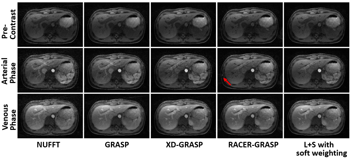
Both two datasets were subdivided into 11 time frames with the temporal resolution of 100 spokes/frame. Phantom dataset was reconstructed by GRASP, XD-GRASP, RACER-GRASP, L+S decomposition and proposed L+S with soft weighting respectively with a matrix size of 512*512*11. The RMSE value between reconstructed results and reference was regarded as the standard to quantify motion correction performance. The liver dataset was reconstructed by GRASP, XD-GRASP, RACER-GRASP with GROG acceleration and L+S with soft weighting with a matrix size of 384*384*11. All the reconstructions were performed using MATLAB 2020b (MathWorks, Natick, MA) on an Intel Core i7-10700 PC with a 2.9 GHz processor.
Results & Discussion
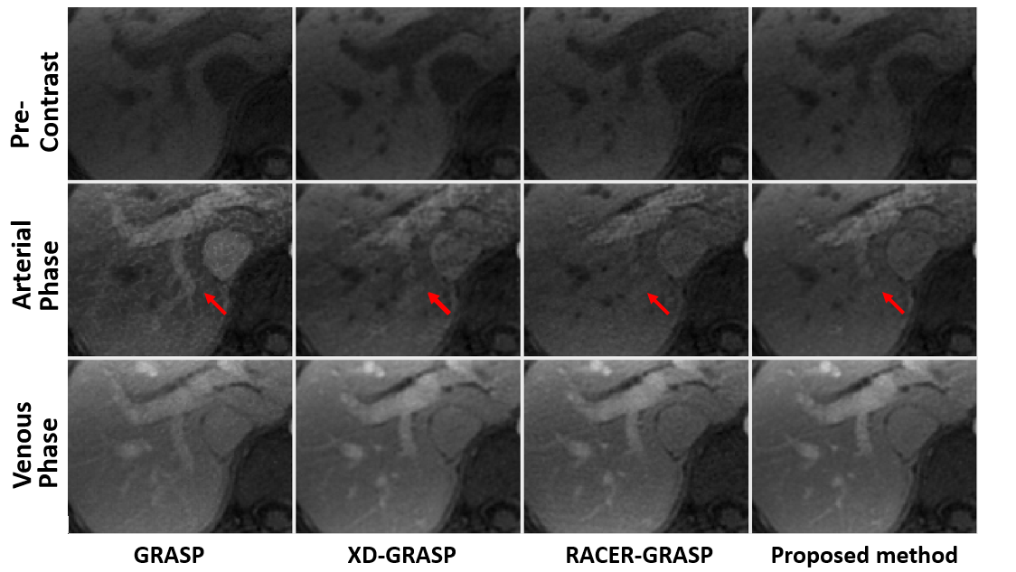
Figure 2 and Figure 3 show a comparison of five reconstruction schemes in the simulated phantom dataset. Obvious motion artefacts were observed in GRASP and L+S decomposition. Motion blurring was compressed effectively in XD-GRASP, RACER-GRASP and L+S with soft weighting. Some residual streaking artefacts were obtained in XD-GRASP. Using the selected motion section in reference as the benchmark, the RMSE value of GRASP, L+S decomposition, RACER-GRASP and L+S with soft weighting was 0.037, 0.038, 0.033, 0.024 and 0.02 respectively, demonstrating the feasibility of the proposed computer simulation framework to evaluate the motion correction quantitatively. Minimum motion error was achieved by proposed method, showing the robustness of soft weighting. Compared with GRASP, additional motion dimension significantly degraded the reconstruction efficiency in XD-GRASP and RACER-GRASP. Our proposed method and standard L+S achieved similar reconstruction efficiency, much higher than GRASP based techniques. The reconstruction period of L+S decomposition and proposed method was 273.2s, and 275.3s respectively. Figure 4 and Figure 5 show a comparison of different reconstruction schemes in liver dataset in end-expiration phase. Residual streaking artefacts were obtained in XD-GRASP. Less motion blurring was observed in RACER-GRASP. Unexpected convolution artefacts were obtained in RACER-GRASP due to the GROG acceleration. Best motion compression and tissue detail were observed in the proposed method especially in arterial phase. GROG improved the reconstruction efficiency of RACER-GRASP significantly while proposed method still achieved the highest reconstruction efficiency without GROG acceleration.Conclusion
A computer simulation framework was developed and validated to quantitatively evaluate the motion correction errors in DCE-MRI. A low rank with soft weighting framework was proposed and evaluated for improved DCE-MRI reconstruction. Soft weighting leads to better motion correction for the L+S decomposition based method. The additional computation cost caused by soft weighting is negligible. Additional feasibility of motion phase can be achieved by shifting the soft weighting matrix.Acknowledgements
No acknowledgement found.References
1. Feng L, Grimm R, Block KT, Chandarana H, Kim S, Xu J, Axel L, Sodickson DK, Otazo R. Golden-angle radial sparse parallel MRI: combination of compressed sensing, parallel imaging, and golden-angle radial sampling for fast and flexible dynamic volumetric MRI. Magn Reson Med. 2014;72(3):707-717.
2. Otazo R, Cande`s E, Sodickson DK. Low-Rank Plus Sparse Matrix Decomposition for Accelerated Dynamic MRI with Separation of Background and Dynamic Components. Magn Reson Med. 2015;73(3):1125–1136.
3. Feng L, Axel L, Chandarana H, Block KT, Sodickson DK, Otazo R. XD-GRASP: golden-angle radial MRI with reconstruction of extra motion-state dimensions using compressed sensing. Magn Reson Med. 2016;75:775–788.
4. Feng L, Huang C, Shanbhogue K, Sodickson DK, Chandarana H, Otazo R. RACER-GRASP: Respiratory-Weighted, Aortic Contrast Enhancement-Guided and Coil-Unstreaking Golden-Angle Radial Sparse MRI. Magn Reson Med 2018;80:77–89.
5. Chen L, Zeng X, Ji B, Liu D, Wang J, Zhang J, Feng L. Improving dynamic contrast-enhanced MRI of the lung using motion-weighted sparse reconstruction: Initial experiences in patients. Magnetic resonance imaging. 2020; 68:36-44.
6. Najeeb F, Zhang J, Wang X, Wang C, Omer H, Gowland P, Francis S, Glover P. Efficient DCE-MRI image reconstruction with feasible temporal resolution in L+S Decomposition model. Proceedings of the ISMRM 30th Annual Meeting & Exhibition 2021. International Society for Magnetic Resonance in Medicine.
Figures