3649
Quantification of fine CSF dynamics using Intra-Voxel Incoherent Motion (IVIM) MRI1Neurosurgery, Shiga University of Medical Science, Otsu, Japan, 2Interfaculty Initiative in Information Studies, Institute of Industrial Science, The University of Tokyo, Tokyo, Japan, 3Radiology, Shiga University of Medical Science, Otsu, Japan, 4Mechanical Science and Bioengineering, Graduate School of Engineering Science, Osaka University, Osaka, Japan
Synopsis
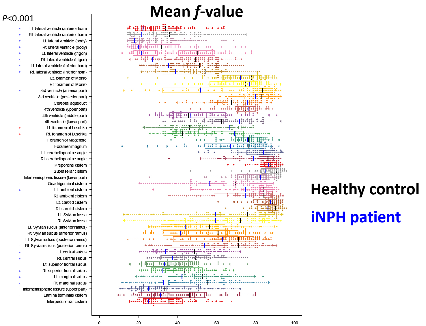
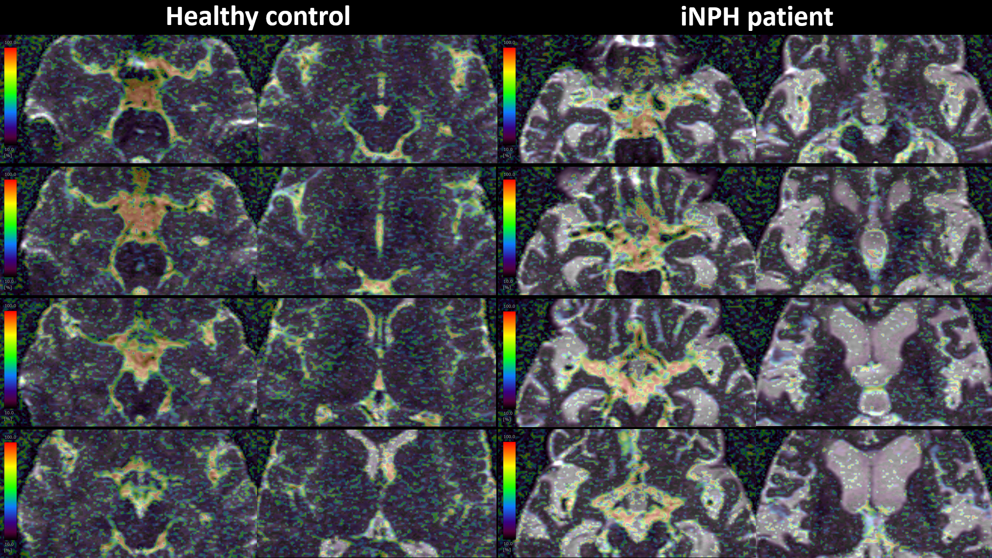
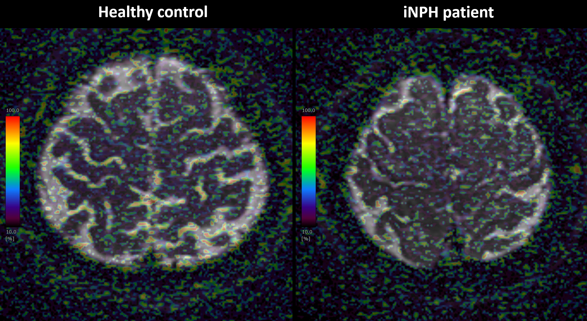
The f value calculated by the Intra-Voxel Incoherent Motion (IVIM) MRI was was high in the areas where the CSF flow was reported to be fast, specifically in the cerebral aqueduct and subarachnoid spaces around the major intracranial arteries. Therefore, f value was considered to reflect flow velocity of CSF, regardless of its direction. The mean f values in the whole lateral ventricles, anterior part of the third ventricle, ambient cisterns, central sulci, and marginal sulci were significantly lower and that in the foramina of Luschka was significantly higher among 28 iNPH patients, compared with 57 healthy volunteers.
Background and Purpose
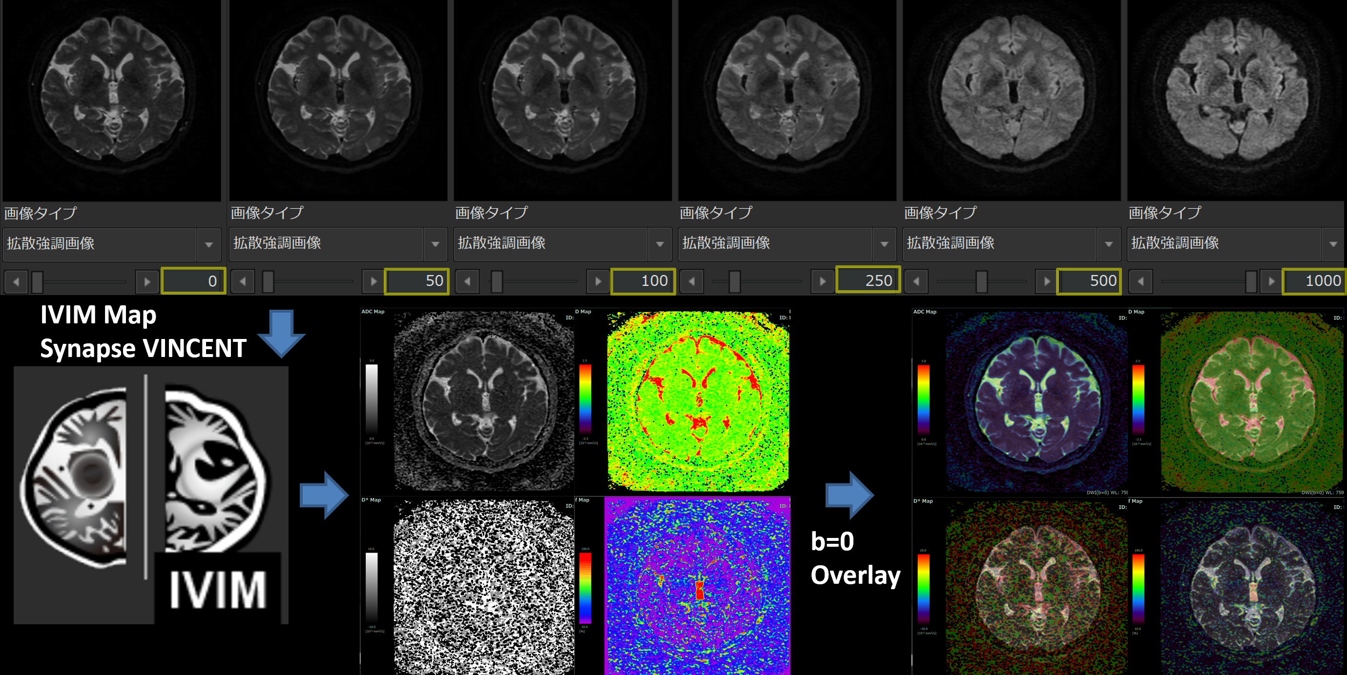
In the glymphatic system, arterial pulsation is considered to be the main driving force for the bulk flow of neurofluid which combined with cerebrospinal fluid (CSF) and interstitial fluid. However, it is difficult to measure the microflow of neurofluid with a flow velocity of less than 1 cm / sec by using the conventional MR imaging methods, such as conventional phase-contrast MRI (1), four-dimensional flow MRI (2), and Time-SLIP MRI (3).A diffusion-weighted image (DWI) with a lower b value could be seen small coherent motion of CSF as a signa attenuation or dephasing. Taoka et al. firstly reported that a b value of 500 s/mm2 in DWI was useful for evaluating CSF motion in the whole cranium (4). In addition, they proposed a new method which combined DWIs with multiple b values of 0, 50, 100, 200, 300, 500, 700, and 1000 s/mm2, named DANDYISM from the acronym “Diffusion ANalysis of fluid DYnamics with Incremental Strength of Motion proving gradient”, which applies the phenomenon that DWIs with higher b value could show the motion-related signal dephasing of CSF in a wider area with small to large CSF motion (5). As an imaging method for measuring nonuniform slow flow utilized multiple low b values of DWIs, however, intravoxel incoherent motion (IVIM) images developed by Le Bihan et al. in 1986 might potentially be used in the quantitative and separate evaluation of diffusion, perfusion, and nonuniform slow flow, and might have a potential usefulness for a CSF flow mapping approach (6).
Therefore, we tried to visualize and quantify the CSF microflow by using IVIM MRI with low multi-b values. Additionally, to investigate glymphatic dysfunction in idiopathic normal pressure hydrocephalus (iNPH), the parameters produced by IVIM were compared between iNPH patients and healthy volunteers.
Methods
Diffusion-weighted sequence with multiple b values of 0, 50, 100, 250, 500, and 1000 sec / mm2 was performed on 3-tesla MRI. In the IVIM analysis, the bi-exponential IVIM fitting method using the Levenberg-Marquardt algorithm was used. The average, maximum, and minimum values of ADC, D, D*, and f produced by IVIM were quantitatively measured in the 45 regions of interests in the whole ventricles and subarachnoid spaces.Results
In the healthy young individuals, f value was high near the ventricular walls of the lateral ventricle, in the third and fourth ventricles, and in the subarachnoid spaces around the relatively large cerebral arteries, such as the basal cistern, Sylvian fossa, central sulcus, and marginal sulcus. The mean f values in the whole lateral ventricles, anterior part of the third ventricle, ambient cisterns, central sulci, and marginal sulci were significantly lower in 28 iNPH patients than in 57 healthy volunteers. On the contrary, only the foramina of Luschka was significantly higher mean f value in iNPH patients compared to healthy individuals, and that in the other parts was no significant difference between the two groups. The ADC, D and D * values were not significantly different between the two groups.Conclusions
The f value calculated by the IVIM method was high in the areas where the CSF flow velocity was considered to be relatively high, specifically in the third ventricle, cerebral aqueduct, fourth ventricle and in the subarachnoid space around the major arteries. Therefore, the f value was considered to reflect fine CSF motion. Furthermore, iNPH patients had significantly lower mean f values in the whole ventricle and high parietal convexity sulci than healthy volunteers, suggesting that the glymphatic system is working poorly.Acknowledgements
This research was supported by research grants from the Japan Society for the Promotion of Science, KAKENHI for 3 years, beginning in 2021(Grant number:21K09098); from the Fujifilm Corporation for 4 years, beginning in 2019; from the G-7 Scholarship Foundation in 2020; and from the Taiju Life Social Welfare Foundation in 2020. The funders had no effect or involvement in the writing of this article.References
1. Bradley WG, Queralt SD. Normal-pressure hydrocephalus: evaluation with cerebrospinal fluid flow measurements at MR imaging. Radiology 1996;198.
2. Yamada S, Ishikawa M, Ito H, Yamamoto K, Yamaguchi M, Oshima M, Nozaki K. Cerebrospinal fluid dynamics in idiopathic normal pressure hydrocephalus on four-dimensional flow imaging. Eur Radiol 2020;30(8):4454-4465.
3. Yamada S, Miyazaki M, Kanazawa H, Higashi M, Morohoshi Y, Bluml S, McComb JG. Visualization of cerebrospinal fluid movement with spin labeling at MR imaging: preliminary results in normal and pathophysiologic conditions. Radiology 2008;249(2):644-652.
4. Taoka T, Naganawa S, Kawai H, Nakane T, Murata K. Can low b value diffusion weighted imaging evaluate the character of cerebrospinal fluid dynamics? Jpn J Radiol 2019;37(2):135-144.
5. Taoka T, Kawai H, Nakane T, Abe T, Nakamichi R, Ito R, Sato Y, Sakai M, Naganawa S. Diffusion analysis of fluid dynamics with incremental strength of motion proving gradient (DANDYISM) to evaluate cerebrospinal fluid dynamics. Jpn J Radiol 2021.
6. Le Bihan D, Breton E, Lallemand D, Grenier P, Cabanis E, Laval-Jeantet M. MR imaging of intravoxel incoherent motions: application to diffusion and perfusion in neurologic disorders. Radiology 1986;161(2):401-407.
Figures