3643
Altered neurochemical cerebral metabolism in patients with gluten ataxia: A pilot in-vivo proton MRS study1Department of NMR, All India Institute of Medical Sciences (AIIMS), New Delhi, India, 2Department of Pathology, All India Institute of Medical Sciences (AIIMS), New Delhi, India, 3Department of Neurology, All India Institute of Medical Sciences (AIIMS), New Delhi, India, 4Department of Gastroenterology and Human Nutrition, All India Institute of Medical Sciences (AIIMS), New Delhi, India
Synopsis
Present study evaluated the neurochemical profile of vermis and right cerebellum of GA patients using 1H MRS. The concentration of N-acetyl aspartate (NAA), glycerophosphocholine (GPC)+phosphocholine (PC), creatine (Cr) + phosphocreatine (PCr) and glutamine (Gln)+glutamate (Glu) were significantly lower in the right cerebellum of patients with GA compared to HC. The concentration of NAA, Cr and Glu were significantly lower in vermis region of patients with GA compared to HC indicating altered cerebral metabolism in GA patients that would have contributed to cerebral damage. The metabolites NAA, GPC+PC, Glu+Gln may have the potential to serve as early indicators of neuronal damage.
Introduction
Gluten sensitivity related disorders have wide range of immune-mediated clinical manifestations including neurological abnormalities like ataxia.1 Gluten ataxia (GA) is the commonest of gluten related neurological disorders, characterized by damage to cerebellum. The diagnosis of GA is not straightforward as antibodies are present in only up to 38% of patients. Moreover, the symptoms of ataxia may be mild at the onset but if remain untreated, may lead to permanent damage. Therefore, there is a need to understand the pathophysiology of GA to determine the biomarkers for early diagnosis and initiate the treatment for better management of GA. In-vivo proton magnetic resonance spectroscopy (MRS) offers the information on altered levels of neurochemicals, thereby providing an insight into neuronal metabolism, physiology and energy state in a non-invasive manner. However, only a limited number of in-vivo MRS studies have been reported suggesting a significant decrease in the ratio of N-acetyl aspartate (NAA)/creatine (Cr) in the cerebellum of patients with GA2,3. Present study therefore evaluated the neurochemical profile of vermis and right cerebellum of GA patients to identify biomarkers for early diagnosis of GA and understand its pathophysiology using in-vivo 1H MRS.Objective: To measure the absolute concentration of neurochemicals in the two brain regions (vermis and right cerebellum) in the patients with GA and compared it with healthy controls using in-vivo MRS to gain an insight into underlying biochemical abnormalities associated with GA and to determine potential biomarkers of cerebral damage.
Materials and methods
Fifteen subjects, five patients with GA (age range, 40-65 yrs) and ten healthy controls (age range, 20-65 yrs) were enrolled. The study was approved by Institute Ethics Committee and written informed consent was obtained from each subject. GA patients were recruited from the Ataxia clinic of our Institute. The control subjects had no neurological/psychiatric disease/no medical or family history of celiac disease or ataxia. The diagnosis of GA was made on the basis of clinical features, positive antigliadin (IgA and IgG) antibodies and genetic testing. Single voxel 1H-MRS was carried out on 3 T (Ingenia, Philips). Prior to MRS, multislice T2-weighted images of whole brain in all three planes, axial, coronal and sagittal were acquired using standard spin-echo pulse sequence which served as reference for positioning of voxel in the two regions; (i) vermis and (ii) right cerebellum hemisphere. MRS was performed using point-resolved spin-echo pulse sequence using the parameters: voxel size=15 mmx15mmx15mm; TE/TR = 35/2000 ms; number of averages = 256, TA = 9 minutes 8 seconds. Spectrum without water suppression was acquired for using water signal as internal reference for estimation of absolute concentration of neurochemicals. Metabolites concentrations were estimated using user-independent frequency domain fitting program (LC Model) version 6.1-4A. The concentration of neurochemicals were compared between GA patient and HC group using student t-test (SPSS, Chicago, Illionois). The level of significance was set at P<0.05.Results
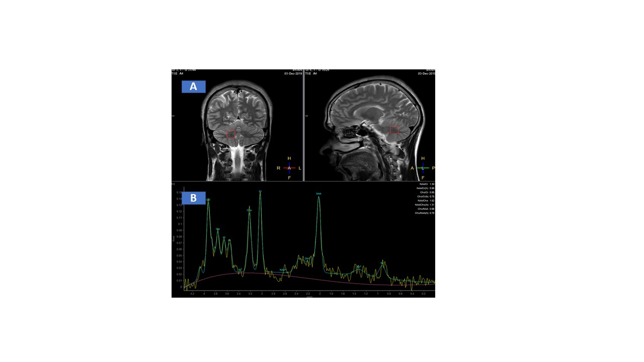
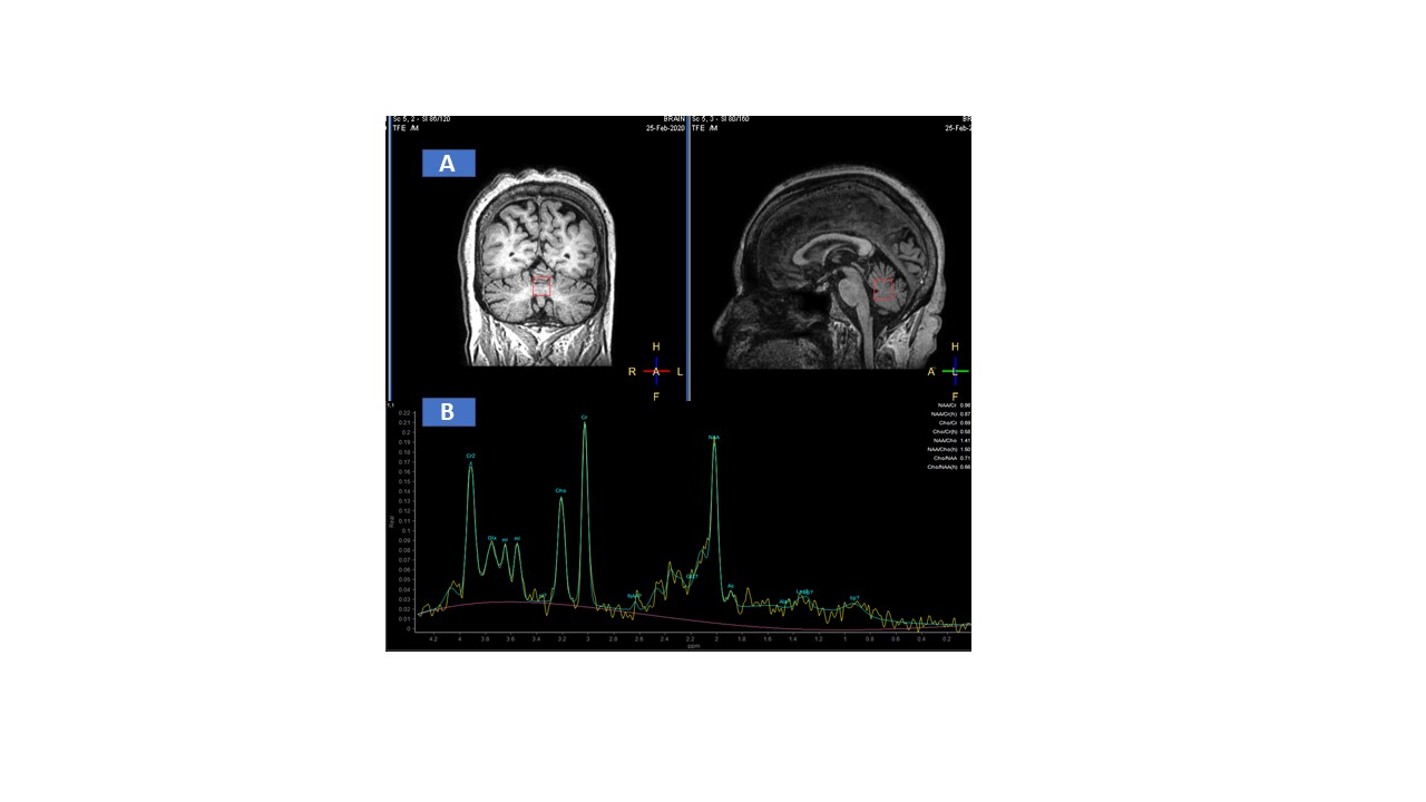
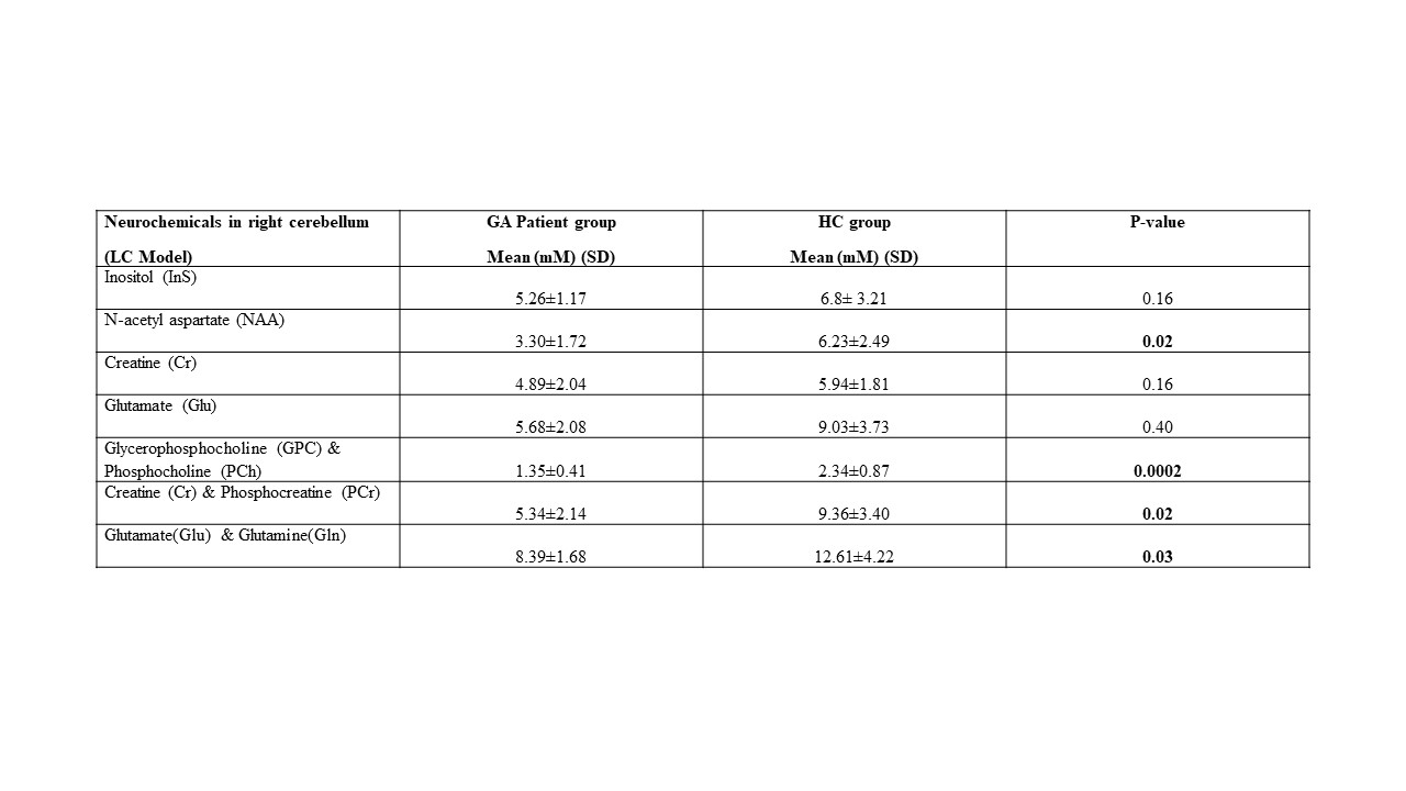
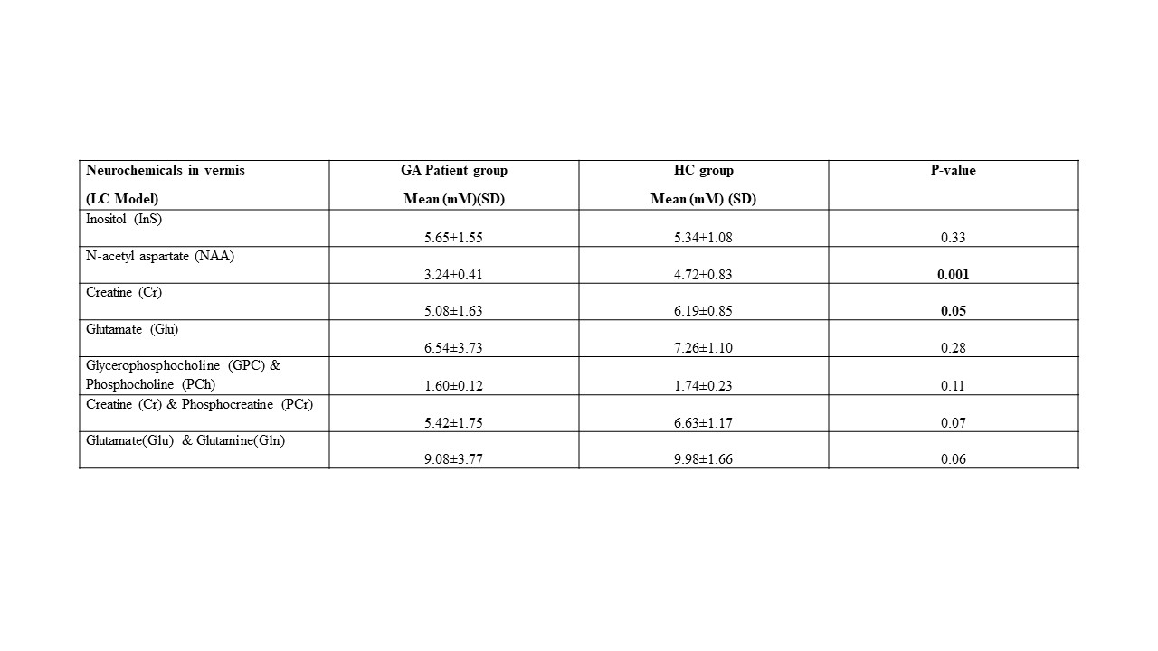
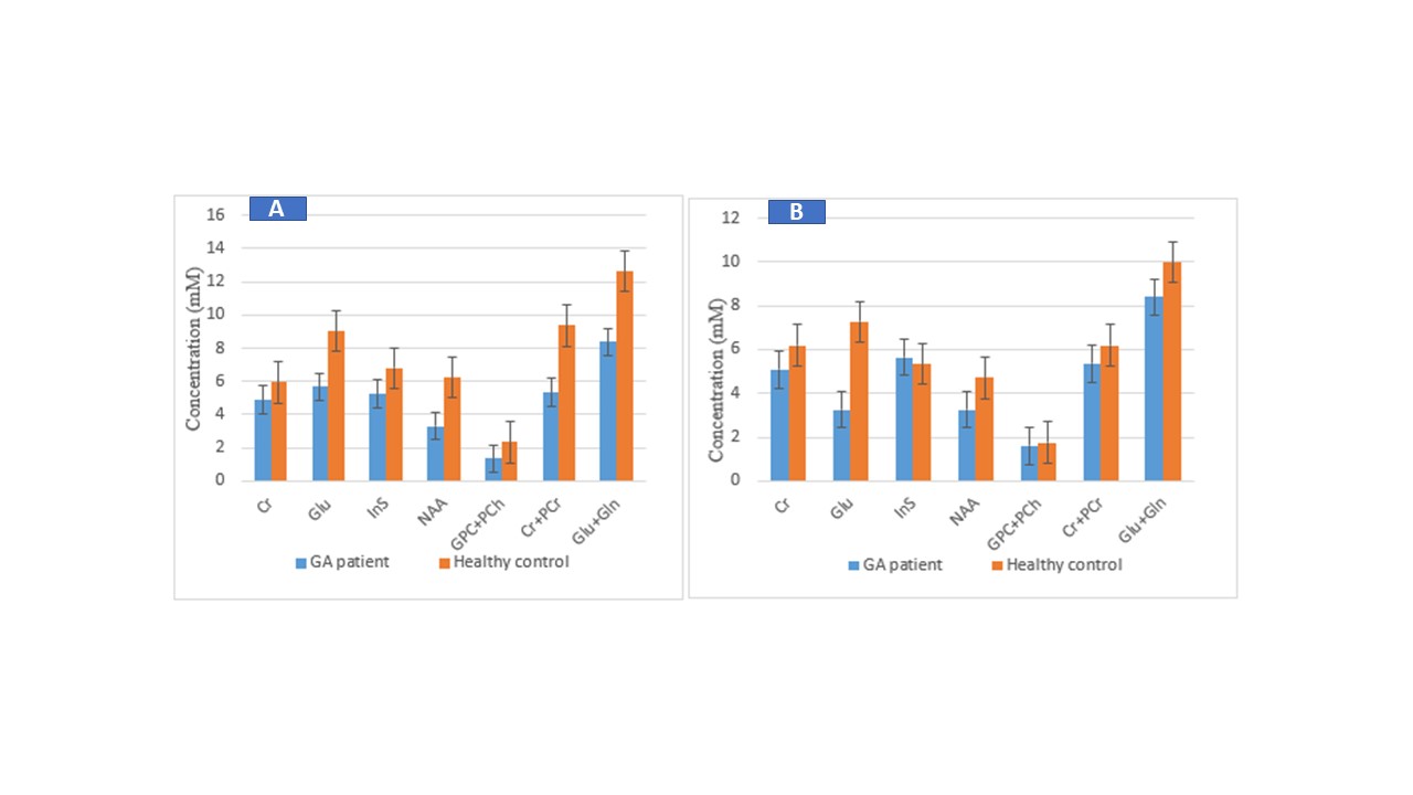
Figure 1 and 2 show the 1H MRS of right cerebellum and vermis of patient with GA. Table 1 and 2 presents the absolute concentration of neurochemicals measured in right cerebellum and vermis in the patients with GA and HC. The concentration of N-acetyl aspartate (NAA), glycerophosphocholine (GPC)+phosphocholine (PC), creatine (Cr) + phosphocreatine (PCr) and glutamine (Gln)+glutamate (Glu) were significantly lower in the right cerebellum of patients with GA compared to HC (Figure 3). The concentration of NAA, Cr and Glu were significantly lower in vermis region of patients with GA compared to HC (Figure 3).Discussion
The present study revealed several significant differences in the neurochemical profile of patients with GA compared to HC. Our data showed significantly lower level of NAA in both cerebellum and vermis of patients with GA compared to HC. The NAA is present at high concentrations and it was documented that its level represent the neuronal health.4 NAA has been implicated to serve as osmolyte, acts as a precursor for the synthesis of N-acetylaspartylglutamate, provides acetate for formation of myelin sheath of oligodendrocytes.5 Thus lower level of NAA may be attributed to degeneration of nerve cells and decreased neuronal density in the cerebellum of GA patients which is in agreement with the earlier findings.2,3 Further patients with GA showed reduced levels of Glu+Gln in the cerebellum. Glu is produced from Gln and serves as a neurotransmitter. In a preclinical study Gln supplementation has been reported to improve symptoms of ataxia-telangiectasia.6 The concentration of membrane precursors GPC and PC and energy metabolites Cr+PCr were lower in the cerebellum of GA patients. It has been reported that GPC metabolises into choline which is a precursor to acetylcholine and glycerophosphate which supports many cognitive functions and are responsible for healthy cell membrane integrity.7,8 Thus lower level of these metabolites may have affected the synthesis of membrane lipids and contributed to neuronal loss in these patients.Conclusion
Our preliminary study provides an insight into altered cerebral metabolism in GA patients that would have contributed to cerebral damage. The metabolites NAA, GPC+PC, Glu+Gln may have the potential to serve as early indicators of neuronal damage.Acknowledgements
The authors would like to acknowledge the intramural funding from (A676) from AIIMS, New Delhi.References
1. Hadjivassiliou M, Sanders DD, Aeschlimann DP. Gluten-related disorders: gluten ataxia. Dig Dis. 2015;33(2):264-8.
2. Wilkinson ID, Hadjivassiliou M, Dickson JM, Wallis L, Grünewald RA, Coley SC, Widjaja E, Griffiths PD. Cerebellar abnormalities on proton MR spectroscopy in gluten ataxia. J Neurol Neurosurg Psychiatry. 2005;76:1011-3.
3. Hadjivassiliou M, Grünewald RA, Sanders DS, Shanmugarajah P, Hoggard N. Effect of gluten-free diet on cerebellar MR spectroscopy in gluten ataxia. Neurology. 2017;89(7):705-709.
4. Moffett JR, Ross B, Arun P, Madhavarao CN, Namboodiri AM. N-Acetylaspartate in the CNS: from neurodiagnostics to neurobiology. Prog Neurobiol. 2007;81(2):89-131.
5. Signoretti S, Marmarou A, Tavazzi B, Lazzarino G, Beaumont A, Vagnozzi R. N-Acetylaspartate reduction as a measure of injury severity and mitochondrial dysfunction following diffuse traumatic brain injury. J Neurotrauma. 2001;18:977-991.
6. Chen J, Chen Y, Vail G, et al. The impact of glutamine supplementation on the symptoms of ataxia-telangiectasia: a preclinical assessment [published correction appears in Mol Neurodegener. 2017 Jan 12;12 (1):4]. Mol Neurodegener. 2016;11(1):60.
7. Jan Krzysztof Blusztajn, Barbara E. Slack, Tiffany J Mellott, et al. Neuroprotective Actions of Dietary Choline. Nutrients. 2017;9(8):815.
8. Khosrow Tayebati S, Tomassoni D, Ejike Nwankwo I, et al. Modulation of Monoaminergic Transporters by Choline-Containing Phospholipids in Rat Brain. CNS & Neurological Disorders - Drug Targets. 2013;12(1):94-103
Figures