3641
Gray matter difference associated with hormone replacement therapy in menopausal women: a voxel-based morphometry (VBM) study1Medical Convergence Research Center, Wonkwang University, Iksan, Korea, Republic of, 2Obstetrics and Gynecology, Wonkwang University School of Medicine and Hospital, Iksan, Korea, Republic of, 3Radiology, Wonkwang University School of Medicine and Hospital, Iksan, Korea, Republic of
Synopsis
Hormone replacement therapy (HRT) in menopausal women can reduce troublesome menopause symptoms and prevent cognitive decline. Several structural and volumetric researches reported that localized morphologic changes associated with estrogen meditation. However, it is still unclear how to minimize menopausal symptoms and to protect a woman’s brain from decline. This study investigated the potential effects of HRT on sex hormone level and brain morphology using an optimized voxel-based morphometry (VBM) method.
Introduction
Hormone replacement therapy (HRT) in menopausal women can reduce troublesome menopause symptoms such as sleep & sexual problems, chills, night sweats, mood changes, and hot flushes and sweats [1]. Also, a number of studies reported that HRT can reduce the incidence of Alzheimer’s disease (AD) by 64% and even more and prevent age-related cognitive decline, provided that it has been used in the past or for a sufficient period of time [2,3]. Goto et al. [4] reported accelerated volume reduction in the hippocampus for postmenopausal women using voxel-based morphometry (VBM) and atlas-based methods. Lord et al. [5] reported larger hippocampal volumes in estrogen therapy users in a VBM study. Other findings have been reported, including a positive association between estrogen therapy use and gray matter density in the inferior parietal lobules and the precuneus [6]. However, it is still unclear how to minimize menopausal symptoms and to protect a woman’s brain from decline.For this study, we investigated the gray matter difference associated with HRT on sex hormone level and whole brain morphology using an optimized VBM method.
Subjects and Methods
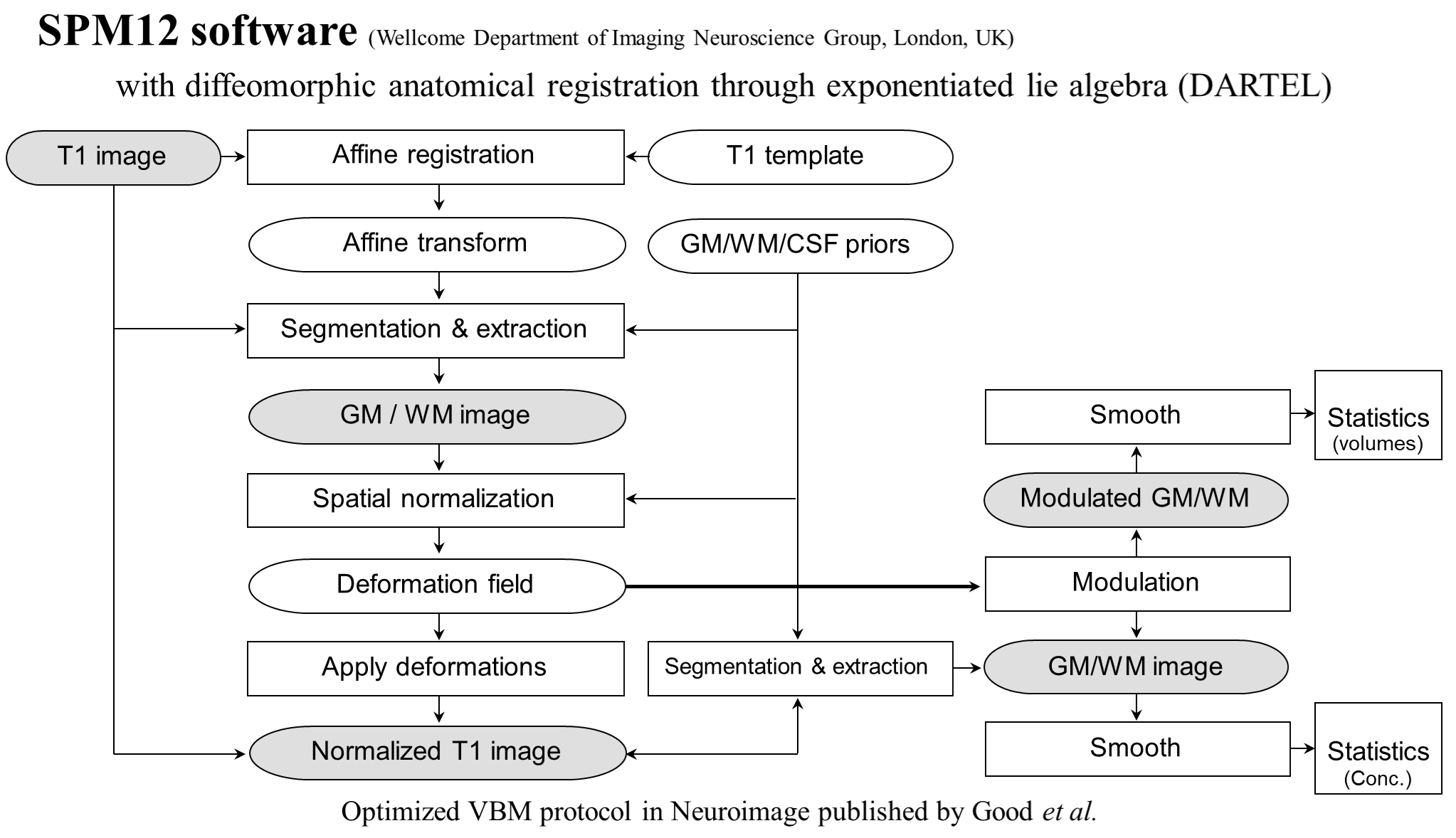
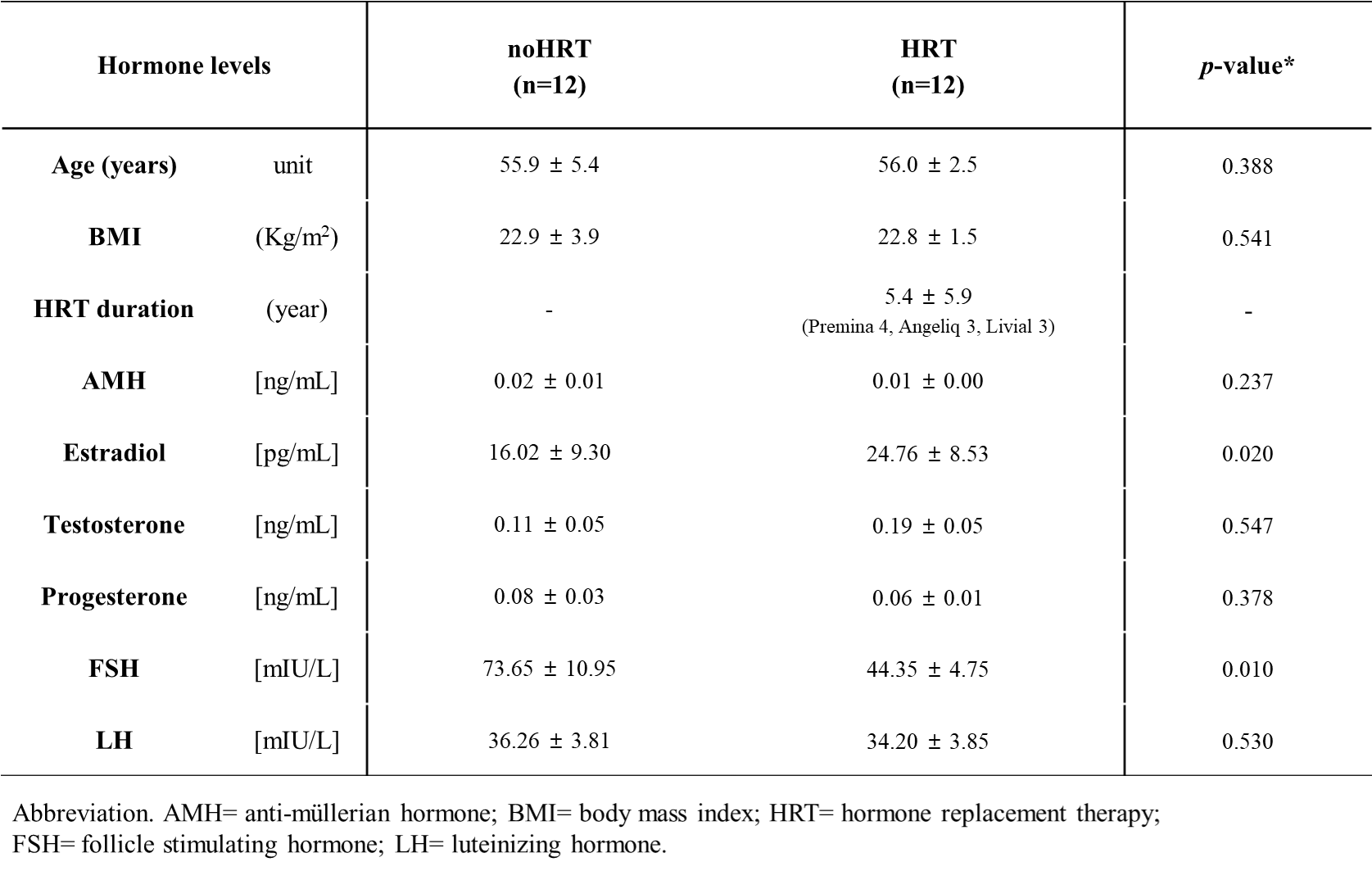
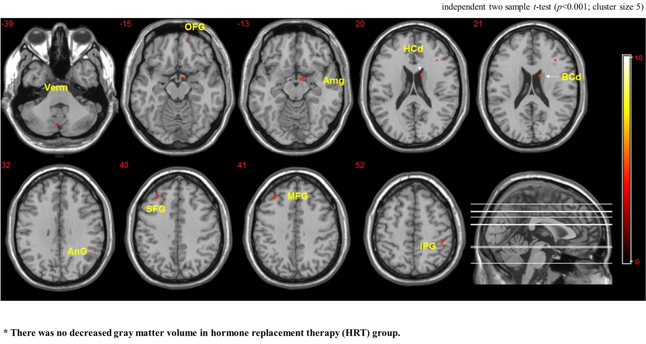
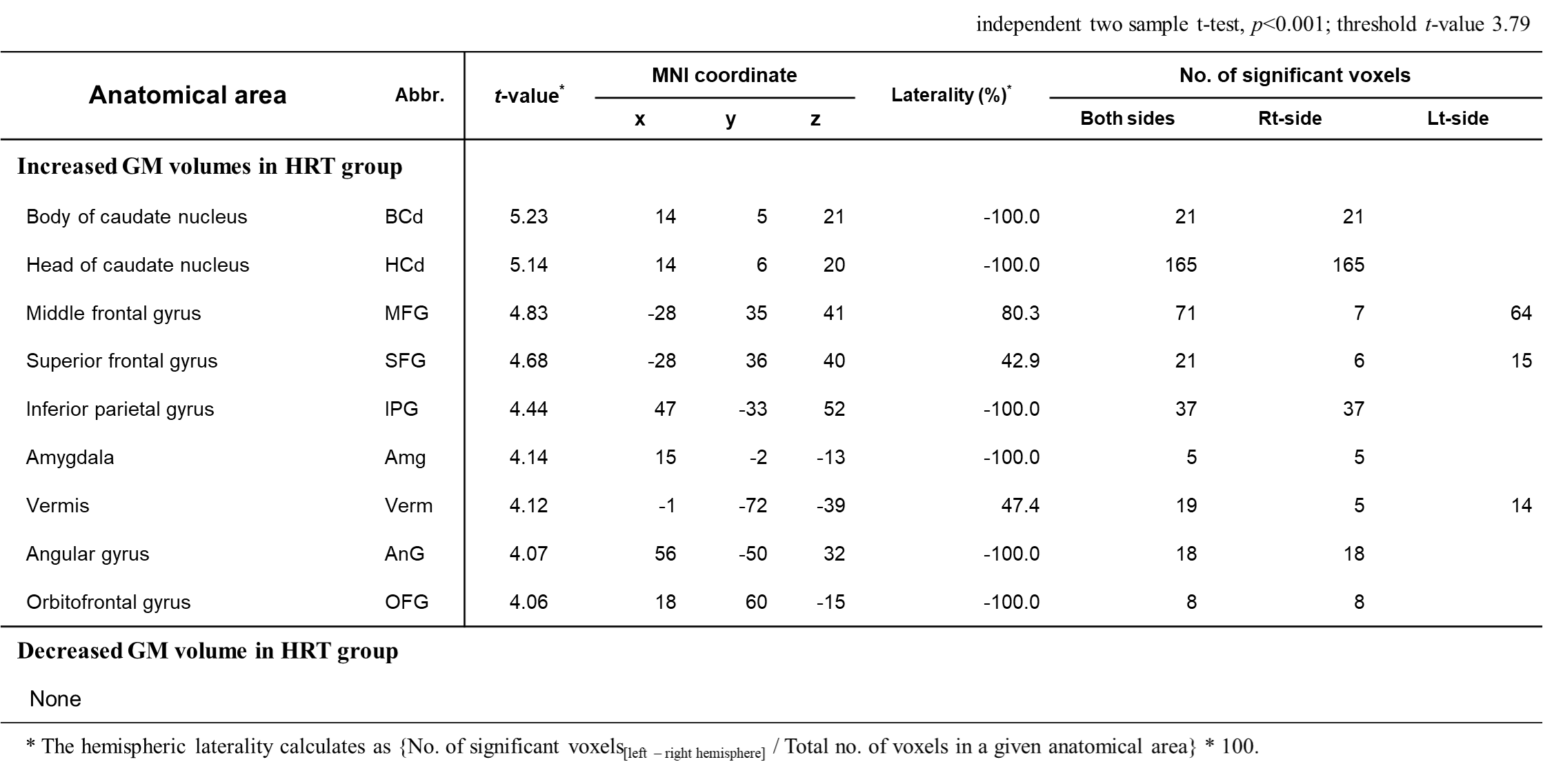
Twenty-four menopausal women including of 12 women without HRT (noHRT group) and age-matched 12 women with HRT (HRT group) were enrolled in the department of obstetrics and gynecology. Both groups have no history of neurological illness. All participants underwent blood testing for comparing the sex hormone levels. The measured sex hormones included anti-mullerian hormone, estradiol, testosterone, progesterone, follicle-stimulating hormone (FSH), and luteinizing hormone (LH).MRI examinations were obtained using a 3T MRI scanner (Achieva; Philips) with standard 8-channel adult head coil. Brain images were acquired using the 3-dimensional T1-weighted acquisition (MP-RAGE) and the scan parameters were as follows: TR/TE = 8.1/4.6 ms, FOV = 256 × 256 × 180 mm2, flip angle = 8°, slices = 180, voxel size = 1×1×1 mm3, acquisition time = 4 min 32 seconds. Voxel-based morphometry (VBM) was used to evaluate changes in gray matter volume between the two groups by using SPM12 with DARTEL procedure (see Fig. 1): gray matter segmentation; multiplication with the non-linear components derived from the normalization matrix (modulation); followed by smooth with a Gaussian kernel of 6 mm FWHM. The variation of brain volume between noHRT and HRT groups was analyzed by independent two sample t-test. Significant brain areas were quantified using customized QUBA software.
Results
Table 1 compares the mean sex hormonal levels and hormone therapeutic information in two groups. In HRT group, the duration of HRT (Premina, Angeliq and Livial) is 5.4 ± 5.9 years, the estradiol level showed higher than that of noHRT group, whereas the FSH level is lower. These sex hormonal levels were significantly different between both groups (p<0.05).Figure 2 shows the significantly increased GM volumes in HRT group (summarized in Table 2). Voxel-based morphometry indicated that HRT was associated with greater GM volumes in the treated women group, which included the body and head of caudate nucleus, middle frontal gyrus, superior frontal gyrus, inferior parietal gyrus, amygdala, vermis, angular gyrus, and orbitofrontal gyrus. However, there was no greater GM volume in the noHRT group.
Conclusion
HRT-treated women might slow down the GM volume reductions compared to menopausal women without HRT. The anatomical structures that showed greater volume in association with HRT included the deep brain areas (caudate and amygdala), cerebellar vermis, and cerebral cortices (frontal and parietal gyrus).Acknowledgements
This study was supported by the grants of the NRF (2020R1I1A1A01073871) and KHIDI, funded by the Ministry of Health &Welfare (HI18C1216).References
[1] Hickey M, et al., BMJ, 2012; 344: e763.
[2] Song Y, et al., Front Neurosci, 2020; 14: 157.
[3] Ibrahim AW, et al., J Neurosci Behav Health, 2018; 10(1): 1-8.
[4] Goto M, et al., J Magn Reson Imaging, 2011; 33: 48-53.
[5] Lord C, et al., Neurobiol Aging, 2008; 29: 95-101.
[6] Lord C, et al., Menopause 2010; 17: 846-851.
Figures