3635
Postnatal age-related change of brain volume and its association with neurobehavior outcome in term neonates1The First Affiliated Hospital of Xi’an Jiaotong University, Xi'an, China, 2Department of Research and Develpoment, Shanghai United Imaging Intelligence Co., Shanghai, China, 3Xiaocheng Wei, GEHealthcare, Beijing, China
Synopsis
Neonatal period is an important stage of brain development, exploring the development and evolution of brain region at this stage is significant for studying brain development and disease mechanisms. We analyzed the relationship between segmented brain region volume and postnatal age in term neonates, the results showed that, after controlling for gestational age, birth weight, body length, head circumference and gender factors, the volume of most brain regions of neonates was positively correlated with postnatal age. In addition, there is no correlation between the volume of most brain regions and neurobehavioral scores.
Main Findings
After controlling for factors such as gestational age, birth weight, head circumference, body length and gender, the volume of most brain regions of neonates are correlated with the postnatal age. Among them, the volume of bilateral anterior temporal lobe, right inferior temporal lobe, right occipital lobe, left caudate nucleus, and bilateral lenticular nucleus of gray matter are strongly correlated with postnatal age. Most brain regions volume had no association with neurobehavioral scoresIntroduction
The brain development in the neonatal period is characterized by rapid and dynamic development, and each brain area has a different maturation trajectory. Previous study had demonstrated that in the first several weeks after birth, total gray matter grew robustly compared with myelinated and unmyelinated white matter and the occipital and parietal regions grew significantly faster than the prefrontal region.[1] After segmenting the MRI images of healthy term neonates into 87 brain regions, we studied the relationship between brain region volume and postnatal age, and further explored the correlation between volume and neurobehavioral scores.Methods
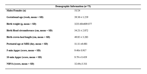
This study was permitted by the local Internal Review Board and written informed consent was obtained from all children’s parents before MRI examination. Patients 75 subjects (51 male, 24 female) who were born at full term without abnormalities in brain MR images are collected. (Table 1) MRI examination images were acquired on a 3.0T scanner (Signa HDxt, GE Healthcare, Milwaukee, WI) with an 8-channel head coil. The imaging parameters were as follow: (1) 3D T1WI: TR/TE=10ms/4.6ms; matrix=256×256; section-thickness=1mm; FOV=240mm); (2) T2WI: TR/TE=4200ms/120ms; matrix=256×256; section-thickness=4mm; FOV=240mm). Based on the dHCP brain parcellation criteria, using the V-net with bottleneck layer to segment the brain into 87 regions. After segmentation, the average volume of 87 brain regions were obtained. All statistics were analyzed by Matlab2016a and SPSS 26.0 software.Result
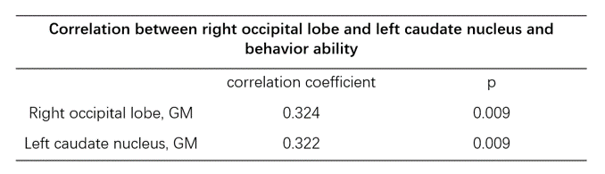
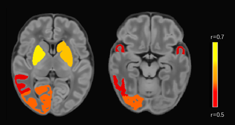
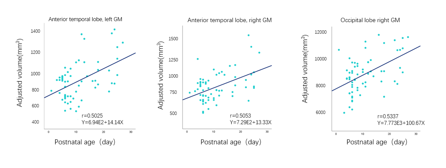
The result indicated that between brain volume and postnatal age after correcting factors such as gestational age, head circumference, body length and gender, 29 of 87 regions had no correlation(p>0.05); 58 of 87 regions had a statistically significant and positive correlation with postnatal age (p<0.05) among them, the bilateral anterior temporal lobe, right inferior temporal lobe, right occipital lobe, left caudate nucleus and bilateral lenticular nucleus of gray matter have the strongest correlation with postnatal age, the correlation coefficient ranged from 0.5 to 0.7. (Figure 1) Select the bilateral anterior temporal lobe and the right occipital lobe to draw a scatter plot and linear fitting line, the draw shows that the volume increases with postnatal age, and the slope of the right occipital lobe is the largest.(Figure 2) In the mean time, term neonatal gyri parahippocampalis of white matter is negatively correlated with neurobehavioral scores, the correlation coefficient is -0.297; the right occipital lobe and left caudate nucleus are positively correlated with behavioral ability, and the volume of the remaining brain regions are not related to neurobehavioral scores (behavior ability and active muscle tone). (Table 2)Discussion
The results of this study show that most of the brain volume of term neonates is correlated with postnatal age, the bilateral anterior temporal lobe, right inferior temporal lobe, right occipital lobe, left caudate nucleus, and bilateral lenticular nucleus has the strongest correlation with postnatal age. The above shows that the environment after birth has an impact on brain development. Some studies have suggested that the human brain grows significantly during the first few years of life and regional brain and cortical growth is significantly associated with brain maturation. [2-3] The linear fitting curve showed that the bilateral anterior temporal lobe and the right occipital lobe had a significant upward trend with the increase of postnatal age, indicating that since birth, neonates begin to receive external stimuli, and the development of audiovisual functions starts. Most of the brain volume is not related to neurobehavioral scores, which may indicate that there is no causal relationship between neurobehavioral development and brain volume. The positive correlation between the right occipital lobe and behavioral ability may be that visual stimuli can affect behavioral ability, but this view remains to be verified. This study is of great significance for the objective evaluation of neonatal brain development.Acknowledgements
This study was supported by National Natural Science Foundation of China (82101815,81901516,81901823,81971581), Shaanxi Provincial Innovation Team (2019TD-018).
* Correspondence : Chao Jin, Ph.D., Professor Department of Radiology The First Affiliated Hospital of Xi’an Jiaotong University, Xi’an, Shaanxi, China E-mali: jinny.369@163.com
References
[1] John H. Gilmore., et. al., Regional Gray Matter Growth, Sexual Dimorphism, and Cerebral Asymmetry in the Neonatal Brain. The Journal of Neuroscience, 2007 27(6): p. 1255-1260
[2] Myong-sun Choe., et. al., Regional Infant Brain Development: An MRI-Based Morphometric Analysis in 3 to 13 Month Olds. Cerebral Cortex, 2013 23(9): p. 2100–2117
[3] Antonios Makropoulos., et. al., Regional growth and atlasing of the developing human brain. NeuroImage, 2016 125: p. 456–478
Figures