3634
Brain structure and volume reveal neurodevelopmental abnormalities in preterm infants with low-grade intraventricular hemorrhage1Department of Radiology, The Third Affiliated Hospital of Zhengzhou University, Zhengzhou, China, 2GE Healthcare, MR Research China, Beijing, China
Synopsis
Premature infants with low-grade intraventricular hemorrhage (IVH) are accompanied by neurodevelopmental abnormalities. Synthetic MRI is an emerging technology that can provide brain volume quantification. Diffusion kurtosis imaging (DKI) can be sensitive to assess brain microstructure changes. Smaller myelin volume, brain parenchymal fraction, lower radial kurtosis value in cerebellum were associated with lower neonatal behavioral neurological assessment. DKI and Synthetic MRI are able to quantitatively evaluate the abnormality of the brain development in preterm infants with low-grade IVH.
Introduction
Intraventricular hemorrhage (IVH) is a characteristic form of brain injury occurring in premature neonates[1]. There is increasing evidence of abnormal neurodevelopmental outcomes in preterm infants with low-grade intraventricular hemorrhage (IVH)[2]. DKI have showed a potential advantage in detecting the normal brain development of infants [3]. Synthetic MRI can quantitatively measure gray matter, white matter and other brain volume, providing a fast and robust volume measurement method[4]. Neonatal behavioral neurological assessment (NBNA) is designed to test neonatal behavior, and allow early detection of mild brain damage for early intervention[5]. At present, it is unclear whether there are structure-function related abnormalities in low-grade IVH. Therefore, the aim of the current study was to explore whether GM and WM microstructure and brain volume are associated with developmental outcomes in low-grade IVH infants by using DKI and Synthetic MRI.Material and Methods
According to Papile classification system standard [3], we include preterm infants with low-grade IVH (grade I-II). Finally, 25 cases in low-grade IVH group and 40 cases in healthy group were included. All studies were performed on a 3.0 T MR imaging scanner (Pioneer, GE Healthcare, Milwaukee, WI). DKI sequence parameters were: TR= 2000 ms, TE = 2.32 ms, directions=30, b value =0, 1000, 2000 mm²/s, slice thickness = 3.0 mm without a gap, field of view = 256 mm × 256 mm, and acquisition time = 7.5 minutes. Synthetic MRI with parameters as follows: slice thickness = 3.0 mm, field of view= 220 mm ×186 mm, scan time = 4 minutes. The mean kurtosis (MK), radial kurtosis (RK), axial kurtosis (AK), fractional anisotropy (FA), mean diffusivity (MD) maps were generated from DKI images. Synthetic MRI data were processed to obtain the brain volumes (gray matter (GM); white matter (WM); myelin (Myc); cerebrospinal fluid; and brain parenchymal fraction (BPF)) in with SyMRI (vesion 8.0 Linköping, Sweden). Two professional radiologists outlined 7 regions of interest (ROIs) including posterior limbs of the internal capsule (PLIC), anterior limbs of internal capsule (ALIC), genu of the corpus callosum (GCC), splenium of the corpus callosum (SCC), thalamus (TH), globus pallidum (GP), and cerebellum (Cere) in DKI parametric maps to get the quantification in each brain region. The neurodevelopmental assessment was performed at 40 weeks of corrected gestational age by using neonatal behavioral neurological assessment[6]. Statistical methods include: the chi-square test for qualitative variables, student’s t-test for normal distribution quantitative variables, the Mann–Whitney U-test and Spearman correlation analysis.Results
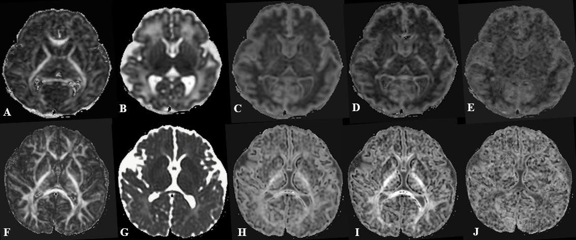
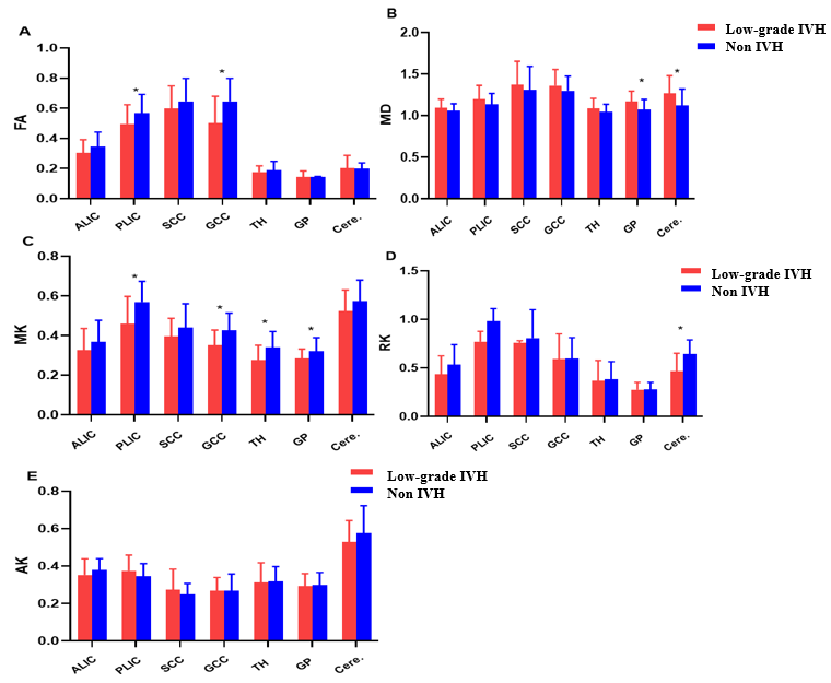
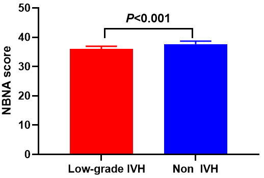
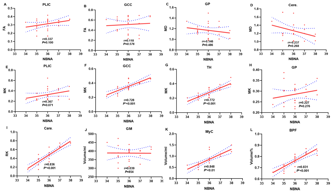
Figure 1 displayed FA, MD, MK, RK and AK images of preterm infant brain with low-grade IVH(A-E) and without IVH (F-G). As shown in Figure 2, significant lower values of the MK and FA values in PLIC, and GCC and RK values in cerebellum were found between low-grade IVH group and control group (all P<0.05) As shown in Table 1. The GM volume, myelin volume and BPF in the low-grade IVH group was also lower than those in the control group (P<0.05). The NBNA score of the low-level group also decreased, as shown in Figure 3. RK in Cerebellum, Myc and BPF were all positively correlated with NBNA (r=0.836,0.848 and 0.831, respectively, P<0.001), as shown in Figure 4.Discussion
Our findings indicate decreased MK and FA values in PLIC and GCC, RK values in cerebellum in preterm infants with low grade IVH. The blood products activate cytotoxic and inflammatory pathways, which may lead to tissue damage, or delays axons or myelin sheaths formation[2]. The decrease of MK and RK means that the neurons density decreased when brain damaged, and cell connectivity is weakened [7]. At the same time, FA reflects that the integrity of IVH children's fiber bundles is affected. Relatively low FA and MK in GCC which mean that damage to this specific corpus callosum area (reflected by the decrease in MK and FA) may further explain the potential movement problems low-grade IVH[8]. Another result was different volume changes of GM, MyC and BPF between the two group. When IVH occurs, a large amount of inflammatory cytokines and free iron are produced, which will damage the original cells of the germinal matrix and stop the development[2]. This may directly damage the cerebral cortex, also hinder myelination and the later white matter development[9]. Myc and BPF are associated with NBNA score outcomes in preterm infants with low-grade IVH. smaller volumes of whole brain and Myc were associated with poorer cognitive and movement outcomes in low-grade IVH[10]. We also found that RK in cerebellum were positively correlated with NBNA. The influence of low-grade IVH on neurodevelopmental outcome in preterm infants is associated with cerebral palsy, visual impairment and mental dysfunction[11]. Smaller myelin volume, brain parenchymal fraction, lower RK value in cerebellum were associated with lower neonatal behavioral neurological assessment. DKI and Synthetic MRI can quantitatively evaluate the brain injury of premature infants with low-grade IVH.Acknowledgements
The National Natural Science Foundation of China (grant 81870983). At the same time, it was also funded by Scientific and technological key project of Henan Province (grant No. 212102310742).References
1. Ramenghi L, Fumagalli M, Groppo M, Consonni D, Gatti L, Bertazzi P, Mannucci P, Mosca F: Germinal matrix hemorrhage: intraventricular hemorrhage in very-low-birth-weight infants: the independent role of inherited thrombophilia. Stroke 2011, 42(7):1889-1893.
2. Volpe J: The encephalopathy of prematurity--brain injury and impaired brain development inextricably intertwined. Seminars in pediatric neurology 2009, 16(4):167-178.
3. Shi J, Chang L, Wang J, Zhang S, Yao Y, Zhang S, Jiang R, Guo L, Guan H, Zhu W: Initial Application of Diffusional Kurtosis Imaging in Evaluating Brain Development of Healthy Preterm Infants. PloS one 2016, 11(4):e0154146.
4. Granberg T, Uppman M, Hashim F, Cananau C, Nordin L, Shams S, Berglund J, Forslin Y, Aspelin P, Fredrikson S et al: Clinical Feasibility of Synthetic MRI in Multiple Sclerosis: A Diagnostic and Volumetric Validation Study. AJNR American journal of neuroradiology 2016, 37(6):1023-1029.
5. Yan C, Zhang B: Clinical significance of detecting serum melatonin and SBDPs in brain injury in preterm infants. Pediatrics and neonatology 2019, 60(4):435-440.
6. Tam E, Miller S, Studholme C, Chau V, Glidden D, Poskitt K, Ferriero D, Barkovich A: Differential effects of intraventricular hemorrhage and white matter injury on preterm cerebellar growth. The Journal of pediatrics 2011, 158(3):366-371.
7. Shi J, Yang S, Wang J, Huang S, Yao Y, Zhang S, Zhu W, Shao J: Detecting normal pediatric brain development with diffusional kurtosis imaging. European journal of radiology 2019, 120:108690.
8. Counsell S, Edwards A, Chew A, Anjari M, Dyet L, Srinivasan L, Boardman J, Allsop J, Hajnal J, Rutherford M et al: Specific relations between neurodevelopmental abilities and white matter microstructure in children born preterm. Brain : a journal of neurology 2008, 131:3201-3208.
9. Reubsaet P, Brouwer A, van Haastert I, Brouwer M, Koopman C, Groenendaal F, de Vries L: The Impact of Low-Grade Germinal Matrix-Intraventricular Hemorrhage on Neurodevelopmental Outcome of Very Preterm Infants. Neonatology 2017, 112(3):203-210.
10. Vasileiadis G, Gelman N, Han V, Williams L, Mann R, Bureau Y, Thompson R: Uncomplicated intraventricular hemorrhage is followed by reduced cortical volume at near-term age. Pediatrics 2004, 114(3):e367-372.
11. Srinivasan L, Allsop J, Counsell S, Boardman J, Edwards A, Rutherford M: Smaller cerebellar volumes in very preterm infants at term-equivalent age are associated with the presence of supratentorial lesions. AJNR American journal of neuroradiology 2006, 27(3):573-579.
Figures