3631
Comparison of Brain Metastasis Detection Capability among 3D FFE with Parallel Imaging and CS and FASE MPV with Fast 3D Wheel1Joint Research Laboratory of Advanced Medical Imaging, Fujita Health University School of Medicine, Toyoake, Japan, 2Radiology, Fujita Health University School of Medicine, Toyoake, Japan, 3Canon Medical Systems Corporation, Otawara, Japan, 4Radiology, Fujita Health University Hospital, Toyoake, Japan
Synopsis
We hypothesize that the newly developed wheel encoding order (Fast 3D wheel: i.e. Fast 3Dw) method can reduce examination time as well as compressed sensing (CS) with parallel imaging (PI) technique (Compressed SPEEDER) and obtain contrast-enhanced MR examination without any degradation of image quality as compared with conventional PI technique in suspected brain metastases patients. The purpose of this study was to compare the capability for examination time reduction and image quality and diagnostic performance improvements among conventional PI, CS and Fast 3Dw methods on contrast-enhanced MR examination for brain metastases screening.
Introduction
The early and accurate identification of brain metastases affects therapeutic strategies, which depend on the size, number, location of lesions1, 2. Contrast-enhanced (CE-) 3D T1-weighted gradient-echo imaging is the most common imaging modality and suggested as useful for brain metastasis surveillance in routine clinical practice. However, high signals from vessels in this sequence cause misleading into enhancing metastases. CE- T1-weighted 3D fast spin-echo sequence can avoid these blood vessel signals and is suggested as useful for brain metastases detection, although it is obtained with long acquisition time3, 4. Recently, compressed sensing (CS) with parallel imaging (PI) technique (Compressed SPEEDER) is introduced by Canon Medical Systems for reducing examination time without degradation of image quality5, 6. Wheel encoding order (Fast 3D wheel: i.e. Fast 3Dw) technique, which is one of the techniques for k-space based acceleration technique and applied with parallel imaging, is also introduced. However, no major reports have been evaluated the capability of Fast 3Dw method for brain metastasis surveillance. The purpose of this study was to compare the capability for examination time reduction and image quality and diagnostic performance improvements among conventional parallel imaging (PI), compressed sensing (CS) and Fast 3Dw methods on contrast-enhanced MR examination in suspected brain metastasis patients.Materials and Methods
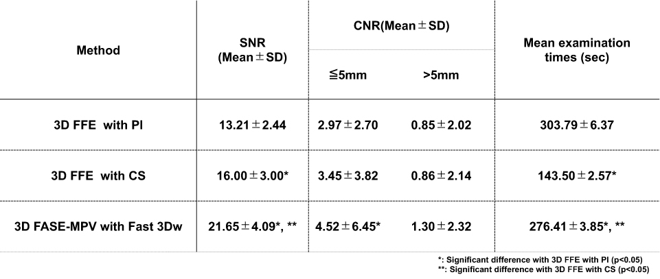
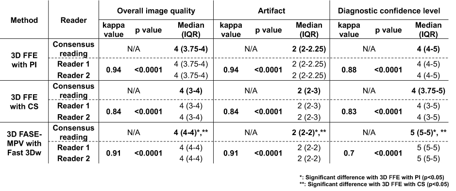
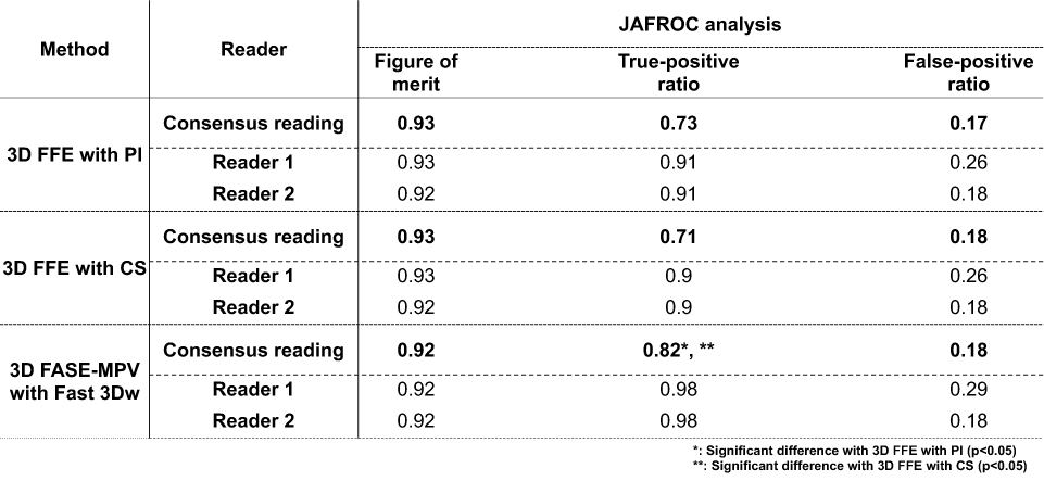
This study included 34 consecutive patients suspected of brain metastases (15 men, 19 women; mean age: 65.6 years; range=40-84 years) underwent CE-3D FFE sequence with PI, CS and CE-3D fast advanced spin-echo (FASE) multi planar voxel (MPV) sequence with Fast 3Dw methods and reconstructed. All MR examinations were performed at a 3T MR scanner (Vantage Centurian: Canon Medical Systems Corporation) with 32ch Head SPEEDER. Each examination time was also recorded. For quantitative image quality assessment, signal-to-noise ratios (SNRs) of normal white matter and contrast noise ratios (CNRs) between lesion and normal white matter were calculated by ROI measurements. For qualitative assessment, overall image quality was also evaluated by visual scoring systems by a 5-point scale by two investigators, and final score was determined as consensus of two readers. To compare detection performance of brain metastasis on CE-3DT1WI among all methods, probability of lesion detection at each lesion was assessed by 5-point scale, and final score was determined by consensus of two readers. To compare examination time, Tukey’s HSD test was performed. To compare quantitative image quality improvements, SNR and CNR were compared among three methods by Tukey’s HSD test. For comparison of each qualitative index, inter-observer agreement for qualitative assessment was determined by κ statistics with χ2 test, and all qualitative indexes were compared by Wilcoxon signed–rank test. To compare detection capability among all data sets, Jackknife alternative free-response receiver operating characteristic (JAFROC) analysis were performed. Finally, detection rates were also compared among three methods by McNemar’s test. A p value less than 0.05 was considered as significant at each statistical analysis.Results
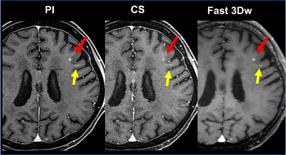
Representative case is shown in Figure 1. Compared results of each quantitative image quality index, mean examination times and qualitative index on CE-3DT1WI obtained with PI, CS and Fast 3Dw are shown in Figure 2 and 3. Mean examination times of CS and Fast 3Dw were significantly shorter than that of PI (p<0.05). SNR of Fast 3Dw was significantly higher than those of PI and CS (p<0.05). Moreover, SNR of CS was significantly higher than that of PI (p<0.05). CNR of Fast 3Dw with the lesion less than 5mm in diameter was significantly higher than that of PI (p<0.05). Inter-observer agreement of each index by all methods were determined as substantial or excellent (0.70≤κ≤0.94, p<0.0001). Image quality index of Fast 3Dw was significantly higher than those of PI and CS (p<0.05). Compared results of JAFROC analysis for brain metastasis detection are shown in Figure 4. True-positive ratio (TPR) of Fast 3Dw had significantly higher than those of PI and CS (p<0.05), although figure of merit (FOM) and false-positive ratio (FPR) of all methods had no significant differences (p>0.05).Conclusion
Fast 3Dw method has equal to or superior potentials for improving examination time, image quality or diagnostic performance as compared with conventional PI and CS on contrast-enhanced MR examination for brain metastases screening.Acknowledgements
This work was technically and financially supported by Canon Medical Systems Corporation.References
1. Linskey ME, Andrews DW, Asher AL, et al. The role of stereotactic radiosurgery in the management of patients with newly diagnosed brain metastases: a systematic review and evidence-based clinical practice guideline. J Neurooncol 2010;96(1):45-68. doi: 10.1007/s11060-009-0073-4
2. Patchell RA. The management of brain metastases. Cancer treatment reviews 2003;29(6):533-540. doi: 10.1016/s0305-7372(03)00105-1
3. Kato Y, Higano S, Tamura H, et al. Usefulness of contrast-enhanced T1-weighted sampling perfection with application-optimized contrasts by using different flip angle evolutions in detection of small brain metastasis at 3T MR imaging: comparison with magnetization-prepared rapid acquisition of gradient echo imaging. AJNR Am J Neuroradiol 2009;30(5):923-929. doi: 10.3174/ajnr.A1506
4. Komada T, Naganawa S, Ogawa H,et al. Contrast-enhanced MR imaging of metastatic brain tumor at 3 tesla: utility of T(1)-weighted SPACE compared with 2D spin echo and 3D gradient echo sequence. Magn Reson Med Sci 2008;7(1):13-21. doi: 10.2463/mrms.7.13
5. Ueda T, Ohno Y, Yamamoto K, et al. Compressed sensing and deep learning reconstruction for women's pelvic MRI denoising: Utility for improving image quality and examination time in routine clinical practice. Eur J Radiol 2021;134:109430. doi: 10.1016/j.ejrad.2020.109430
6. Ikeda H, Ohno Y, Murayama K, et al. Compressed sensing and parallel imaging accelerated T2 FSE sequence for head and neck MR imaging: Comparison of its utility in routine clinical practice. Eur J Radiol 2021;135:109501. doi: 10.1016/j.ejrad.2020.109501
Figures