3629
Neuroimaging-based brain age prediction of first-episode schizophrenia and the alteration of brain age after early medication1Xi'an People's Hospital (Xi'an Fourth Hospital), Xi'an, China
Synopsis
Schizophrenia is related to accelerated brain ageing, while individualized biomarkers are scarce. We established a brain age prediction model based on diffusion tensor imaging sequence. The difference between brain age and chronological age of first-episode schizophrenia patients and healthy controls were compared, and the longitudinal studies were performed in follow-up patients after a short-term antipsychotic treatment. The results verified the theory of accelerated brain ageing and medication have an effect on ameliorating it. Structural brain changes in schizophrenia could reveal the accelerated aging and quantification of age discrepancy could contribute to clinical decision-making and find new targeted therapies.
Introduction
Neuroimaging-based brain-age prediction of schizophrenia is well established. However, the diagnostic significance and the effect of early medication on first-episode schizophrenia remains unclear. To explore whether predicted brain age can be used as a biomarker for schizophrenia diagnosis, and the relationship between clinical characteristics and brain-predicted age difference (brain-PAD), and the effects of early medication on predicted brain age.Methods
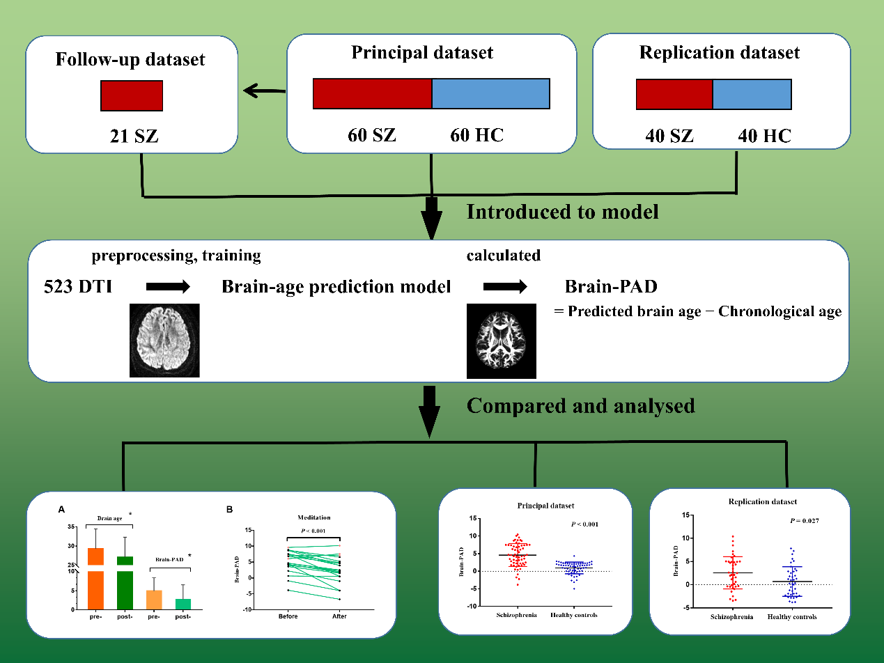
The predicted model was built on 523 diffusion tensor imaging scans from healthy controls. Relevance vector regression was introduced for model training of the relationship between fractional anisotropy values and chronological age. First, the brain-PAD of 60 patients with first-episode schizophrenia, 60 healthy controls in the principal data-set and 40 pairs of individuals in the replication data-set were calculated. Next, the brain-PAD between groups were compared and the correlations between brain-PAD and clinical measurements were analyzed. Last, after about four months of medication, the brain-PAD of 21 follow-up patients were calculated and compared with baseline, meanwhile, the differences in fiber characteristics before and after treatment were analyzed.Results
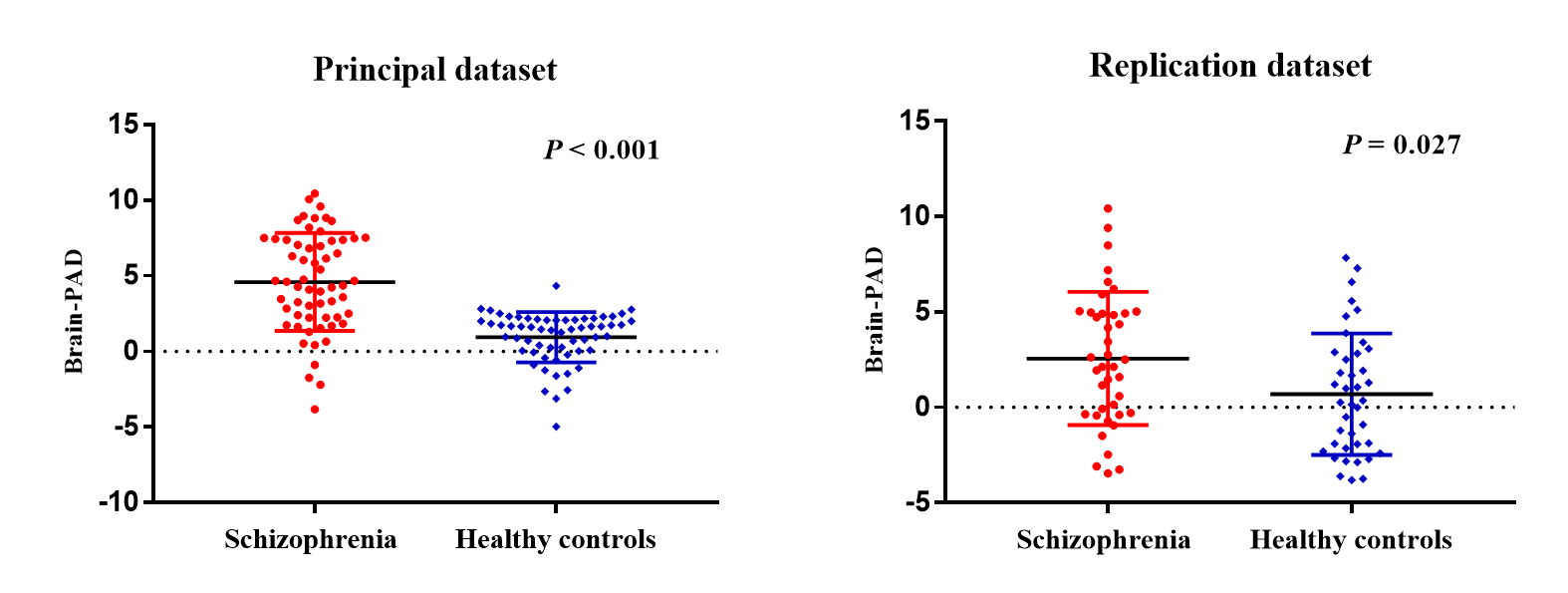
The patients of first-episode schizophrenia showed a significant increase in brain-PAD compared with healthy controls in the principal data-set (4.6 ± 3.2 vs. 0.9 ± 1.7 years) and in the replication data-set (2.6 ± 3.5 vs. 0.7 ± 3.2). The brain-PAD was negatively associated with the chronological age of the patients in the principal and pooled data-sets (P< 0.05), but not significantly in the replication data-set.After short-term medication, the brain-PAD of patients decreased significantly compared with baseline (4.932 ± 3.486 vs. 2.718 ± 3.818, P< 0.001). The fractional anisotropy value of 31/33 white matter tract features, which related to the brain-PAD scores, had significantly statistical differences between baseline and follow-up measurements (P< 0.05).
Correlation analysis showed that the brain-PAD was negatively associated with the positive score on the Positive and Negative Syndrome Scale in the principal data-set (r=−0.326, P= 0.014). In addition, there was a negative correlation between brain-PAD and semantic verbal fluency after medication (r = −0.632, P = 0.015).
Discussion
In the schizophrenia group, the difference in the estimated brain age and their chronological age appeared to be 4.6 years older in the principal data-set and 2.6 years older in the replication data-set, which represented statistically significant differences compared with healthy controls. In the follow-up data-set, we found an average difference of 2.2 years between brain and chronological age at baseline after early medication. However, Schnack and colleagues reported that the brain age of patients with schizophrenia was progressively increased at a rate of about 1.24 years annually over the 3.5-year follow-up period 1. The difference between our findings with Schnack et al may be the result of dynamic changes with clinical course and long-term therapy, and further evidence is needed to verify the findings.Impaired white matter bundle connectivity between the brain regions plays a critical role in the neuropathology of schizophrenia 2. In this study, there was a trend to an increase in brain-PAD with younger age in principal and pooled data-sets, while not significant in the replication data-set. It has been reported that younger psychiatric patients could show more severe accelerated brain ageing, which indicates that abnormal white matter integrity may occur at a younger age rather than with a progressive development with age 3, 4. In other words, early-onset schizophrenia is more symptomatic and has a worse prognosis. Therefore, quantitative studies based on brain age might be conducive in revealing the pathological mechanism involved.
Cognitive impairments are commonly seen in individuals with schizophrenia, which might be in line with accelerated or premature ageing 5. The brain-PAD might have potential correlation with cognitive impairments, and there might be no correlations between the age gap and some aspects of cognition impairments 6, or the correlation was negative, which indicated worse performance with higher brain age gap 7. In the present study, we observed negative correlations between brain-PAD scores and semantic verbal fluency after medication, as well as the scores on the positive scale of the PANSS in the principal data-set. It has been reported that the FA value of white matter fibres are negatively associated with total PANSS scores in a never treated schizophrenia group 4. Nevertheless, no consistent results were found in replication group and pooled group of our study. Overall, patients with schizophrenia might have accelerated brain age and cognitive impairments that exist from an early stage of their illness and then follow an age-related changes, which needs to be validated in larger longitudinal studies.
Conclusion
The brain age of patients with first-episode schizophrenia may be older than their chronological age. The results support the theory of accelerated ageing in patients with schizophrenia from the perspective of brain structural changes. Early medication holds promise for improving the patient’s brain ageing. Quantification of age discrepancy could be useful for screening for schizophrenia and searching for new targeted therapies. Given these findings, measuring brain ageing based on neuroimaging holds promise for establishing biomarkers that could contribute to clinical decision-making of psychiatric disorders.Acknowledgements
The authors would like to thank the people living with schizophrenia and the healthy controls who participated in this study for giving up so much of their time.References
1. Schnack HG, van Haren NE, Nieuwenhuis M, Hulshoff Pol HE, Cahn W, Kahn RS. Accelerated Brain Aging in Schizophrenia: A Longitudinal Pattern Recognition Study. Am J Psychiatry. 2016;173(6):607-16.
2. Xiang B, Wang Q, Lei W, Li M, Li Y, Zhao L, et al. Genes in immune pathways associated with abnormal white matter integrity in first-episode and treatment-naive patients with schizophrenia. Br J Psychiatry. 2019;214(5):281-7.
3. Masuda Y, Okada G, Takamura M, Shibasaki C, Yoshino A, Yokoyama S, et al. Age-related white matter changes revealed by a whole-brain fiber-tracking method in bipolar disorder compared to major depressive disorder and healthy controls. Psychiatry Clin Neurosci. 2021;75(2):46-56.
4. Xiao Y, Sun H, Shi S, Jiang D, Tao B, Zhao Y, et al. White Matter Abnormalities in Never-Treated Patients With Long-Term Schizophrenia. Am J Psychiatry. 2018;175(11):1129-36.
5. Harvey PD, Rosenthal JB. Cognitive and functional deficits in people with schizophrenia: Evidence for accelerated or exaggerated aging? Schizophr Res. 2018;196:14-21.
6. Jonsson BA, Bjornsdottir G, Thorgeirsson TE, Ellingsen LM, Walters GB, Gudbjartsson DF, et al. Brain age prediction using deep learning uncovers associated sequence variants. Nat Commun. 2019;10(1):5409.
7. Richard G, Kolskar K, Sanders AM, Kaufmann T, Petersen A, Doan NT, et al. Assessing distinct patterns of cognitive aging using tissue-specific brain age prediction based on diffusion tensor imaging and brain morphometry. PeerJ. 2018;6:e5908.
Figures