3627
Diffusion–Based Virtual MR Elastography of the asymmetric features of neonatal white matter1Department of Radiology, The First Affiliated Hospital of Xi’an Jiaotong University, xi’an, China, 2Department of Radiology, The First Affiliated Hospital of Xi’an Jiaotong University, xi'an, China
Synopsis
Exploring the asymmetry of the neonatal brain white matter is of great significance for exploring the developmental rules and disease pathogenesis. Virtual MR elastography (VMRE) as a non-invasive technique has also enabled to relate maturation of cerebral structures to neonatal neurodevelopment in addition to conventional techniques.This study aims to explore the capability of diffusion–based VMRE in the characterization of the asymmetric of the neonatal white matter stiffness.The stiffness of neonatal white matter existed asymmetry, and there was a correlation between it and PMA. VMRE may enable to explore the developing brain at several levels.
Introduction
Within white matter, the number of axons, axonal density, axon size, myelin thickness, etc. vary between different tracts. The asymmetry of brain white matter at birth provides a new perspective for understanding the pathogenesis of genetic factors, fetal growth and development restriction, perinatal diseases, other diseases, assessing early neonatal brain damage and judging prognosis. With advancements in the brain MR elastography acquisition and post-processing techniques, one now has the opportunity to measure the regional variation in brain stiffness between specific structural components. Le Bihan et al proposed a method of virtual MR elastography(VMRE) to evaluate the viscoelasticity of the tissue without mechanical motion[1]. Against the limitations of conventional MRE, VMRE do not increase special equipments[2], avoid increasing operational complexity and can make use of the existing sequence. Recent work has suggested that brain MRE could be used to characterize changes in brain tissue viscoelasticity[3].The purpose of this study is to explore the capability of diffusion–based VMRE in the characterization of the asymmetric of the neonatal white matter shear stiffness.Materials and Methods
The Institutional Review Broad of the first author’s affiliation approved this study and written informed consent was obtained from parents of the neonates. This study enrolled 61 preterm and term subjects without abnormalities on magnetic resonance imaging from the First Affiliated Hospital of Xi’an Jiaotong University. Subjects were divided into different groups according to the gestational age: early-preterm neonates, near-preterm neonates, and term neonates. Neonates who were confirmed or suspected to have congenital malformation of central nervous system,congenital infections,metabolic disorders, abnormal appearances in MRI were excluded. All subjects were examined by using a 3.0T scanner (Signa HDxt, General Electric Medical System, Milwaukee, WI, USA) with an 8-channel head coil. DKI was performed by using a single shot echo planar imaging sequence with following parameters: b values = 0, 50, 200, 500, 1000, 2000, 2500 s/mm2; 18 gradient directions per nonzero b value; NEX = 1; repetition time/echo time = 11000/91.7 ms; slice thickness = 4 mm; field of view = 180 × 180 mm2; acquisition matrix =128 × 128; the acquisition voxel size = 1.4 × 1.4 × 4 mm3. Diffusion kurtosis images of the lower b-value (Slow, b value = 200 s/mm2) and those of the higher b-value (Shigh, b value = 1000 s/mm2) were used to estimate the virtual shear stiffness [1,4]: virtual shear stiffness = a·ln (Slow/Shigh) + b. The scaling (a) and the shift (b) factors were separately set to −9.8 and 14 according to the previous calibration studies [4,5].The region of interests of white matter structures included left and right regions of 3 projection tracts:Posterior limb of internal capsule(PLIC), Anterior corona radiata(ACR), Corticospinal tract(CST); 1 commissural tracts:Corpus callosum(CC); left and right regions of 2 association tracts:Uncinate fasciculus(UF), Superior longitudinal fasciculus(SLF). And External capsule(EC), Cingular gyrus(CGG). Asymmetry index of the shear stiffness was calculated by the following equation: Asymmetry index(AI) = (Left-Right) /0.5*(Left+Right).Inter-group differences in demographics and regional values of metrics were evaluated using Kruskal-Wallis rank sum test. Correlations between postmenstrual age(PMA) and Asymmetry index were performed using Spearman correlation after postnatal age at scan. p<0.05 was considered as statistically significant difference.Results
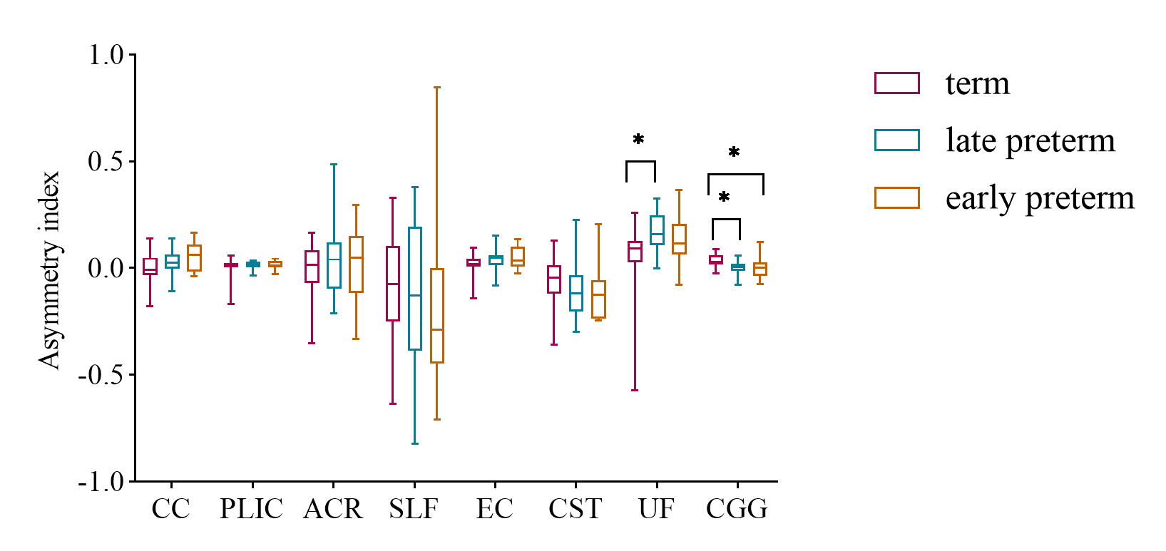
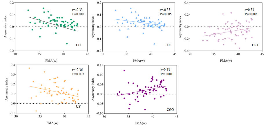
A total of 61 neonates were enrolled, including 9 early-preterm neonates, 15 late-preterm neonates and 37 term neonates. No significant differences in postnatal age at MRI scan were found between groups (Table 1). Comparison of the asymmetry index of the shear stiffness of white matter fiber bundles in the early-preterm neonates group, late-preterm neonates group and term neonates group, UF and CGG have statistical difference between the groups (Figure 1), no statistical difference between the remaining fiber bundles. The asymmetry indices of the shear stiffness of CGG of term neonates were higher than that of early-preterm and late-preterm neonates.The asymmetry indices of the shear stiffness of UF of term neonates are higher than that of late-preterm neonates. PMA related changes were observed in CC, EC, CST, UF, CGG.(Figure 2),while no significant correlation between the remaining fiber bundles and PMA could be found.Discussion
This study found that the stiffness of neonatal cerebral white matter already existed asymmetry, and the asymmetry existed in both sides.The distribution of asymmetry index of white matter stiffness in term neonates was wider than that of premature neonates. There was a correlation between PMA and asymmetry index of white matter stiffness in partial bundles.Previous study suggested myelin content was also asymmetrical in multiple WM regions.Preterm infants present with less gray matter / white matter differentiation and myelination in comparison with full-term newborns.[5] DTI studies in neonates have found the general pattern of age-related decrease in mean diffusivity and increase in anisotropy in different WM regions (corpus callosum).Our results suggested that the asymmetry of shear stiffness of white matter existed difference in premature and term neonates, this reflects asymmetric structural development in white matter,which provides structural evidence for asymmetry during neonates. However, VMRE has not been verified by theory. This is the limitation of this study and needs to be confirmed in the future.Conclusions
The stiffness of neonatal cerebral white matter existed asymmetry, and there was a correlation between it and PMA. It may enable to explore the developing brain at several levels.Acknowledgements
This study was supported by the National Natural Science Foundation of China ( 81971581, 81901823 and 81901516), the Innovation Team Project of Natural Science Fund of Shaanxi Province (2019TD-018). Please address correspondence to Jian Yang, e-mail: yj1118@mail.xjtu.edu.cn.References
[1] Ota, T., Hori, M., Le Bihan, D., Fukui, H., Onishi, H., Nakamoto, A., Tsuboyama, T., Tatsumi, M., Ogawa, K., & Tomiyama, N. (2021). Diffusion-Based Virtual MR Elastography of the Liver: Can It Be Extended beyond Liver Fibrosis?. Journal of clinical medicine, 10(19), 4553.
[2] Yin, Z. , Romano, A. J. , Manduca, A. , Ehman, R. L. , & Huston, J. . (2018). Stiffness and beyond: what mr elastography can tell us about brain structure and function under physiologic and pathologic conditions. Topics in Magnetic Resonance Imaging, 27(5), 305-318.
[3] Dubois J, Dehaene-Lambertz G, Kulikova S, Poupon C, Hüppi PS, Hertz-Pannier L. The early development of brain white matter: a review of imaging studies in fetuses, newborns and infants. Neuroscience. 2014 Sep 12;276:48-71. doi: 10.1016/j.neuroscience.2013.12.044. Epub 2013 Dec 28. PMID: 24378955.
[4] Le Bihan D, Ichikawa S, Motosugi U. Diffusion and intravoxel incoherent motion mr imaging-based virtual elastography: a hypothesis-generating study in the liver. Radiology, 2017, 285(2):609–619.
[5] Lagerstrand K, Gaedes N, Eriksson S, et al. Virtual magnetic resonance elastography has the feasibility to evaluate preoperative pituitary adenoma consistency. Pituitary, 2021, 24:530–541.
Figures