3607
Disrupted small-world networks in children with attention-deficit/hyperactivity disorder: a DTI-based network analysis1Diagnostic Radiology, The First Affiliated Hospital of Sun Yat-sen University, Guangzhou, Guangdong, China, 2MR Research, GE Healthcare, Beijing, China
Synopsis
Attention-deficit/hyperactivity disorder (ADHD) is the most common neurodevelopmental disorder in school-age children, the pathogenesis is still unclear. In current study, 49 pediatric with ADHD and 51 age- and gender-matched typically developing (TD) children were included. Diffusion tensor imaging (DTI) and graph theory approaches was used. At the global level, patients with ADHD showed increased characteristic Lp, γ, σ and decreased Eglob; regionally, altered nodal profiles were mainly in the default mode, central executive network and basal ganglia. In particular, nodal betweenness of left caudate was negatively associated with the clinical symptom severity.
Introduction
Attention-deficit/hyperactivity disorder (ADHD) is the most common neurodevelopmental disorder with an estimated prevalence of up to 9% in school-age children1. Although the exact etiology and neurobiological substrate of ADHD remain unclear, the existing neuroimaging studies have showed brain structural and functional abnormalities in ADHD2,3.By using graph theory analysis, brain structure can be interpreted as an integrated network which quantify the whole brain as a single graph comprising nodes linked by edges. To be specific, the small-world network pattern, which seems to have evolved to mediate high-efficiency parallel information transfer4, has been used to define pathologic abnormalities in neurodevelopmental disorders, including ADHD5. In this study, we use diffusion tensor imaging (DTI) and graph theory approaches to investigate the brain structural connectome in pediatric ADHD and explain the potential cause of the developmentally inappropriate behavioral symptoms.Methods
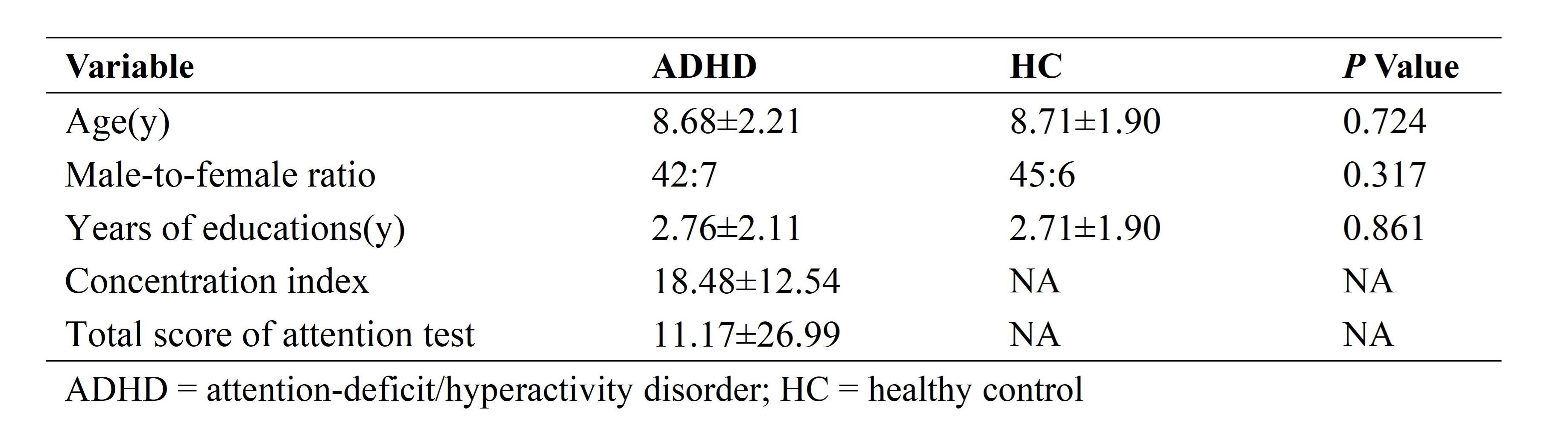
A total of 49 pediatric with clinical diagnosis of ADHD based on the criteria of DSM-5 and 51 age- and gender-matched typically developing (TD) children were included. The average age was 9 year (6-14year). The structural connectome was constructed by using DTI tractography and thresholding the mean fractional number of 90 brain regions to yield 90×90 partial correlation matrixes based on AAL90 atlas. Graph theory analysis was used to examine the group-specific topologic properties, and nonparametric permutation tests were used for between-group comparisons of topologic metrics. For all tests, a P-value < 0.05 was considered statistically significant. Importantly, a false-discovery rate (FDR) was performed to control the error of multiple comparisons. What’s more, the relationship between clinical symptom severity and altered nodal profiles was assessed.Results
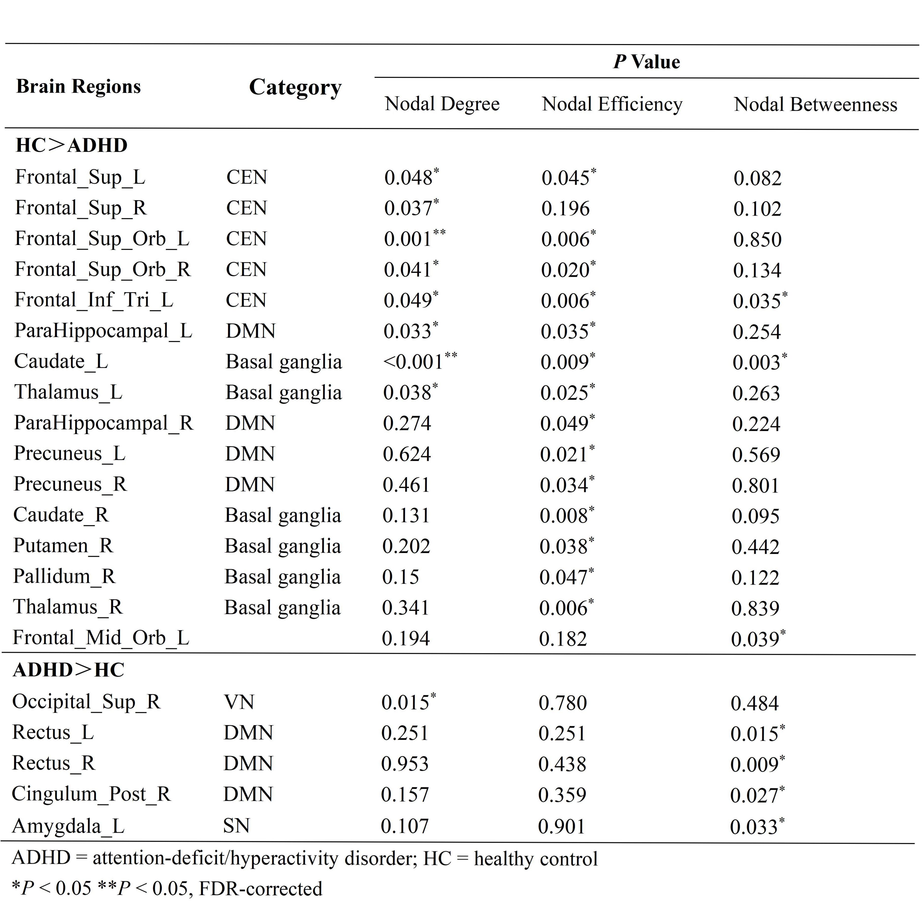
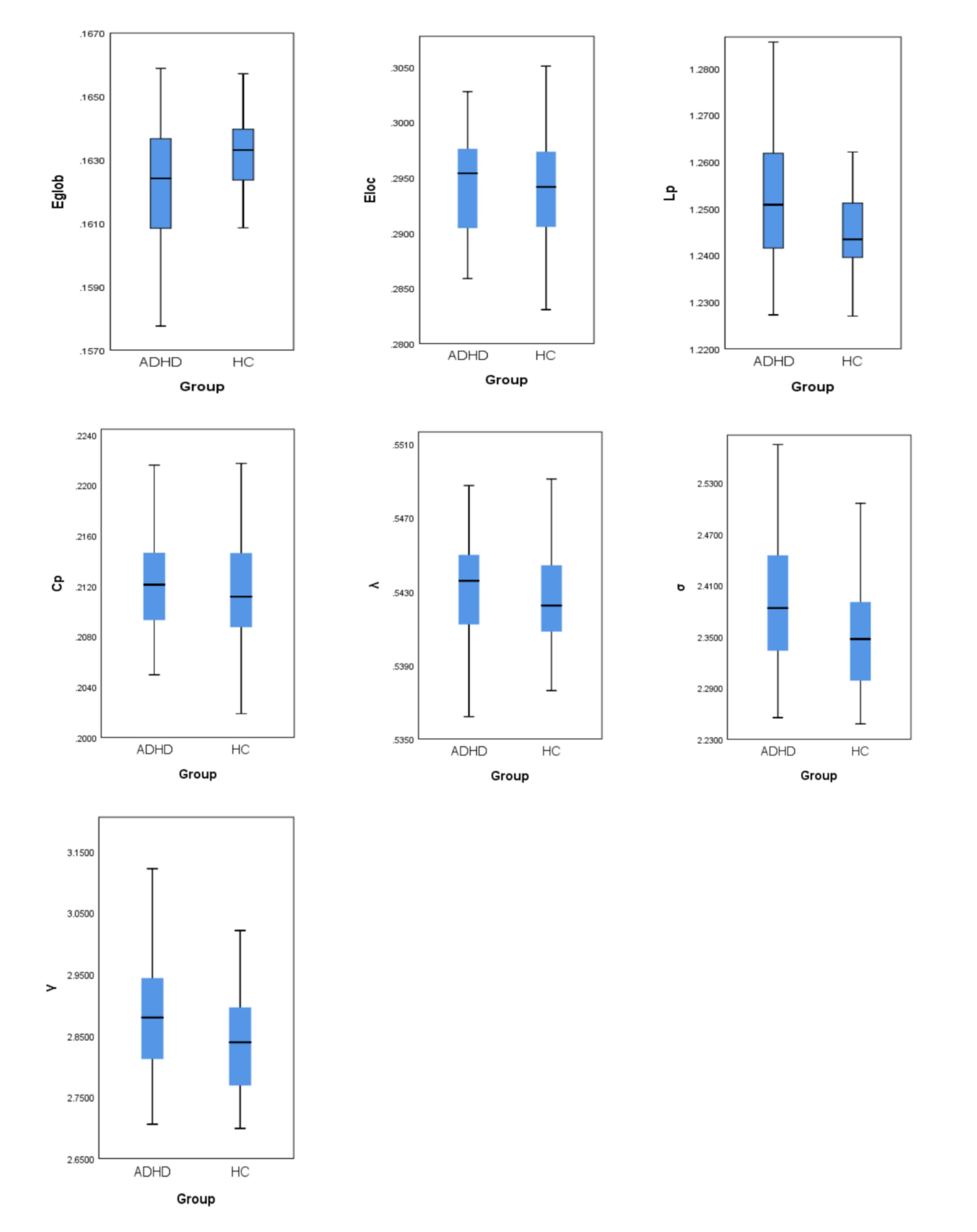
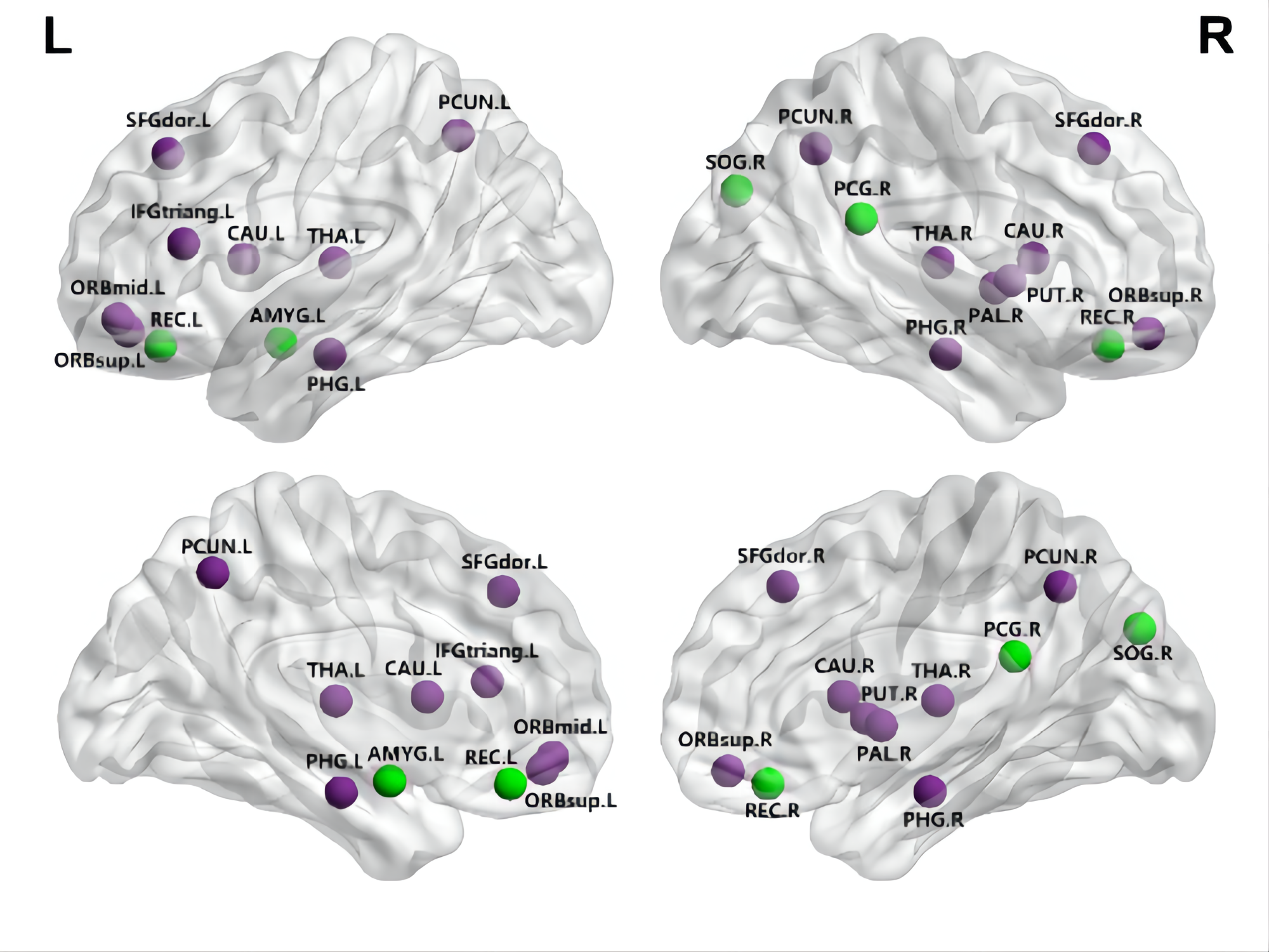
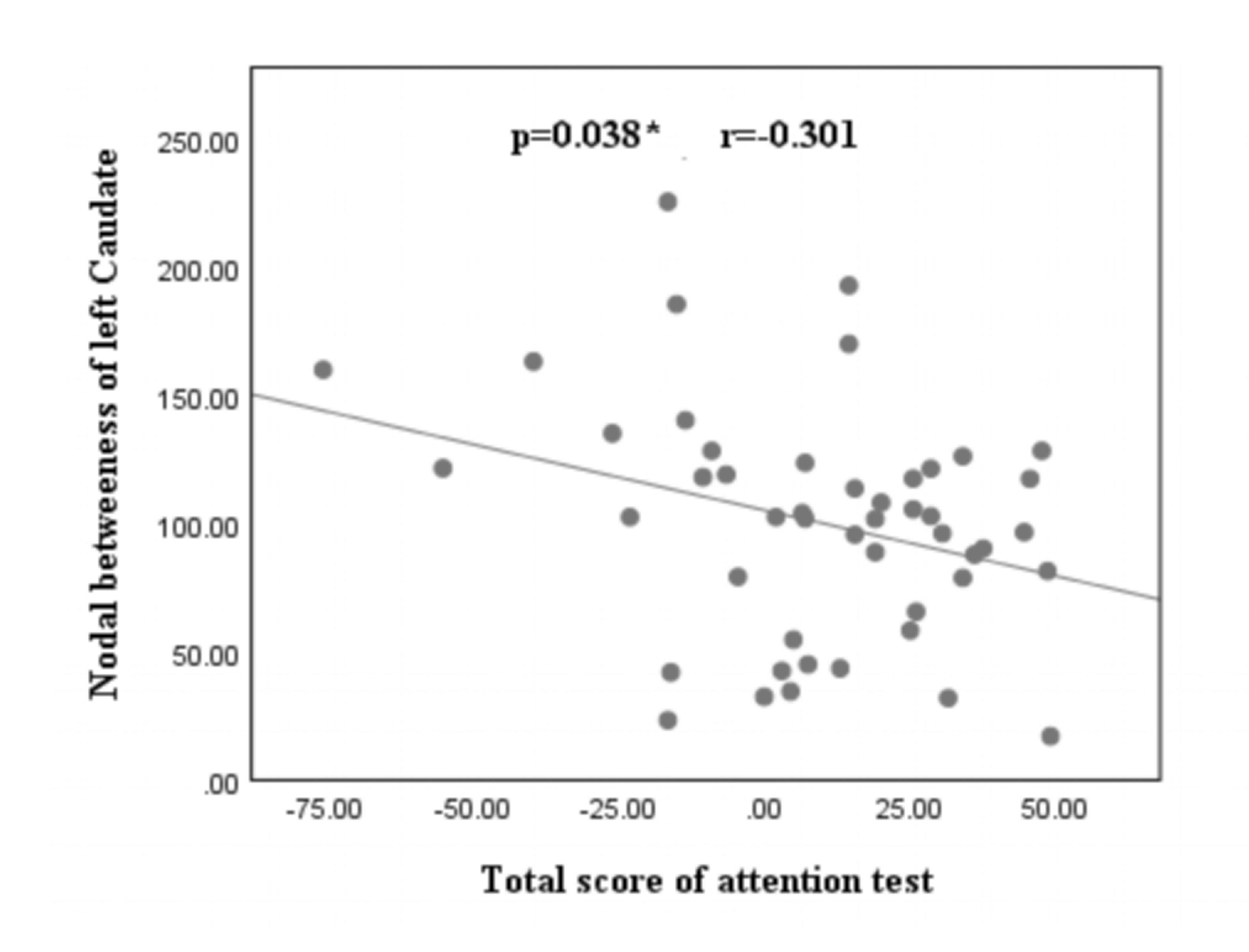
Both the ADHD and the TD groups showed small-world topology. However, patients with ADHD showed an increase in the characteristic path length (Lp, P=0.019), normalized clustering coefficient (γ, P=0.024), small-worldness (σ, P=0.029) and a decrease in global efficiency (Eglob, P = 0.019) (Fig 1). Furthermore, patients with ADHD showed altered nodal profiles, mainly in the default mode network (DMN), central executive network (CEN), and basal ganglia (P < 0.05, FDR-corrected). (Table 2)The nodal betweenness of left caudate in the ADHD patients was negatively associated with the concentration index (r=-0.29 P=0.048) and the total score of attention test (r=-0.301, P=0.038).(Fig 3)Discussion
Deterministic tractography based on DTI confirmed small-world architecture in both groups, but patients with ADHD showed a variety of differences relative to TD subjects: at the global level, increased characteristic Lp, γ, σ and decreased Eglob were observed; regionally, altered nodal profiles were found mainly in the default mode, central executive network and basal ganglia.Interestingly, in our study we found that ADHD showed hyper and hypoconnectivity of the DMN, which appeared in different models in previous studies6. Although DMN shows higher activity and stronger functional connectivity during resting-state, it’s abnormal transition can lead to momentary lapses in attention7. Hence, we consider the alternation of the DMN may cause the wrong modulation of its activity, which may interfere with task-relevant attention networks. Reduced nodal efficiency in ADHD was observed in CEN and basal ganglia, which are key structures of the cingulo-fronto-parietal (CFP) attention network and cortico-striato-thalamo-cortical (CSTC) loops. The CFP and CSTC loops are crucial to the attention and executive functions, whose disruption may cause abnormal behaviors of inattention. In particular, nodal betweenness of left caudate was negatively associated with the clinical symptom severity, which indicated a crucial role of left caudate in the pathogenesis of ADHD. We speculate that the observation of the increase nodal betweenness may reflect more severe clinical symptoms, but this need to be further determined in the future.Conclusion
Our results indicated that children with ADHD showed a shift toward weaker small-worldness pattern, providing a structural basis for functional alterations of pediatric ADHD. These abnormalities suggest that the pathogenesis of ADHD can be explained by the dysfunction of large-scale spatially distributed neural networks.Acknowledgements
None.References
1. Visser SN, Danielson ML, Bitsko RH, et al. Trends in the parent-report of health care provider-diagnosed and medicated attention-deficit/hyperactivity disorder: United States, 2003-2011. J Am Acad Child Adolesc Psychiatry. 2014;53(1):34-46.e2.
2. van Ewijk H, Heslenfeld DJ, Zwiers MP, Buitelaar JK, Oosterlaan J. Diffusion tensor imaging in attention deficit/hyperactivity disorder: a systematic review and meta-analysis. Neurosci Biobehav Rev. 2012;36(4):1093-1106.
3. De La Fuente A, Xia S, Branch C, Li X. A review of attention-deficit/hyperactivity disorder from the perspective of brain networks. Front Hum Neurosci. 2013;7:192. Published 2013 May 15.
4. Watts DJ, Strogatz SH. Collective dynamics of 'small-world' networks. Nature. 1998;393(6684):440-442.
5. Chen Y, Huang X, Wu M, et al. Disrupted brain functional networks in drug-naïve children with attention deficit hyperactivity disorder assessed using graph theory analysis. Hum Brain Mapp. 2019;40(17):4877-4887.
6. Konrad K, Eickhoff SB. Is the ADHD brain wired differently? A review on structural and functional connectivity in attention deficit hyperactivity disorder. Hum Brain Mapp. 2010;31(6):904-916.
7. Weissman DH, Roberts KC, Visscher KM, Woldorff MG. The neural bases of momentary lapses in attention. Nat Neurosci. 2006;9(7):971-978.
Figures