3591
Associations of gestational age, birth indicators, and brain volume in full-term newborns1The First Affiliated Hospital of Xi’an Jiaotong University, Xi’an, China, 2Shanghai United Imaging Intelligence Co., Shanghai, China, 3GE Healthcare, Beijing, China
Synopsis
Newborn gestational age (GA) and birth indicators (birth weight, birth length and head circumference) have been used as clinical indicators to assess the level of brain development. Cerebral changes are particularly intense during the last weeks of gestation. Previous studies have shown that the total brain volume at birth is about one third of the brain volume of healthy adults[1], but it remains unclear about the effect factors of brain volume. Our studies have shown that compared with other birth indicators, the gestational age of full-term newborns is strongly correlated with brain volume.
Summary of Main Findings:
We found that gestational age, birth weight, and head circumference are related to brain volume, while in which gestational age has the most significant indicator.Introduction:
Current research on abnormal neonatal brain volume development is mainly based on premature and low birth weight newborns. Also, neonatal brain MR images present reduced tissue contrast that renders the segmentation difficult. Few studies investigated the correlation between the segmented volume of full-term neonatal brain and the clinical indicators. In this study, the neonatal brain was divided into 87 anatomical regions, and we applied Pearson correlation and multiple linear regression to evaluate the correlation between birth indicators of full-term neonates and the volume of each brain region.Methods:
This study has been approved by the institutional IRB of the First Affiliated Hospital of Xi'an Jiaotong University and written informed consents were obtained from the children’s parents. From November 2010 to September 2017, we retrospectively collected 75 full-term newborns with no abnormalities in routine MRI (Table 1). All newborns were scanned with GE Signa HDxt 3.0 T MRI equipment. The neonates were given 10% chloral hydrate 0.5 ml/kg about 30 minutes before the MRI examination. After the scan, the doctor in charge and parents escorted the neonates back to the neonatal care unit. The acquired MRI sequence and parameters are as follows: 3D-T1WI: TR=10.28 ms, TE=4.62 ms, slice thickness=1 mm, matrix acquisition=240×240, FOV=24 cm, imaging resolution=1×1×1 mm3; T2WI: TR =4200 ms,TE =118.91 ms,slice thickness=4 mm, matrix acquisition=256×256, FOV=18 cm, imaging resolution=0.7×0.7×4 mm3. With the DHCP brain parcellation criteria, a deep learning model was trained to segment the brain into 87 regions by using the V-Net with bottleneck layer and the volume of each brain area was calculated. Multiple linear regression was used to analyze the correlation between each brain area and birth indicators with controlling postnatal age and sex. The standardized regression coefficients were used to analyze the weight of the impact of birth indicators on each brain area. All analyses were performed using SPSS software (version 20.0) and MATLAB software (version R2016b). P<0.05 indicates that the correlation is statistically significant.Results:
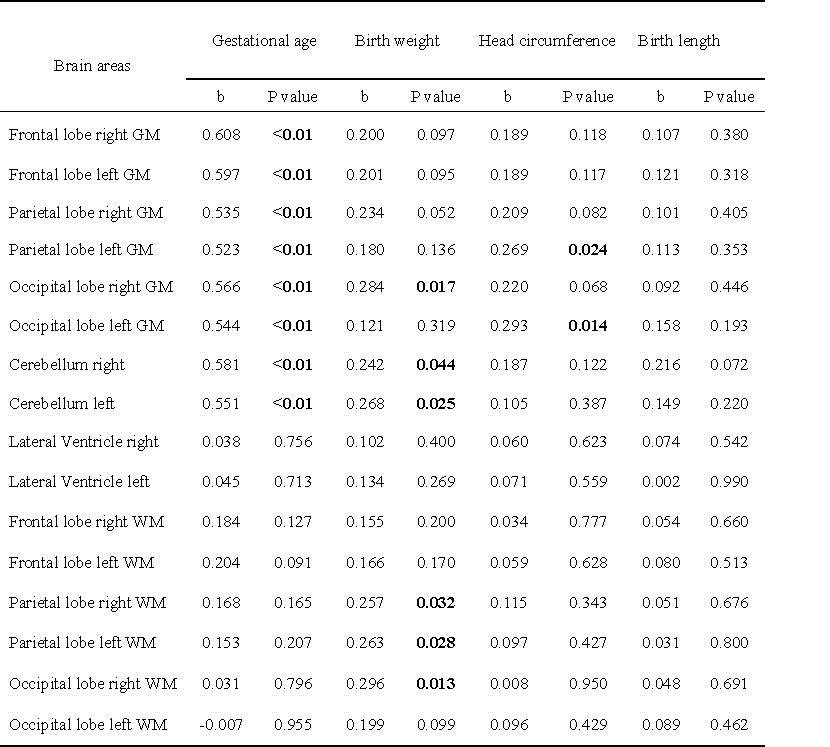
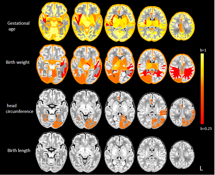
Compared with other birth indicators, the standardized regression coefficients (b) between gestational age and brain volume is the highest among full-term newborns, and there is no correlation between birth length and brain volume (p>0.05) (Figure 2). Compared with other brain regions, the gray matter of the frontal, parietal, and occipital cortex has a higher correlation (Table 2).Discussion and conclusion:
Previous studies have pointed out that preterm newborns (GA<37) may have atypical brain volumes and micro-structures when adjusted to term [2]. Our study using full-term neonates shows that gestational age and cerebral cortex gray matter (such as frontal, parietal, occipital lobe) volume have a significantly positive correlation, suggesting that full-term newborns that leave the uterus later can lead to more mature gray matter development, while the white matter volume is less affected gestational age. In addition, related studies have pointed out that the correlation between birth weight and brain development may be related to nutritional intake during pregnancy [3]. The brain volume of premature newborns with low birth weight and head circumference would correspondingly reduce [4][5]. The full-term newborns in this study also had a consistent correlation. We found that the correlation between the occipital lobe and frontal lobe is more significant, suggesting that larger birth weight and head circumference may indicate better visual-motor function. In addition, it also shows that the correlation between birth weight, head circumference, and brain volume has a hemispherical asymmetric distribution. The results of the study show that in full-term newborns, gestational age was significantly positively correlated with cerebral cortex gray matter. Birth weight, head circumference, and brain volume had a weak correlation, while body length was not correlated with brain volume.Acknowledgements
This study was supported by National Natural Science Foundation of China (81901516, 81901823, 81971581, 82101815), Shaanxi Provincial Innovation Team (2019TD-018).
* Correspondence: Chao Jin, Ph.D., Professor Department of Radiology The First Affiliated Hospital of Xi’an Jiaotong University, Xi’an, Shaanxi, China E-mail: jinny.369@163.com
References
1.Dominic Holland, Linda Chang, Thomas M Ernst et al. Structural growth trajectories and rates of change in the first 3 months of infant brain development.JAMA Neurol. 2014 Oct;71(10):1266-74.
2.Deanne K. Thompson, Claire E. Kelly et al. Characterisation of brain volume and microstructure at term-equivalent age in infants born across the gestational age spectrum.Neuroimage Clin. 2019;21:101630.
3.Victoria A Power, Alicia J Spittle, Katherine J Lee, Peter J Anderson et al. Nutrition, Growth, Brain Volume, and Neurodevelopment in Very Preterm Children.J Pediatr. 2019 Dec;215:50-55.e3.
4. Nehal A Parikh , Robert E Lasky et al.Perinatal factors and regional brain volume abnormalities at term in a cohort of extremely low birth weight infants.PLoS One. 2013 May 9;8(5):e62804.
5. Yukako Kawasaki, Taketoshi Yoshida et al. Clinical Factors That Affect the Relationship between Head Circumference and Brain Volume in Very-Low-Birth-Weight Infants.J Neuroimaging. 2019 Jan;29(1):104-110.
Figures