3590
The structural and functional change of glymphatic system in children with ADHD1First Affiliated Hospital, Sun Yat-sen University, Guangzhou, China, 2GE Healthcare, Beijing, China, 3the first affliated hospital of Xi'an Jiaotong University, Xi'an, China
Synopsis
The newly found structure – glymphatic system, may offer a new point for exploring the pathogenesis of attention deficit hyperactivity disorder (ADHD),. Our study investigated the change of glymphatic system in the treatment-naïve ADHD children by quantitatively measuring the VRS volume and using DTI-ALPS method. As the results showed, ADHD children have enlarged VRS, and the diffusivities along the VRS and ALPS-index were significantly lower in children with ADHD than in TD subjects, suggesting the impaired glymphatic drainage. Our study suggested that the glymphatic system alternation may play a role in the pathogenesis of ADHD and deserves further investigation.
Introduction
Attention deficit hyperactivity disorder (ADHD) is the most common childhood-onset neurodevelopmental disorder that may continue through adolescence and adulthood. The gene alteration of ADHD found in the previous genetic analysis was related with perivascular Virchow-Robin space (VRS), which indicated that the glymphatic system alternation may exist in ADHD patients. however, how does the glymphatic system change in ADHD still remains unknown.1The VRS is the visible structure of the glymphatic system in MRI. The enlarge VRS is very common to be seen in the neurodegenerative diseases. To some extent, the enlarge VRS reveals not only the structural change, but also the functional impairment in glymphatic system. The glymphatic system alternation can be revealed by the altered VRS volume. Besides, another non-invasive method called “Diffusion Tensor Image- Analysis aLong the Perivascular Space (DTI-ALPS)” has been introduced for glymphatic assessment. For the VRS in the periventricular area usually go vertically along the ventricular wall, by analyzing the diffusive ratio along the VRS, this DTI-based technique can quantitatively assess the perivascular flow in this area, which can also reflect the glymphatic system function.2This study aims to investigate both of the structural and functional change of glymphatic system in the treatment-naïve ADHD children by quantitatively measuring the VRS volume and using DTI-ALPS method.Methods
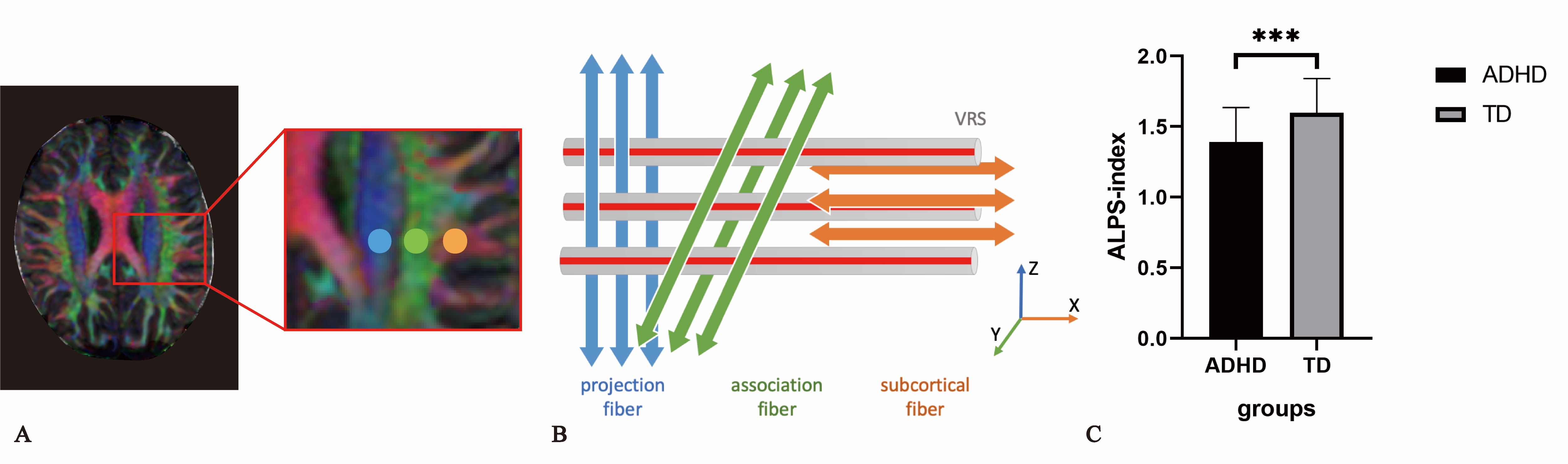
Axial T2WI images and DTI images were prospectively collected for 47 pediatric patients with clinical diagnosis of ADHD and 52 age- and gender matched typically developing children (TD) with age range of 6-13 years. A custom script was used to automate the segmentation of VRS in the white matter of the whole brain. Than manual correction was done to remove the VRS outside the cerebrum. Subsequently, the cerebral VRS volume were obtained. And the white matter (WM) volume and intracranial volume (ICV) was also estimated with FreeSurfer (v6.0.0) to calculate the VRS ratios.To calculate the ALPS-index, two neuro-radiologists, blinded to the clinical findings, independently measured the diffusivities along the x-axis(Dx), y-axis(Dy) and z-axis(Dz) in the projection (Dproj), the association (Dassoc), and the subcortical (Dsubc) neural fiber areas on the diffusivity maps. The ALPS-index was calculated with the following formula:ALPS-index = mean (Dxproj, Dxassoc) / mean (Dyproj, Dzassoc)Differences in VRS volume, VRS ratios, the diffusivities and ALPS-index between the two groups were analyzed by the Mann-Whitney U test.Results
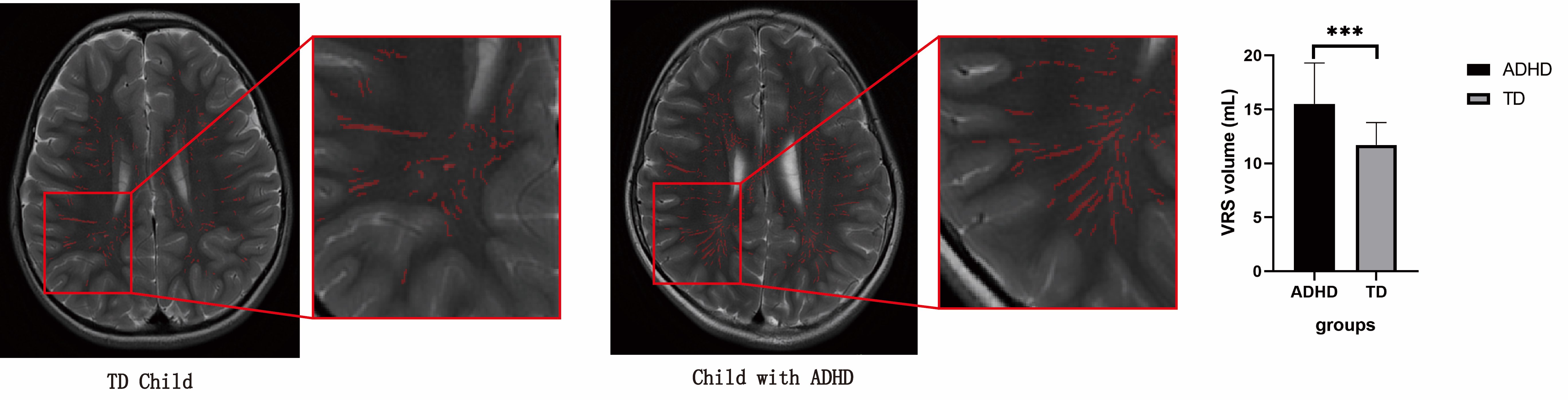
In the VRS analysis, although the ICV was smaller in ADHD group (ADHD: 1316.43 mL± 166.69 vs TD: 1374.37mL ± 137.97, p<0.05), the WM volume did not show any statistical difference between two groups (ADHD: 433.79 ml ± 50.95 vs TD: 439.87mL ± 49.54, p>0.05). And the VRS volume (ADHD: 15.514mL ± 3.773 vs TD: 11.702mL ± 2.068, p<0.001), VRS volume/ cerebral WM volume (ADHD:3.599% ± 0.868 vs TD: 2.692% ± 0.535, p<0.001), and VRS volume/ ICV(ADHD: 1.197% ± 0.326 vs 0.858% ± 0.159, p<0.001) are all significantly larger in the ADHD group than in TD group (Figure 1).In the DTI-ALPS analysis, the Dxproj (ADHD: 0.660×10⁻³mm2/s ±0.101 vs TD: 0.700×10⁻³mm2/s ± 0.069, p<0.05) and Dxassoc (ADHD: 0.740×10⁻³mm2/s ± 0.104 vs TD: 0.817×10⁻³mm2/s ± 0.112, p<0.001) are significantly smaller in ADHD group than in TD group. Correspondingly, the ALPS-index is significantly smaller in ADHD group, comparing with TD group (ADHD: 1.40±0.25 vs TD: 1.59±0.24, p<0.001) (Figure 2)Discussion
In this study, the enlarged VRS was found in ADHD children, which demonstrated the alternation of glymphatic system structure. The Dxproj, Dxassoc and ALPS-index were significantly lower in children with ADHD than in TD subjects, suggesting the impaired perivascular water flow, i.e., the impaired glymphatic drainage. The possible mechanisms to cause the glymphatic system alternation are as following:1) ADHD is proven to be associated with neurotransmitter dysregulation. Among those neurotransmitters, the norepinephrine is established as the primary neurotransmitter that suppresses the brain’s glymphatic system when awake by inhibiting the choroid plexus cerebral spinal fluid flow.32) The impaired arousal state in ADHD reduces the efficacy of glymphatic system. The impairment of arousal regulation was found to be a prominent symptom in ADHD patients. Since the dopaminergic signaling is central to the regulation of arousal, the impairment of dopamine metabolism in ADHD may lead to impairment regulation of sleep and wakefulness.4ADHD symptom is highly correlate with neuroinflammation. As an important immune system, the glymphatic system will be activated to protect against the neuroinflammation. Its clearance rate will be reduced as the permeability of astrocytes and intestinal space changed.5Conclusion
Our study demonstrate the morphological and functional change of glymphatic system in children with ADHD, which suggest that the glymphatic system alternation may play a role in the pathogenesis of ADHD and deserves further investigation. What’s more, the VRS volume and ALPS-index could be used as the metrics in the diagnosis of ADHD.Acknowledgements
We would like to thank the participants and their families as well as the staff at the MRI in the First Affiliated Hospital of Sun Yat-sen University for making this study possible.This study was supported by the National Natural Science Foundation of China [grant numbers 82001439] and the Medical Scientific Research Foundation of Guangdong Province [grant numbers A2020327].References
1. Vilor-Tejedor N, Alemany S, Forns J, et al. Assessment of Susceptibility Risk Factors for ADHD in Imaging Genetic Studies. J Atten Disord 2019; 23(7): 671-81.
2. Taoka T, Masutani Y, Kawai H, et al. Evaluation of glymphatic system activity with the diffusion MR technique: diffusion tensor image analysis along the perivascular space (DTI-ALPS) in Alzheimer's disease cases. Jpn J Radiol 2017; 35(4): 172-8.
3. Jessen NA, Munk AS, Lundgaard I, Nedergaard M. The Glymphatic System: A Beginner's Guide. Neurochem Res 2015; 40(12): 2583-99.
4. Wisor JP. Dopamine and Wakefulness: Pharmacology, Genetics, and Circuitry. Handb Exp Pharmacol 2019; 253: 321-35.
5. Mogensen FL, Delle C, Nedergaard M. The Glymphatic System (En)during Inflammation. Int J Mol Sci 2021; 22(14).
Figures