3588
Assessment of the correlation between blood perfusion and protein contents in brain of healthy subjects1Radiology and Nuclear medicine, Xuanwu Hospital, Capital Medical University, Beijing, China, 2Philips Healthcare, Beijing, China, Beijing, China
Synopsis
The blood perfusion and protein contents in brain of healthy subjects were measured in this study by 3D pseudo continuous arterial spin labeling and 3D amide proton transfer weighted imaging. Results showed a negative correlation between blood perfusion and the content of mobile cellular proteins and peptides in brain of heathy subjects; and the blood perfusion in normal brain, rather than the protein content, showed a significant dependence to age.
Introduction
The human brain undergoes a series of changes in anatomy, function, and organization during the brain development, which are necessary to support the complex adaptive behavior of normal individuals. The capability to measure developmental changes in different brain regions would provide a means to understand the structure-function relationships in both normal and disease states. Arterial spin labeling (ASL) is a magnetic resonance (MR) imaging technique used to assess cerebral blood flow noninvasively by magnetically labeling inflowing blood. [1] Amide proton transfer (APT) weighted imaging is a molecular MRI technique that generates image contrast based predominantly on the amide protons in mobile cellular proteins and peptides that are endogenous in tissue. It been used successfully for imaging of protein content and pH, the latter being possible due to the strong dependence of the amide proton exchange rate on pH. [2] This study aims to assess the correlation between brain perfusion and protein content measured by 3D pseudo continuous ASL (3D pCASL) and 3D APT methods, respectively, in healthy subjects, and explore their dependences to age.Methods
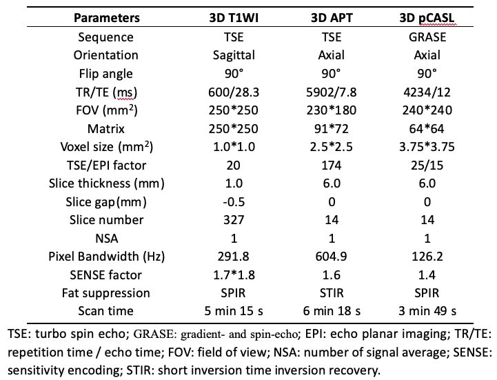
32 subjects (aged 21-66 years) gave written informed consent before participating in this study. Inclusion criteria for the study were as follows: normal results of T1WI, T2WI, fluid-attenuated inversion recovery, diffusion-weighted imaging; no history of head trauma, central nervous system infection, or cerebral structural lesions; and no psychiatric diseases or exposure to psychotropic drugs. MR scans were carried out on a 3.0T scanner (Ingenia, Philips Healthcare, Best, the Netherlands) using a 16-channel head coil. Detailed scan parameters for the 3D T1WI, 3D APT and 3D pCASL sequences were listed in Table 1. Saturation radio-frequency pulses for 3D APT were implemented with an amplitude of 2 µT and a duration of 2 s. And the post label delay (PLD) duration for 3D pCASL was 2000 ms. The MTRasym(3.5 ppm) map by 3D APT and the CBF map by 3D pCASL were reconstructed on the MR console immediately after the data acquisition. The atlas-based MTRasym(3.5 ppm) and CBF analyses were performed using the PMOD Software (Version 3.902). First, the MTRasym(3.5 ppm) and CBF maps were registered to the 3D T1WI image, and then the T1WI was registered to the standard brain. Then, 67 brain regions were separated by using the Anatomical Automatic Labeling (AAL) atlas provided by Montreal Neurological Institute (MNI). Finally, APT and CBF values of the 67 regions and whole brain were automatically calculated. Pearson correlation analyses were performed to assess the correlations among MTRasym(3.5 ppm), CBF values and age. P<0.05 was considered statistically significant.Results
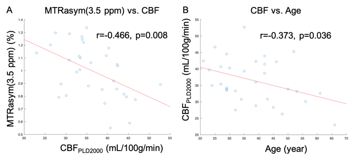
For whole brain analysis, MTRasym(3.5 ppm) was significantly correlated with CBF (r=-0.466, p=0.008, Figure 1A) in all subjects. And CBF, rather than MTRasym(3.5 ppm), showed a significant correlation with age (r=-0.373, p=0.036, Figure 1B). For regional analysis, MTRasym(3.5 ppm) was significantly correlated with CBF in 19 of the 67 brain regions (P<0.05); CBF was significantly correlated with age in 22 brain regions (P<0.05); and MTRasym(3.5 ppm) was significantly correlated with age only in 2 brain regions (P<0.05).Discussion
The negative correlation between MTRasym(3.5 ppm) and CBF indicate that the increased blood perfusion can lead to reduced mobile cellular proteins and peptides in normal brain tissues, which is contrary to the situations in brain tumors. The relationships of CBF and MTRasym(3.5 ppm) to age observed in this study were consistent with results by previous studies [3-4].Conclusion
The blood perfusion was observed to be negatively correlated with mobile cellular proteins and peptides in brain of heathy subjects; and the blood perfusion in normal brain, rather than the protein content, showed a significant dependence to age.Acknowledgements
No acknowledgement found.References
1. Haller S, Zaharchuk G, Thomas D L, et al. Arterial spin labeling perfusion of the brain: emerging clinical applications. Radiology, 2016, 281(2): 337-356.
2. Zhou J, Heo H Y, Knutsson L, et al. APT-weighted MRI: Techniques, current neuro applications, and challenging issues. Journal of Magnetic Resonance Imaging, 2019, 50(2): 347-364.
3. Biagi L, Abbruzzese A, Bianchi M C, et al. Age dependence of cerebral perfusion assessed by magnetic resonance continuous arterial spin labeling. Journal of Magnetic Resonance Imaging: An Official Journal of the International Society for Magnetic Resonance in Medicine, 2007, 25(4): 696-702.
4. Sugawara K, Miyati T, Ueda R, et al. Quantitative Analysis of Mobile Proteins in Normal Brain Tissue by Amide Proton Transfer Imaging: Age Dependence and Sex Differences. Journal of Computer Assisted Tomography, 2021, 45(2): 277-284.
Figures