3529
User-Friendly Application for Consistent Multi-Vendor Hyperpolarized 129Xe MRI Analysis1Center for Pulmonary Imaging Research, Cincinnati Children’s Hospital Medical Center, Cincinnati, OH, United States, 2Department of Biomedical Engineering, University of Cincinnati, Cincinnati, OH, United States, 3Department of Radiology, Cincinnati Children’s Hospital Medical Center, Cincinnati, OH, United States, 4Department of Pediatrics, University of Cincinnati, Cincinnati, OH, United States
Synopsis
Advances hyperpolarized 129Xe production, data acquisition and image analysis have allowed 129Xe MRI to emerge as a leading pulmonary imaging modality. As the number of sites capable of 129Xe MRI and quantitative image-derived metrics grow, the need for an intuitive, organized analysis tool increases. Here, we provide a user-friendly application, designed as MATLAB language-based interface, to analyze HP 129Xe data. This open-source, research tool enables standardized analysis of hyperpolarized 129Xe calibration, ventilation, diffusion, and gas-exchange data. Furthermore, the modular design allows developers to easily implement additional functions and analysis procedures, providing a framework for growth and sharing of analysis techniques.
Introduction
The high signal intensity and unique contrast provided by hyperpolarized (HP) 129Xe MRI enables regional changes in pulmonary ventilation, alveolar microstructure size, and gas exchange to be assessed1-5. Recent years have seen advances in coil hardware6, sequence design7-9, and image reconstruction, and in parallel with ongoing technical advances, the number of sites able to perform routine 129Xe MRI in humans continues to expand. As acquisition options and the number of 129Xe sites grow, it will become increasingly feasible to perform large, multi-site clinical trials using metrics derived from 129Xe MRI as endpoints. For multi-site 129Xe trials to be impactful, however, it will be vital to standardize data acquisition and analysis in manner that can be implemented by sites lacking decades of HP gas experience10. Here, we develop an executable application in MATLAB, to provide a user-friendly, graphical user interface-based pipeline to analyze and visualize 129Xe MRI data, including calibration scans, ventilation, diffusion, and gas exchange images. This application is designed for modular implementation of analysis methods, in an open-source platform to allow new methods and custom analyses to be implemented efficiently as 129Xe technology continues to evolve.Methods
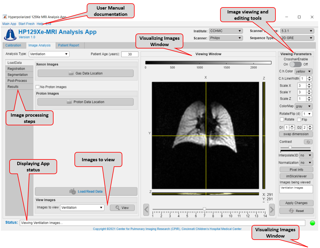
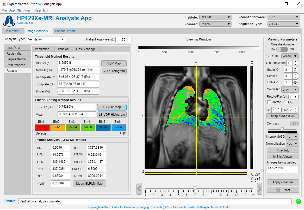
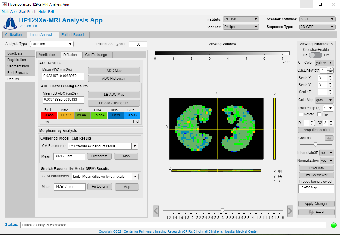
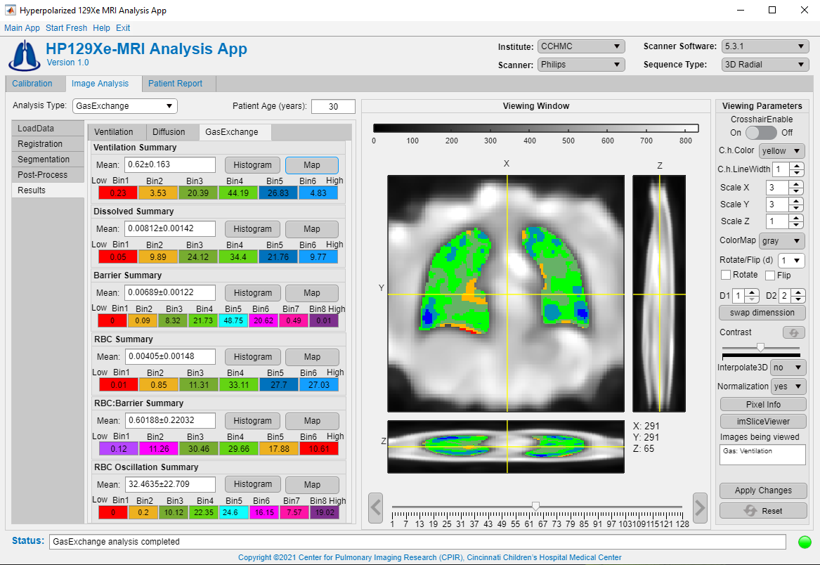
The HP 129Xe MRI analysis application was written in MATLAB R2020b (Mathworks, Natick, MA) using the Application Designer Toolbox. The layout of the compiled application is shown in Figure 1. The application is split into three primary tabs. Tab 1 is used to analyze scan calibration data and calculate the output needed to set scan parameters (flip angles, 129Xe frequency offset, etc.). Tab 2 is used to process and analyze 129Xe image data. This includes loading images generated by the scanner or reconstructing raw k-space data, registering images, segmenting lungs to generate masks for ROI analysis, and performing post-processing. Under post-processing panel, 3 additional tabs contained analysis input parameters for quantifying ventilation, diffusion, or gas-exchange images. Similarly, 3 tabs are built under the Results panel for displaying the main findings for each analysis type. Tab 2 also features a display window and image processing tools for visualizing and editing data. Tab 3 is used to generate patient analysis reports to facilitate the dissemination of data.To analyze ventilation-MRI data, 4 clusters and linear-binning thresholding methods11,12 are implemented to quantify ventilation-defect-percentage (VDP). Texture analysis13 is included as an optional analysis. To analyze diffusion data, basic ADC mapping with multiple fitting options (e.g., linear, weighted-linear, Bayesian)5 is included. A linear binning method (originally developed for ventilation images) 12 was modified to incorporate the expected age-dependence of ADC, and this capability was implemented to aid in ADC mapping—particularly for pediatric subjects. Methods to perform diffusion morphometry based on the cylindrical14 and stretched-exponential15 models are included. To analyze gas-exchange data, generalized linear binning16 is implemented. After processing, images and results can be exported in standard, image outputs and data presentation formats (.dcm, .nii, .png, .tif, .pptx, .xlsx, etc.). User manual documentation will also be made available as a pdf with the application.
The figures displaying 129Xe images were collected from healthy volunteers on a Philips-Achieva 3T MRI scanner. IRB approval and informed consent were obtained for all human studies.
Results
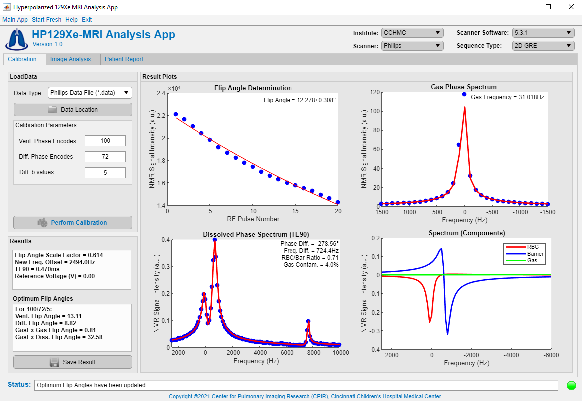
Figure 2 shows an example of processing calibration data and recommended scanner input parameters for the Philips MRI platform with the corresponding data fittings. An example of ventilation image processing is showing in Figure 3, which illustrates the main findings (mean ± standard deviation) of the thresholding methods and texture analysis. Figure 4 shows data processing for diffusion images and displays ADC and morphometry mapping summaries. Figure 5 displays gas-exchange data processing and shows summary findings alongside a binned colored map, based on reference data from healthy subjects.Discussion
This HP 129Xe MRI Analysis Application provides user-friendly interface tools to reconstruct and analyze 129Xe data. The application is designed in MATLAB, and therefore enables rapid implementation of new functions and code changes. These MATLAB applications are easily compiled as stand-alone installers that require no MATLAB license.This ongoing development of this application aims to reduce redundant effort and enable the streamlined analyses required to implement new techniques across multiple sites by providing an open-source framework for developers to implement and share methods and best practices. This optimize improve workflow across sites and reduced the training required for new and inexperienced users to perform analyses.
Finally, the functionality of the application can readily be expanded to include data processing for different sequences and scanners vendors; customized analysis reports that can be interpreted easily by clinicians; and new types of MRI contrast such as pulmonary capillary dynamics7. Of particular utility for higher through-put scanning, a 129Xe hyperpolarization model based on fundamental polarization physics and practical polarizer engineering considerations 17 will also be implemented to allow polarizer operators to assess tradeoffs between 129Xe polarization, subject dosing, and production rate.
Conclusion
A user-friendly interface application has been developed to aid in the implementation and sharing of HP 129Xe analysis methods across multiple HP 129Xe MRI sites and scanner platforms. This will enable consistent analysis of 129Xe MRI data between sites.Acknowledgements
The authors acknowledge the following sources for research funding and support: NIH(R01HL151588, R01HL143011, R01HL126771).References
1. Walkup LL, Woods JC. Translational applications of hyperpolarized 3He and 129Xe. NMR in Biomedicine 2014;27:1429-1438.
2. Goodson BM. Nuclear magnetic resonance of laser-polarized noble gases in molecules, materials, and organisms. Journal of Magnetic Resonance Imaging: An Official Journal of the International Society for Magnetic Resonance in Medicine 2002;155:157-216.
3. Ruppert K. Biomedical imaging with hyperpolarized noble gases. Reports on Progress in Physics 2014;77:116701.
4. Mugler III JP, Altes TA. Hyperpolarized 129Xe MRI of the human lung. Journal of Magnetic Resonance Imaging 2013;37:313-331.
5. Bdaiwi AS, Niedbalski PJ, Hossain MM, et al. Improving hyperpolarized 129Xe ADC mapping in pediatric and adult lungs with uncertainty propagation. NMR Biomed 2021:e4639.
6. Loew W, Thomen R, Pratt R, Cleveland Z, Dumoulin C, Woods J, Giaquinto RO. A dual loop t/r -xenon coil for homogenous excitation with improved comfort and size ISMRM2016.
7. Niedbalski PJ, Bier EA, Wang Z, Willmering MM, Driehuys B, Cleveland ZI. Mapping cardiopulmonary dynamics within the microvasculature of the lungs using dissolved 129Xe MRI. 2020;129:218-229.
8. Willmering MM, Niedbalski PJ, Wang H, Walkup LL, Robison RK, Pipe JG, Cleveland ZI, Woods JC. Improved pulmonary 129Xe ventilation imaging via 3D‐spiral UTE MRI. Magnetic Resonance in Medicine 2020;84:312-320.
9. Abdullah S. Bdaiwi, Matthew M. Willmering, Hui Wang, Cleveland ZI. 2D and 3D spiral for diffusion weighted MRI with hyperpolarized 129Xe. International Society for Magnetic Resonance in Medicine. Virtual 2021.
10. Niedbalski PJ, Hall CS, Castro M, et al. Protocols for multi-site trials using hyperpolarized 129Xe MRI for imaging of ventilation, alveolar-airspace size, and gas exchange: A position paper from the 129Xe MRI clinical trials consortium. 2021;86:2966-2986.
11. Roos JE, McAdams HP, Kaushik SS, Driehuys B. Hyperpolarized gas MR imaging: Technique and applications. Magn Reson Imaging Clin N Am 2015;23:217-229.
12. He M, Zha W, Tan F, Rankine L, Fain S, Driehuys B. A comparison of two hyperpolarized 129Xe MRI ventilation quantification pipelines: The effect of signal to noise ratio. Academic radiology 2019;26:949-959.
13. Galloway MM. Texture analysis using gray level run lengths. Computer graphics and image processing 1975;4.2 (1975): 172-179.
14. Sukstanskii A, Yablonskiy D. Lung morphometry with hyperpolarized 129Xe: Theoretical background. Magnetic resonance in medicine 2012;67:856-866.
15. Chan HF, Stewart NJ, Norquay G, Collier GJ, Wild JM. 3D diffusion‐weighted 129Xe MRI for whole lung morphometry. Magnetic resonance in medicine 2018;79:2986-2995.
16. He M, Ziyi Wang, Leith Rankine, Sheng Luo, John Nouls, Rohan Virgincar, Joseph Mammarappallil, and Bastiaan Driehuys. Generalized linear binning to compare hyperpolarized 129Xe ventilation maps derived from 3D radial gas exchange versus dedicated multislice gradient echo MRI. Academic radiology 2020;27, no. 8 (2020): e193-e203.
17. Plummer JW, Emami K, Dummer A, Woods JC, Walkup LL, Cleveland ZI. A semi-empirical model to optimize continuous-flow hyperpolarized 129Xe production under practical cryogenic-accumulation conditions. Journal of Magnetic Resonance 2020;320:106845.
Figures