3477
Combination of Multichannel Blind Deconvolution (MALBEC) and GRAPPA for Highly Accelerated 3D Imaging1Department of Biomedical Engineering, Department of Electrical Engineering, University at Buffalo, Buffalo, NY, United States, 2Program of Advanced Musculoskeletal Imaging (PAMI), Cleveland Clinic, Cleveland, OH, United States
Synopsis
Most parallel imaging methods require calibration data for reconstruction. Low-rank-based methods allow calibration-free reconstruction from randomly undersampled, multi-channel data. This abstract presents a novel reconstruction method to combine multichannel blind deconvolution (MALBEC), a calibration-free method, and GRAPPA, a calibration-based method for highly accelerated imaging. The method sequentially performs MALBEC and GRAPPA with specially designed sampling masks such that the benefits of low-rank structure and the availability of calibration data can be utilized jointly. Our results demonstrate that the proposed method can achieve an acceleration factor that is the product of the factors achieved by MALBEC and GRAPPA alone.
Introduction
In parallel MRI1, calibration-based methods(e.g., GRAPPA2-3) have been widely used, which requires fully sampled center k-space for calibration. However, the acceleration factor is usually limited to two in clinical scans. Some calibration-free reconstruction methods (e.g., MALBEC4-8) have been proposed to achieve higher acceleration. These methods require random undersampling in 2D to formulate the reconstruction problem as a low-rank matrix recovery problem without knowledge of calibration data or coil sensitivities. Despite their potential, the calibration-free methods alone cannot achieve very high acceleration factors either. In this study, we take advantage of the benefits of both calibration-free and calibration-based methods and propose a novel k-space reconstruction method, which integrates MALBEC and GRAPPA to achieve an acceleration factor that is the product of the factors achieved individually. Among many calibration-free methods, we choose MALBEC due to its superior reconstruction quality, ease of parameter selection, and fast computation. The sampling pattern is specially designed such that MALBEC and GRAPPA are performed in a sequential manner. The performance of the method is evaluated using 3D Dual-Echo Steady-State (DESS) knee images.Method
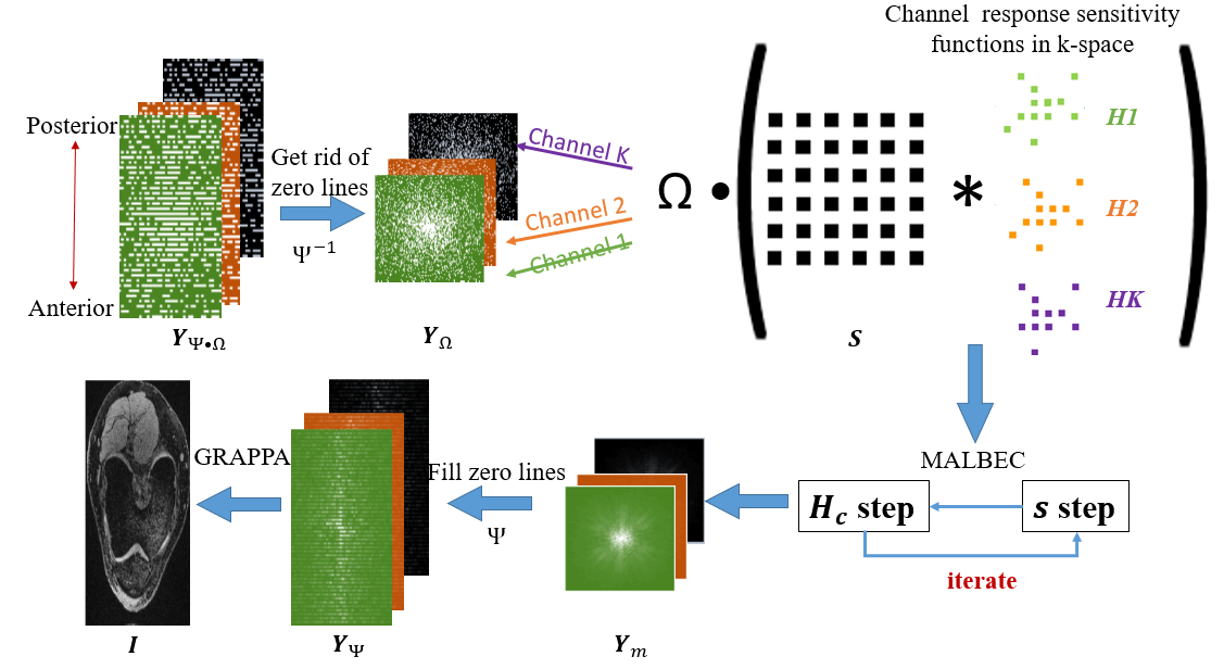
Our proposed method performs MALBEC and GRAPPA sequentially, as shown in Figure 1. The undersampled k-space data $$$Y_{\Psi,\Omega }$$$ are acquired with a specially designed pattern, which can be represented as an integration of two undersampling operators $$$\Psi$$$ and $$$\Omega$$$. The first operator $$$\Psi$$$ uniformly undersamples k-space by a factor of $$$\psi$$$ in the phase-encoding direction (GRAPPA-like undersampling), and the second operator $$$\Omega$$$ randomly undersamples the remaining data by a factor of $$$\omega$$$ after $$$\Psi$$$ in both phase- and slice-encoding directions. As a result, the total acceleration reduction is $$$\psi\times\omega$$$. In the first part of reconstruction, the undersampled k-space data $$$Y_{\Omega }$$$ (generated by eliminating the zero-valued phase-encoding lines in $$$Y_{\Psi,\Omega }$$$ from all channels) are reconstructed using MALBEC. Specifically, denoting $$$y_{\Omega,c}[p,q]$$$ as the k-space data at location $$$(p,q)$$$ for channel $$$c$$$, each data point can be represented as the convolution of the desired unknown intermediate-reconstructed k-space data $$$s[m,n]$$$ and the k-space of the coil sensitivities for the corresponding channels $$$H_{c}[m,n]$$$, $$y_{\Omega,c}[p,q]=\sum_{m=0}^M\sum_{n=0}^NH_{c}[m,n]s[p-m,q-n](1)$$where $$$c=1,2 \cdots C,(p,q)\in\Omega$$$. MALBEC formulates the reconstruction of $$$s$$$ from the undersampled data as a low-rank matrix recovery problem4-6 without knowledge of $$$Y_{ m}$$$ or $$$H_{c}$$$. First, we initialize the estimated and $$$H_{c,0}$$$ as $$$s_{0}=F \big(sos\big( F^{-1}\big(T \bullet y_{ \Omega,c}\big)\big)\big), H_{c,0}=F^{-1}\big(T \bullet y_{\Omega,c}\big)/s_{0}(2)$$$. where $$$T$$$ represents a smooth turkey window, $$$sos$$$ represents the sum-of-square operator. The method then solves for $$$s$$$ and channel responses $$$H_{c}$$$ alternatively based on Eq.(1). Specifically, in the $$$s$$$-step and $$$H$$$-step, $$$s$$$ and $$$H_{c}$$$ are solved by using: $$s=argmin_{s}\|\sum_{k=1}^K\Omega(s \ast H_{c})\ast\overline{H_{c}}-\sum_{k=1}^K y_{\Omega,c}\ast\overline{H_{c}}\|^{2}(3)$$
$$H_{c}=argmin_{H_{c}}\|\Omega(Hankel(s) \bullet H_{c})-y_{\Omega ,c}\|^{2}(4)$$
We generate the Hankel structured data matrix to represent the convolution problem. The two steps are performed iteratively and the iteration usually converges fast (~ 2 iterations). After both $$$s$$$ and $$$H_{c}$$$ are obtained, the multi-coil intermediate-reconstructed k-space data $$$Y_{m}$$$ can be obtained from their convolution. Adding previously eliminated zero-valued phase-encoding lines transforms $$$Y_{m} $$$ to $$$Y_{\Psi }$$$, the uniformly undersampled data. Finally, GRAPPA is performed on $$$Y_{\Psi }$$$ to obtain the final desired image.
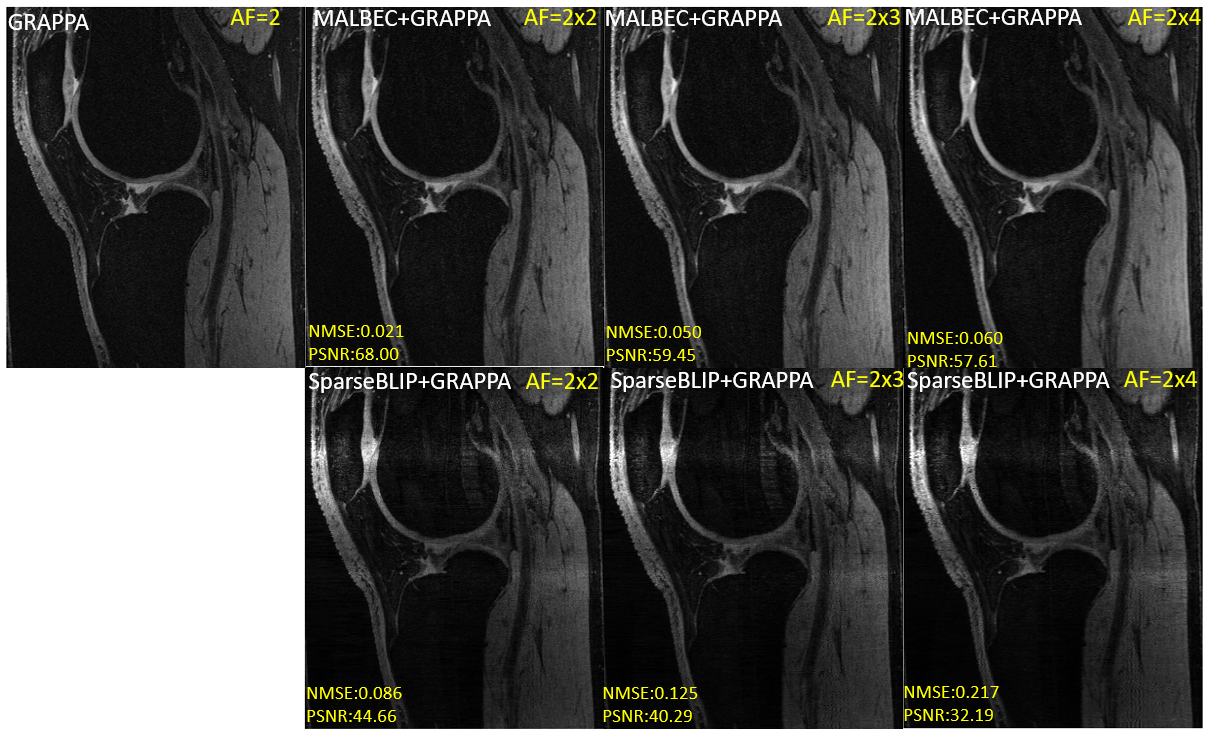
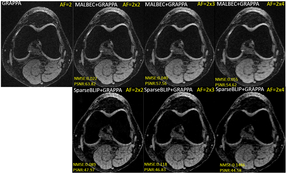
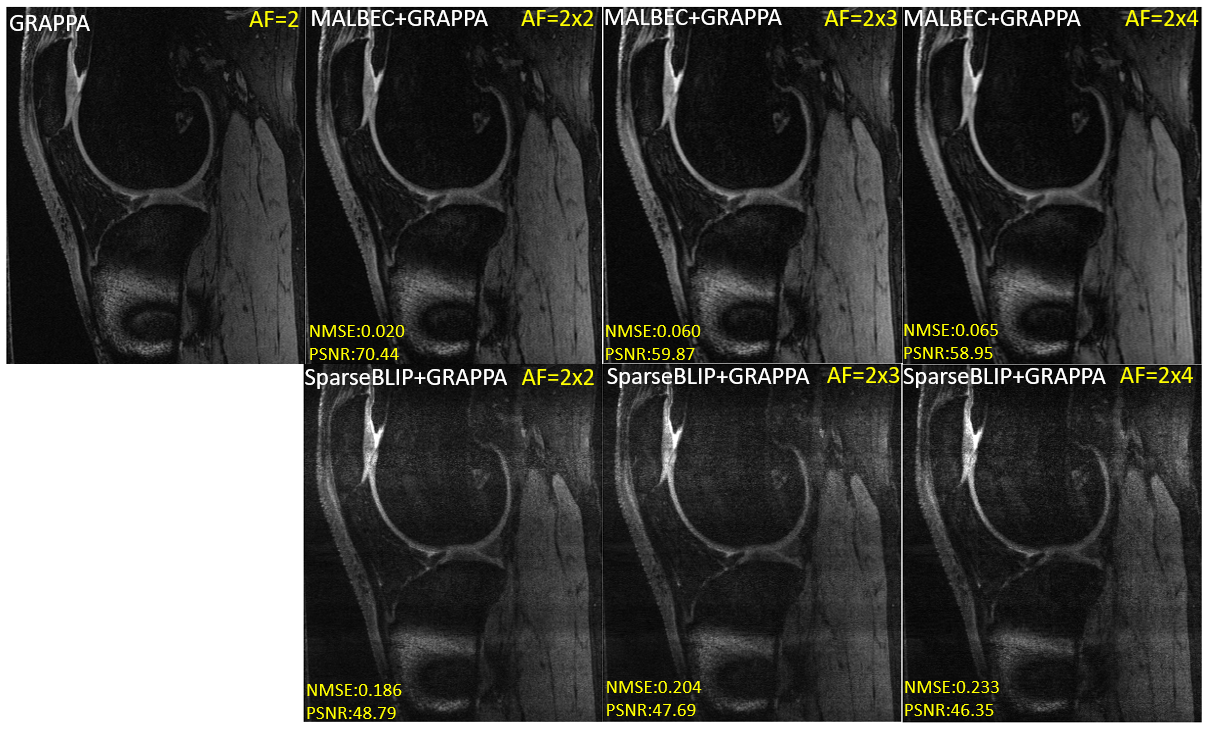
Total 10 in-vivo knees including 5 knees with metal artifact from ACL reconstruction were scanned on a 3T Siemens scanner with a 15-channel knee coil. A 3D DESS sequence was scanned with scan parameters as follows; TE = 6.02ms, TR = 17.55ms, and flip angle=20°. FOV= 140 mm, slice thickness 0.7mm, matrix size of 384x307x160 with GRAPPA factor of 2 $$$ (\psi =2) $$$. We further manually undersampled the GRAPPA-undersampled data with 2D random undersampling patterns $$$\Omega$$$ $$$ (\omega =2,3,4)$$$ to simulate combined acceleration factors of $$$2\times2$$$, $$$2\times3$$$, $$$2\times4$$$.
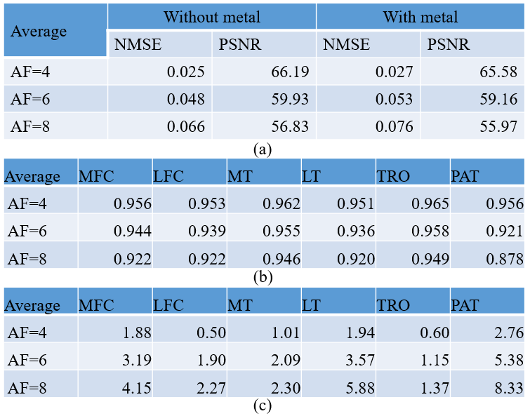
PSNRs and NMSE were calculated for reconstructions, using the 2× undersampled data using GRAPPA as the reference. For knees without metal artifacts, cartilage was automatically segmented into six compartments: MFC/LFC, MT/LT, TRO, and PAT cartilages9. Two metrics were used to compare the reference and accelerated images: DICE coefficients of segmented cartilage, and the absolute difference of average cartilage thickness of each compartment10.
Results and Discussion
Fig2 shows representative reconstructed images in sagittal and axial views. For comparison, we replaced MALBEC with SparseBLIP in our combined method. The MALBEC+GRAPPA showed superior results compared to SparseBLIP+GRAPPA in terms of reconstruction quality as well as PSNRs and NMSE shown in each image, especially when metal artifact was present. The performance of MALBEC+GRAPPA reconstruction does not degrade with metal artifact, as shown in Table1(a). In knees without metal artifacts, all DICE were higher than 0.9 except for patellar cartilage with acceleration factor(AF=8), all absolute differences of cartilage thickness were smaller than 6%, except for patellar cartilage with AF=8, Table1(b). Not unexpected, DICE decreases with an increase in AF, and the difference in cartilage thickness increases with an increase in AF.Conclusion
In this abstract, we developed a novel acceleration method combining MALBEC and GRAPPA. Experimental results in 3D knee DESS images show that the proposed method can further accelerate the acquisition time by a factor of 4 on top of the GRAPPA factor of 2, which reduces the scan time from 6 minutes to 1.5 minutes. More data sets will be used for evaluating tissue quantification accuracy (cartilage thickness and composition, with/without metal artifacts) in future studies.Acknowledgements
This work is supported in part by NIH/NIAMS R01 AR077452.References
[1] Ying L, Liang ZP. Parallel MRI using phased array coils (2010) IEEE Signal Processing Magazine, 27 (4), pp. 90-98.
[2] Griswold MA, Jakob PM, Heidemann RM, Nittka M, Jellus V, Wang J, Kiefer B, Haase A. Generalized autocalibrating partially parallel acquisitions (GRAPPA). Magn Reson Med. 2002 Jun;47(6):1202-10.
[3] Lustig M, Pauly JM. SPIRiT: Iterative self-consistent parallel imaging reconstruction from arbitrary k-space. Magn Reson Med. 2010 Aug;64(2):457-71.
[4] Lyu J, Nakarmi U, Zhou Y, Zhang C, and Ying L. Calibration-free Parallel Imaging Using Randomly Undersampled Multichannel Blind Deconvolution (MALBEC). Proc. Intl. Soc. Mag. Reson. Med. 24 (2016)
[5] Shin PJ, Larson PE, Ohliger MA, Elad M, Pauly JM, Vigneron DB, Lustig M. Calibrationless parallel imaging reconstruction based on structured low-rank matrix completion. Magn Reson Med. 2014 Oct;72(4):959-70.
[6] Haldar JP, Zhuo J. P-LORAKS: Low-rank modeling of local k-space neighborhoods with parallel imaging data. Magn Reson Med. 2016 Apr;75(4):1499-514.
[7] She H, Chen RR, Liang D, DiBella EV, Ying L. Sparse BLIP: BLind Iterative Parallel imaging reconstruction using compressed sensing. Magn Reson Med. 2014 Feb;71(2):645-60.
[8] Trzasko, JD, Manduca A. Calibrationless parallel MRI using CLEAR. Signals, Systems and Computers (ASILOMAR), 2011 Conference Record of the Forty Fifth Asilomar Conference on. IEEE, 2011.
[9] Gaj S, et al. Automated cartilage and meniscus segmentation of knee MRI with conditional generative adversarial networks. Magnetic resonance in medicine 84.1 (2020): 437-449.
[10] Maier J, Black M, Bonaretti S, Bier B, Eskofier B, Choi JH, Levenston M, Gold G, Fahrig R, Maier A. Comparison of Different Approaches for Measuring Tibial Cartilage Thickness. J Integr Bioinform. 2017 Jul 28;14(2):20170015.
Figures