3436
Brain Connectomics in a Mouse Model of Early and Late Stage Pruritus
Talaignair N Venkatraman1, Ouyang Chen2, Allen W Song3, Ru-Rong Ji2, and Chris D Lascola3
1Radiology, Duke University Medical Center, Durham, NC, United States, 2Anesthesiology, Duke University Medical Center, Durham, NC, United States, 3BIAC and Radiology, Duke University Medical Center, Durham, NC, United States
1Radiology, Duke University Medical Center, Durham, NC, United States, 2Anesthesiology, Duke University Medical Center, Durham, NC, United States, 3BIAC and Radiology, Duke University Medical Center, Durham, NC, United States
Synopsis
We performed high-resolution ex-vivo DTI in mice with cutaneous T-Cell lymphoma (CTCL) in a mouse model of pruritus. Our results show significant network changes within and between specific brain regions, namely thalamus and hippocampus, by the later stage of disease. At the chronic stage (55 days), micro-structural changes and enhanced connectivity are established between the hippocampus and thalamus.
Introduction:
Pruritus (Itch) in chronic form results in immense suffering and adverse effects on quality of life. The mechanisms of itch are increasingly understood to overlap with spinal pathways known to be associated with pain, as well as brain regions involved with learning and memory. We performed high-resolution ex-vivo DTI in mice with cutaneous T-Cell lymphoma (CTCL), an established mouse model of pruritus. We looked at regional CNS changes in DTI metrics at early (20 Days) and chronic stages (55 Days) of disease. Our results show significant network changes within and between specific brain regions, namely thalamus and hippocampus, by the later stage of disease.Materials and Methods:
Total Mice n =22 ( Days 20 ; Control = 5, CTCL = 5 and Days 55; Control = 6, CTCL = 6). Control and CTCL were perfused and harvested at 20 days and 55 days after tumor inoculation and stored in fixative solution of 0.5 % ProHance-doped formalin. Image Acquisition: Brains in Fomblin solution were scanned on a Bruker 7.0T system with quadrature volume-transmit and surface-receive coils. MRI protocol: (A) 3D T1-weighted FLASH sequence with FOV = 1.8 x 1.8 x 1.8 cm; matrix = 256 x 256 x 256; resolution = 70 x 70 x 70 μm/pixel; TE/TR = 6/30 ms; averages = 16; flip-angle = 34(B) High resolution spin-echo based 3D DTI with FOV = 1.8 x 1.8 x 1.8 cm; matrix = 128 x 128 x 128; resolution = 141 x 141 x 141 μm/pixel; TE/TR = 25/250 ms; diffusion directions = 60; Ao images = 5; B-value per direction = 1500 S/mm2. Post-Processing: Brains were registered to a reference template from Wake forest University’s Mouse Database and segmented using ITK-SNAP. DTI parameters were calculated using TrackVis.Results:
Figure 1 shows ROI Fractional Anisotropy (FA) analysis of Hippocampus and Thalamus at 20 days and 55 days. It is observed that FA significantly dropped at early stage (20 days) and at chronic stage (55 days) significantly increased. Figure 2 shows un-parcellated fiber tracts through Hippocampus and Thalamus. No significance is noted for Fiber Tract Numbers (FTN) and associated FA at 20 days and at 55 days except for FA in the hippocampus. Figure 3 shows connectomic analysis of Hippocampus-Thalamus fiber tracts. It is found that no significance noted at 20 days whereas at chronic stage (55 days) both FA and FTN increased significantly.Conclusions:
In a CTCL mouse model of pruritus, large, micro-structural changes and enhanced connectivity are established by the chronic stage (55 days) between the hippocampus and thalamus. These change are highly significant and asymmetric, strongly correlating with the somatic location of lymphomatous involvement.Acknowledgements
No acknowledgement found.References
No reference found.Figures
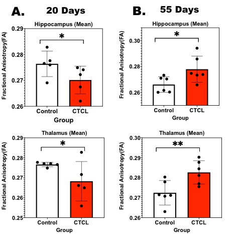
Figure-1: Fractional Anisotropy of Hippocampus (Top) and Thalamus (Bottom) at 20 Days ( A ) and 55 Days ( B ) using ROI analysis. * p<0.01and **p<0.001.
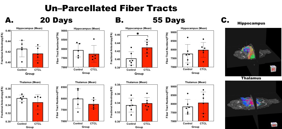
Figure-2: Fractional Anisotropy (FA) and Fiber Tract Numbers (FTN) of Hippocampus (Top) and Thalamus (bottom) at 20 Days ( A ) and 55 Days( B ) and corresponding DTI Tractography illustrated ( C ).
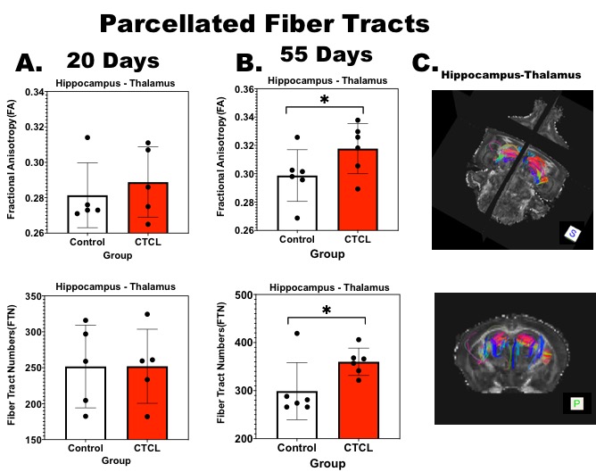
Figure-3: Fractional Anisotropy (FA) [Top] and Fiber Tract Numbers (FTN) [Bottom] of Hippocampus-Thalamus fiber connectivity at 20 Days ( A ) and 55 Days( B ) and corresponding DTI Tractography illustrated ( C ).
DOI: https://doi.org/10.58530/2022/3436