3433
Optimization of a highly sensitivity 19F MRI probe for NK cell immunotherapy tracking1Imaging Physics, The University of Texas MD Anderson Cancer Center, Houston, TX, United States, 2Dmitry Nevozhay, The University of Texas MD Anderson Cancer Center, Houston, TX, United States, 3Pediatrics, The University of Texas MD Anderson Cancer Center, Houston, TX, United States, 4Chemistry, The University of Texas at Austin, Austin, TX, United States, 5The University of Texas at Austin, Austin, TX, United States, 6The University of Texas MD Anderson Cancer Center, Houston, TX, United States
Synopsis
NK cell immunotherapy has great potential as a safe and effective anticancer therapy yet there are significant challenges for immunotherapy in brain tumors. We have developed a 19F probe with enhanced sensitivity for the purposes of 19F labeling of NK cells to monitor NK cell biodistribution and immunotherapy. In this paper, we report on the relaxometric characteristics of the probe, acquisition optimization, and initial ex vivo imaging tests in murine models. The agent displayed improved sensitivity relative to Celsense and was clearly visible in the cerebral cortex in approximately equivalent amounts that will be used for cell tracking experiments.
INTRODUCTION
NK cell immunotherapy has been identified as having great potential as safe and effective anticancer therapy1. However, there are significant challenges in brain tumors and it is anticipated that imaging will play a key role in addressing those challenges2. To help improve the visualization of NK cells as they distribute during the course of therapy, we are developing a novel perfluorocarbon-based contrast agent that may be advantageous as compared to prior appraches3. Our overall goal is to achieve improved 19F sensitivity through improved relaxation characteristics and improved cellular loading during the course of NK cell immunotherapy in murine models of brain cancer and ultimately translation to clinical scanners. We hypothesize that improved 19F sensitivity may be achieved with an iron-containing perfluorocarbon agent. In this paper, we report on the relaxation characteristics of our agent and initial imaging tests to optimize acquisition parameters and initial ex vivo tests.METHODS
For each of the made preparations, the mixture of 18mg 1,2-distearoyl-sn-glycero-3-phosphocholine (DSPC, 94.3% by molarity), 1.36 mg 1,2-distearoyl-sn-glycero-3-phosphoethanolamine-N-methoxy-polyethyleneglycol-2000 (DSPE-PEG-2000, 2%), 0.34 mg 1,2-distearoyl-sn-glycero-3-phosphoethanolamine-N-[maleimide(polyethylene glycol)-2000 (DSPE-PEG-2000-Mal, 0.5%), and 0.3 mg cholesterol (3.2%; all from Avanti Polar Lipids, Inc., Alabaster, AL) in 2 mL chloroform was evaporated to form a lipid cake and then rehydrated in 2 mL deionized (DI) water4-7. Next, 100 µL of each PFC compound (pure PFCE and also iron-doped at 5, 15, and 30 mM) was added to respective vials with rehydrated lipid cakes and suspensions were emulsified using bath sonication (CPX-962-218R; Thermo Fisher Scientific, Waltham, MA) at 50 °C for 7 minutes. Mean size and polydispersity index (PDI) of the resulting nanodroplets preparations were 180-210 nm and 0.100-0.170, respectively, assessed using dynamic light scattering (DLS).To quantify the relaxation properties of the agents, concentrations were determined by integrating an NMR spectrum of each compound relative to a known trifluoroacetic acid reference4. To ascertain sensitivity, three separate phantoms were constructed which had multiple concentration of TFA, Crown (Celsense), and MDA compound. Each solution was placed in a sealed cutoff 5 mM NMR tube that was immersed in an outer cylindrical glass container completely filled with water to minimize susceptibility effects.
All MRI was performed on a 7 Tesla Bruker Biospec AV3HD using either a 35 mm birdcage volume coil (Bruker Biospin Inc., Billerica, MA) for anatomic imaging or a 35 mm 19F birdcage coil for 19F detection (Rapid MR, International, Columbus, Ohio). Scout images and shimming were acquired with 1H and then the coil was switched for 19F acquisition. 1H (19F) images were acquired with 3D RARE sequence using a field of view (FOV) of 32 x 32 x 16 mm3 (32 x 32 x 24 mm3), a matrix size of 128 x 128 x 16 (32 x 32 x 8), a RARE factor = 32 (16), TE = 72 ms (6.0 ms), and TR = 2000 ms (300 ms). T1 and T2 maps and subsequent analyses were computed using in-house written Matlab code. Optimization of imaging parameters was performed for each compound separately.
Ex vivo stereotaxic injection into the cerebral cortex was done below the coronal suture of the right hemisphere and just above the lambdoid suture to minimize the potential for injection site overlap from agent diffusion.
RESULTS
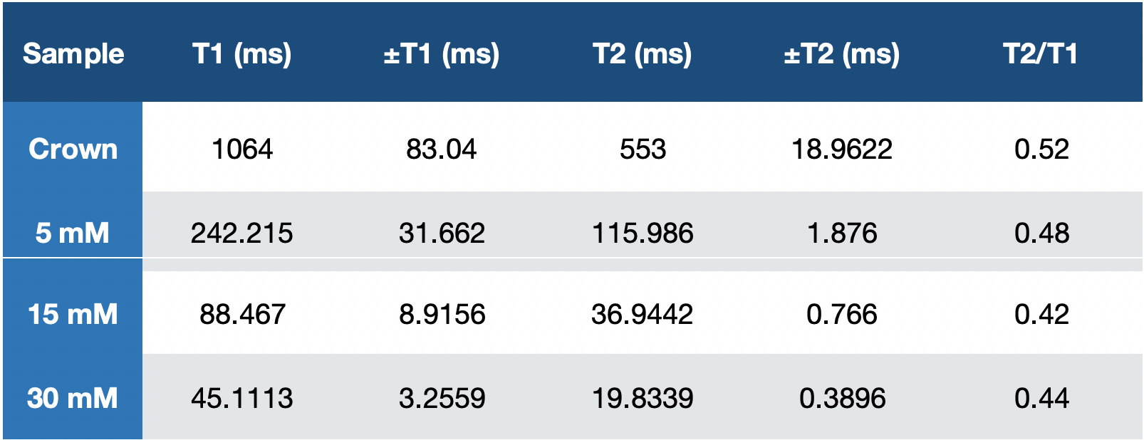
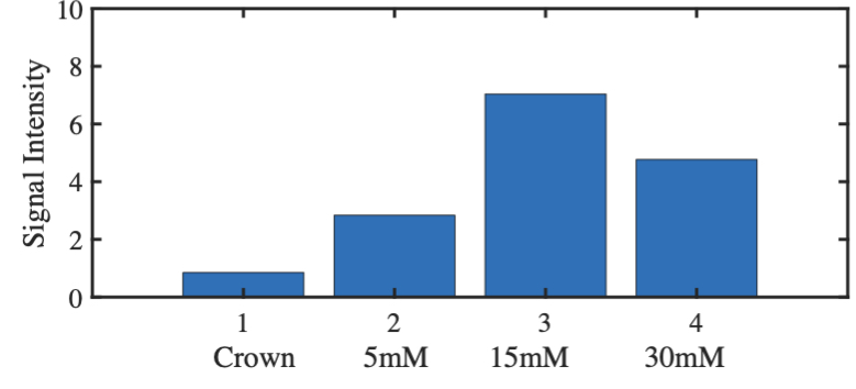
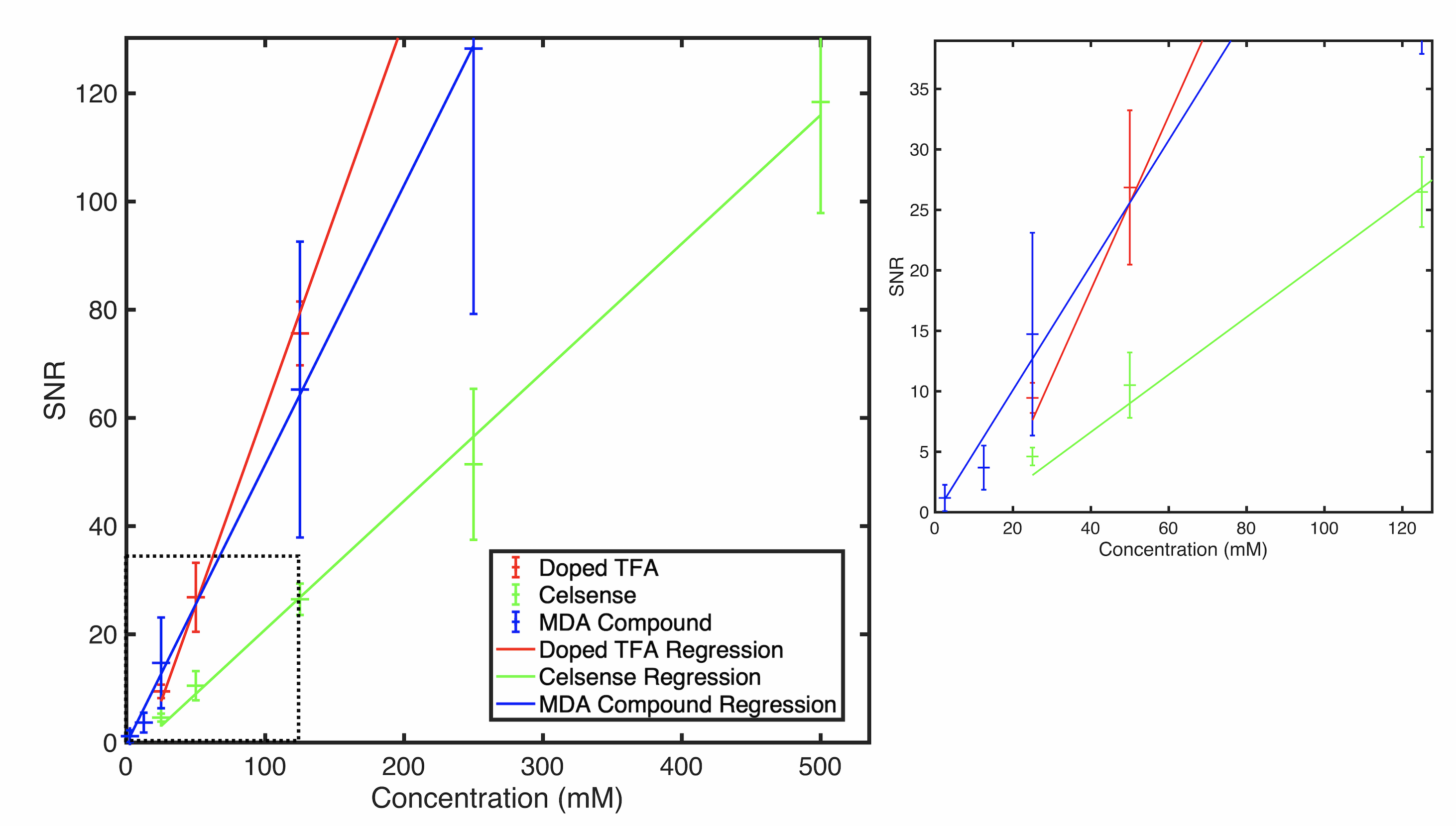
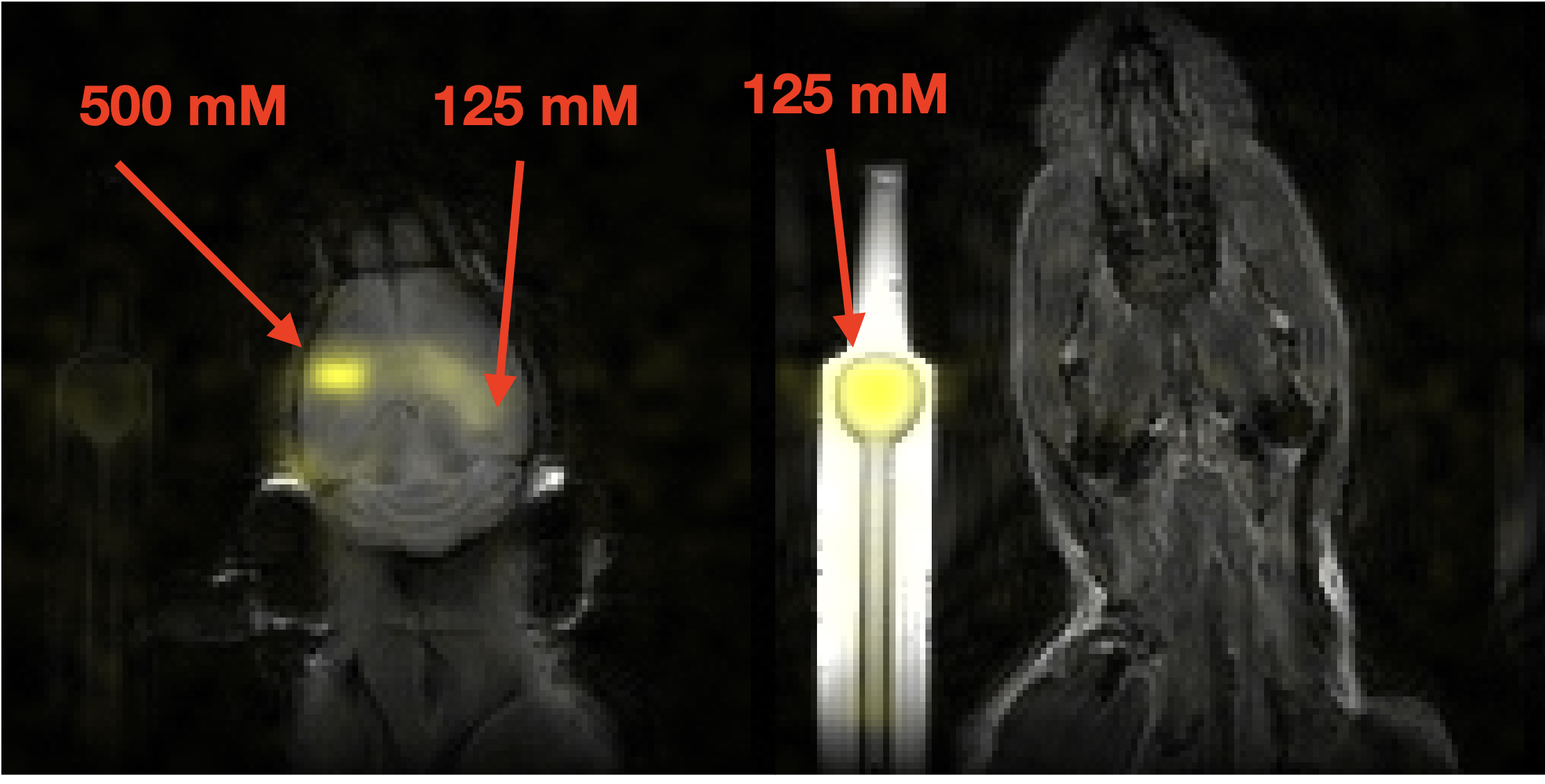
In Table 1, the crown is an analog of Celsense and reduction of relaxation time constants was observed for Fe-containing samples according to their Fe3+ content. Figure 1 shows the results of simulation with a spin echo signal equation for the MDA compound using optimized acquisition parameters, where 15 mM MDA compound displayed the best SNR (TR = 300 ms, TE = 6 ms, centric encoding, 3D RARE).To test the limit of detectability, the various cylindrical phantoms were used. The results are displayed in Figure 2 and demonstrate that the MDA compound is above a threshold SNR of 5 at half the concentration (12.5 mM) of Celsense. Figure 3 shows the agent is clearly visible within the brain resulting from stereotaxic injection.
DISCUSSION
The MDA compound displays shortened T1 and T2 relaxation time-constants that is useful for improved signal intensity per unit time. These relaxation properties provide advantages with regards to enhancement in signal intensity because imaging pulse-sequences may be driven with a larger duty cycle. The fact that the compound shows improved sensitivity relative to Celsense suggests that it has the potential to show a greater degree of NK cell biodistribution during the course of immunotherapy. Furthermore, the 19F visibility within the cerebral cortex suggests that this approach holds promise. Of note, the 19F signal is attenuated, as reported in Figure 3, due to relatively rapid diffusion of compound within the brain. Loading it into cells is expected to keep the agent from diffusing away thereby retaining intensity.CONCLUSION
This agent shows great promise to be used for NK cell tracking of immunotherapy based on its relaxometric characteristics relative to Celsense. However, additional proof on concept imaging in 19F agent-loaded cells directly injected into the brain as well as direct imaging during immunotherapy will be pursued in future studies.Acknowledgements
This work was supported by funding from the National Cancer Institute (P30-CA016672) and the and the Cancer Prevention and Research Institute of Texas (RP200223). The content is solely the responsibility of the authors and does not necessarily represent the official views of the sponsors.References
1 Myers, J. A. & Miller, J. S. Exploring the NK cell platform for cancer immunotherapy. Nat Rev Clin Oncol 18, 85-100, doi:10.1038/s41571-020-0426-7 (2021).
2 Sampson, J. H., Maus, M. V. & June, C. H. Immunotherapy for Brain Tumors. J Clin Oncol 35, 2450-2456, doi:10.1200/JCO.2017.72.8089 (2017).
3 Kennis, B. A. et al. Monitoring of intracerebellarly-administered natural killer cells with fluorine-19 MRI. J Neurooncol 142, 395-407, doi:10.1007/s11060-019-03091-5 (2019).
4 Liu, C. H. et al. Nanobomb optical coherence elastography. Opt Lett 43, 2006-2009, doi:10.1364/OL.43.002006 (2018).
5 Liu, C. H. et al. Longitudinal elastic wave imaging using nanobomb optical coherence elastography. Optics Letters 44, 3162-3165, doi:10.1364/Ol.44.003162 (2019).
6 Nevozhay, D., Weiger, M., Friedl, P. & Sokolov, K. V. Spatiotemporally controlled nano-sized third harmonic generation agents. Biomed Opt Express 10, 3301-3316, doi:10.1364/Boe.10.003301 (2019).
7 Boerner, P. et al. Repetitive optical coherence elastography measurements with blinking nanobombs. Biomed Opt Express 11, 6659-6673, doi:10.1364/Boe.401734 (2020).
Figures