3422
MRI assessment of abscopal effect induced by radiation/immune checkpoint blockade combination therapy1Radiation Biology branch, National Cancer Institute, Bethesda, MD, United States
Synopsis
Radiation therapy (RT) occasionally induces regression of non-irradiated metastatic lesions, which is called abscopal effect. Immune checkpoint inhibitors enhance abscopal effects. In this study we examined the physiological changes induced by abscopal effect and identify using MRI-based imaging biomarkers which predict the successful abscopal effect. Hypoxic fraction < 10 mmHg (HF10), permeability, perfusion, and CD8+ T cell infiltration in unirradiated tumor increased after the combination of RT and PD-1 blockade therapy. Interestingly, higher permeability/perfusion and lower HF10 in irradiated tumor before treatment is associated with slower growth of the unirradiated tumor after treatment of the tumor on the contralateral side.
Introduction
It has been reported that radiation therapy (RT) occasionally causes regression of non-irradiated metastatic lesions, which is called abscopal effect. Abscopal effects arise from systemic anti-tumor immune responses induced by localized RT. The radiation-mediated systemic anti-tumor effects are induced by an activation of tumor-specific CD8+ T cells which are primed by antigen-presenting cells that capture tumor-specific antigens from the collapsed tumor.1 The primed CD8+ T cells can induce apoptosis of tumor cells at distant non-irradiated sites as well as irradiated site through Fas/Fas ligand and/or Perforin/Granzyme B pathways.2 However, the overall occurrence rate of the abscopal effect by RT alone is extremely low because such anti-tumor effects are inhibited by PD-1/PD-L1 pathway.3 Although it has been reported that immunotherapy, especially immune checkpoint blockade (ICB), can enhance abscopal effect, the imaging biomarkers which predict the induction of the abscopal effect have not been investigated to date.4 Hypoxia, a feature of the tumor microenvironment (TME), is reported to cause T cell exhaustion by inducing a mitochondrial defects.5 Increased vessel permeability and perfusion may predict the efficacy of ICB.6 Therefore, in the current study, we investigated pO2 distribution, permeability and perfusion in the primary/metastatic model tumors treated with the combination therapy to explore the physiological changes in the tumors showing abscopal effect.Methods
MC38 colon adenocarcinoma treated with RT and PD-1 inhibitor were used to evaluate the abscopal effect. For in vivo treatment model, 1x10⁶ tumor cells and 2x10⁵ were inoculated subcutaneously into right and left hindlegs of C57BL/6 mice, respectively. Four treatment groups were prepared. In group 1, only right leg tumor was irradiated, and αPD-1 antibody (200 μg) was intraperitoneally injected on days 0, 3, and 7 after treatment. In group 2, mice were injected with IgG isotype antibody 200 μg at the same schedule without radiation therapy. In group 3, right leg tumor was irradiated and IgG isotype antibody 200 μg was injected at the same schedule. In group 4, PD-1 antibody was injected at the same schedule without radiation therapy. Imaging studies were performed on days 0 or 9 on both hindleg tumor and the data were compared among groups. Electron para-magnetic resonance imaging (EPRI) were performed for quantitative intra-tumor pO2 mapping with high resolution (~0.2mm) by observing the linewidth of the exogenously administered trityl radical probe Ox063. Dynamic contrast-enhanced magnetic resonance imaging (DCE-MRI) were performed on a 3 T scanner (Bruker BioSpec 3T). T1-weighted fast low-angle shot (FLASH) images were obtained with TR = 117.2 ms; TE = 6 ms; flip angle = 30˚; two slices; 28 x 28 mm resolution; 15-second acquisition time per image; and 45 repetitions. Gd-DTPA solution (4 mL/g of body weight of 50 mmol/L Gd-DTPA) was injected through a tail vein cannula 1 minutes after the start of the dynamic FLASH sequence. To determine the local concentrations of Gd-DTPA, T1 maps were calculated from three sets of Rapid Imaging with Refocused Echoes (RARE) images obtained with TR = 320, 400, 600, 1,000, 2,000, and 3,000 ms, with the acquisitions being made before running the FLASH sequence. Flowcytometry: To analyze tumor infiltrating lymphocytes (TILs), on day 9 after treatment, single-cell suspensions were prepared from left hind leg tumor by digestion with a mixture of collagenase, DNase, and hyaluronidase. The cell surface phenotypes were determined by direct immunofluorescence staining with aCD3, aCD8, and aCD4 antibody and analyzed using FACS Calibur (BD Biosciences). TILs were identified and gated on a forward scatter versus side scatter plot.Results and Discussion
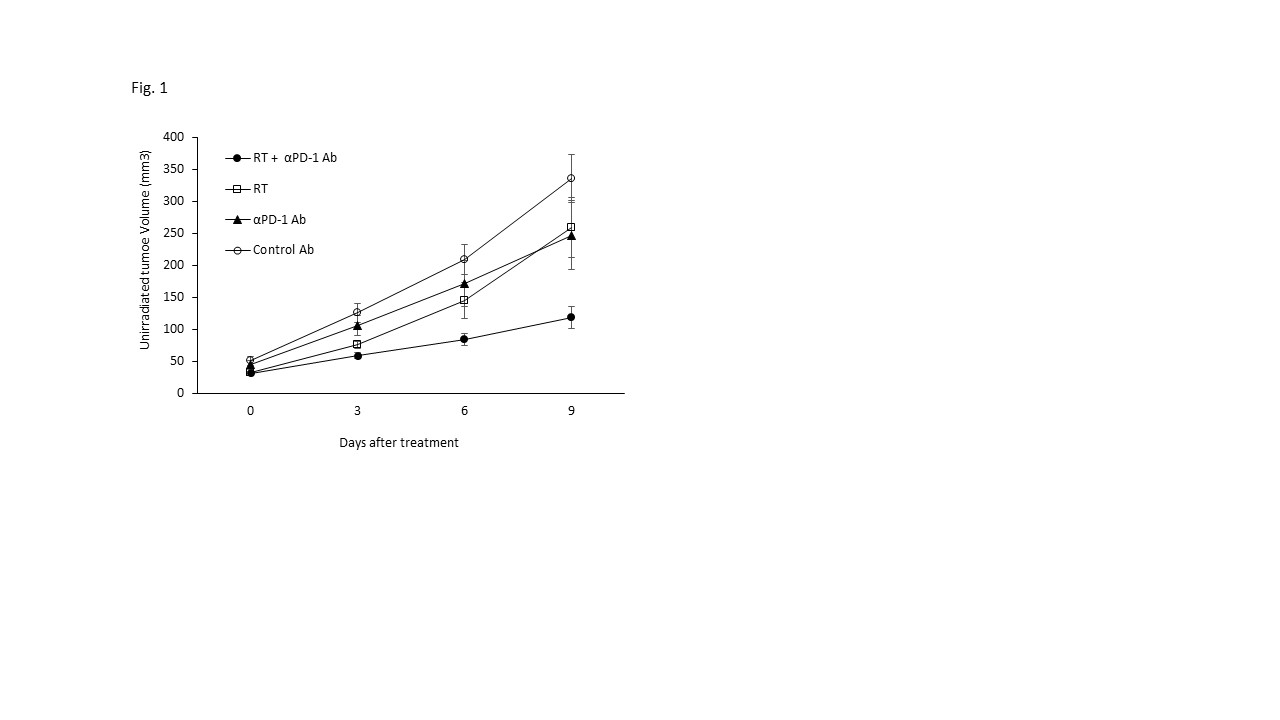
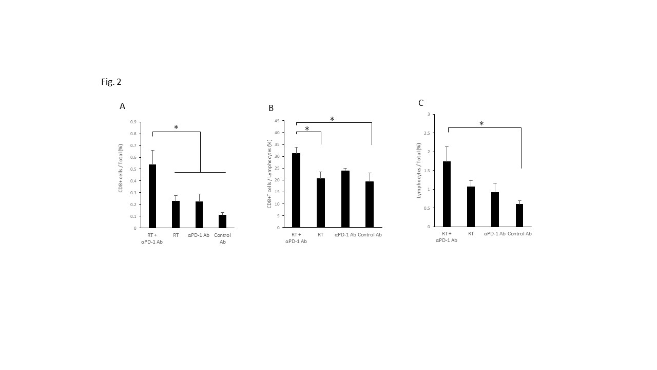
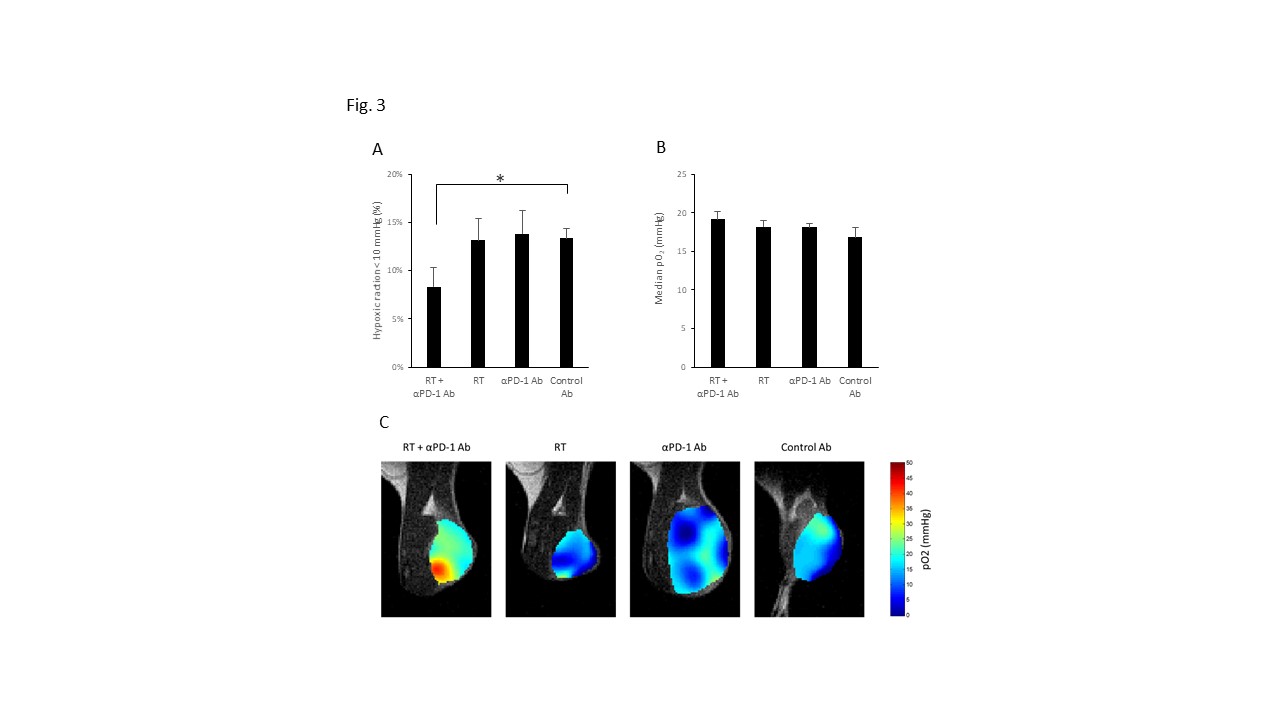
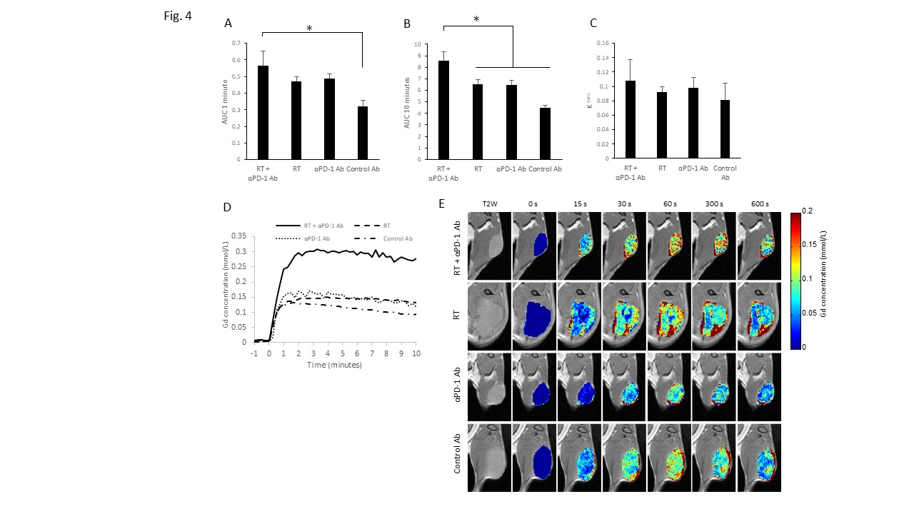
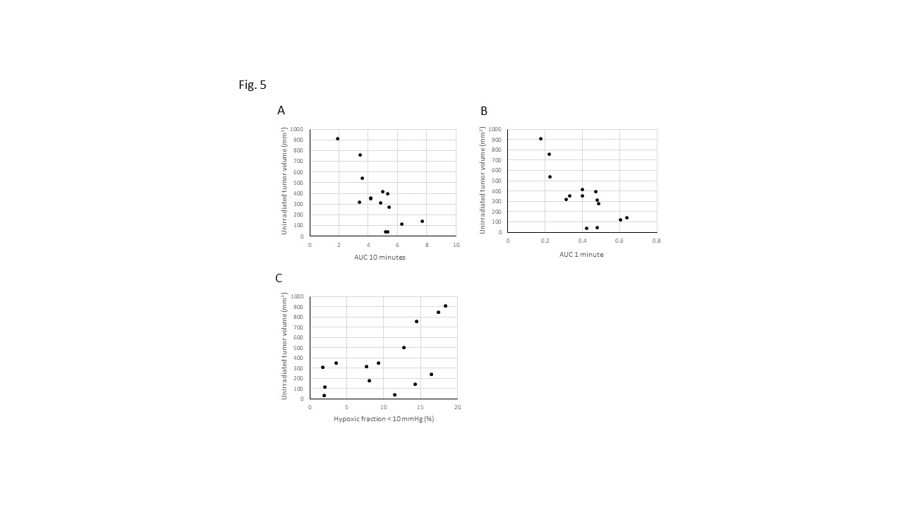
A mouse tumor model exhibiting abscopal effect induced by the combination of RT and PD-1 inhibitor was established. Combination treatment of RT and PD-1 inhibitor showed a synergistic effect on MC38 tumor (Fig.1). Flowcytometry showed that CD8+ T cell infiltration in unirradiated tumor increased after the combination of RT and PD-1 inhibitor (Fig.2), suggesting that in vivo synergistical effect was caused by higher CD8+ T cell infiltration. Hypoxic fraction < 10 mmHg (HF10), permeability, perfusion in unirradiated tumor improved after the combination of RT and PD-1 inhibitor (Fig.3 and 4), suggesting that enhanced CD8+ T cell infiltration by the abscopal effect caused the changes in these imaging biomarkers. Interestingly, these biomarkers in TME (high Permeability, Perfusion, and low HF10) before treatment were found to be associated with the extent of induction of abscopal effect (Fig.5), which may imply the mode of tumor cell death by radiation therapy has a significant impact on the induction of abscopal effect.Conclusion
Hypoxic fraction < 10 mmHg, permeability, perfusion and CD8+ T cell infiltration in unirradiated tumors improved after the combination of RT and PD-1 inhibitor. Higher permeability/perfusion and lower HF10 in irradiated tumor before treatment was associated with slower unirradiated tumor growth after the treatment. These data can provide imaging biomarkers to predict the successful abscopal effect with PD-1 inhibitor.Acknowledgements
No acknowledgement found.References
1. Wang, D.; Zhang, X.; Gao, Y.; Cui, X.; Yang, Y.; Mao, W.; Li, M.; Zhang, B.; Yu, J., Research Progress and Existing Problems for Abscopal Effect. Cancer Manag Res 2020, 12, 6695-6706.
2. Henkart, P. A., Lymphocyte-mediated cytotoxicity: two pathways and multiple effector molecules. Immunity 1994, 1 (5), 343-6.
3. Taube, J. M.; Anders, R. A.; Young, G. D.; Xu, H.; Sharma, R.; McMiller, T. L.; Chen, S.; Klein, A. P.; Pardoll, D. M.; Topalian, S. L.; Chen, L., Colocalization of inflammatory response with B7-h1 expression in human melanocytic lesions supports an adaptive resistance mechanism of immune escape. Sci Transl Med 2012, 4 (127), 127ra37.
4. Liu, Y.; Dong, Y.; Kong, L.; Shi, F.; Zhu, H.; Yu, J., Abscopal effect of radiotherapy combined with immune checkpoint inhibitors. J Hematol Oncol 2018, 11 (1), 104.
5. Liu, Y. N.; Yang, J. F.; Huang, D. J.; Ni, H. H.; Zhang, C. X.; Zhang, L.; He, J.; Gu, J. M.; Chen, H. X.; Mai, H. Q.; Chen, Q. Y.; Zhang, X. S.; Gao, S.; Li, J., Hypoxia Induces Mitochondrial Defect That Promotes T Cell Exhaustion in Tumor Microenvironment Through MYC-Regulated Pathways. Front Immunol 2020, 11, 1906.
6. Zheng, X.; Fang, Z.; Liu, X.; Deng, S.; Zhou, P.; Wang, X.; Zhang, C.; Yin, R.; Hu, H.; Chen, X.; Han, Y.; Zhao, Y.; Lin, S. H.; Qin, S.; Wang, X.; Kim, B. Y.; Zhou, P.; Jiang, W.; Wu, Q.; Huang, Y., Increased vessel perfusion predicts the efficacy of immune checkpoint blockade. J Clin Invest 2018, 128 (5), 2104-2115.
Figures