3376
Motion-Compensated Low-Rank Reconstruction with Iterative Registration for Ventilation Analysis on Ultrashort Echo Time (UTE) Lung MRI1Radiology and Biomedical Imaging, University of California, San Francisco, San Francisco, CA, United States, 2GE Healthcare, Menlo Park, CA, United States
Synopsis
In this abstract, we introduce motion-compensated low-rank constrained reconstruction with iterative registration. This method jointly improves the motion field estimation and the reconstruction, providing both functional and structural images. In addition, we test the effect of the regularization parameter on structural images and ventilation maps. We conclude that the motion-compensated low-rank constrained with iterative registration method is suitable for regional ventilation quantification.
Introduction
Lung imaging is challenging for conventional MRI methods due to its low proton density, short T2*, and respiratory motion (1). UTE MRI is gaining more attention on pulmonary imaging because it can capture the lung parenchyma signal and resolve the respiratory. Our group previously developed motion-resolved reconstruction (2), iterative motion-compensated reconstruction (3), motion-compensated low-rank reconstruction to improve the quality of structural lung image. Based on the advanced reconstructions, we also developed ventilation analysis using motion fields to measure pulmonary function (4). In this abstract, we build on the motion-compensated low-rank constrained reconstruction and introduce iterative registration. By using this approach, we hope to jointly improve the motion field estimation and the reconstruction to get a ventilation map and structural image. In addition, we tested the effect of the regularization parameter on structural images and ventilation maps.Methods
Data acquisitionAll procedures were approved by the University of California, San Francisco Institutional Review Board. Data were acquired on a 3T clinical scanner (MR750, GE Healthcare, Waukesha, WI, USA) with an optimized 3D radial ultrashort echo time (UTE) sequence (5). Key parameters are: flip angle = 4, resolution = 1.56 mm isotropic, # spokes = 80,000. We included one healthy adult dataset for this prototype study.
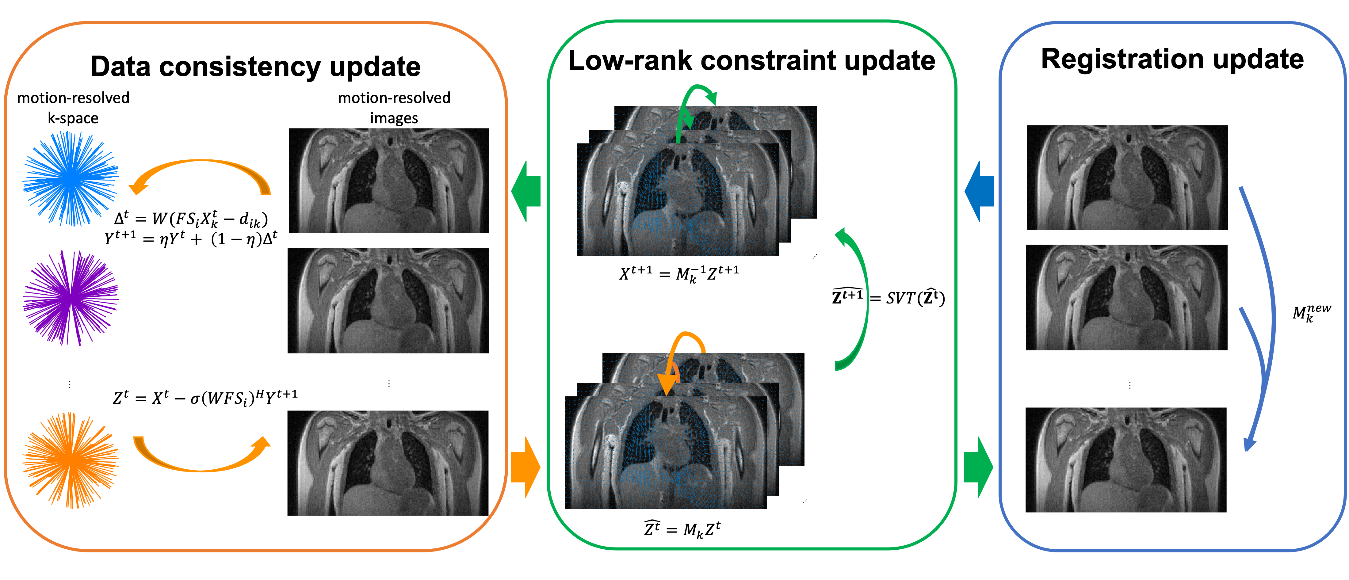
Motion-compensated low-rank with iterative registration
The data were binned into 6 phases according to the respiratory motion position. Trajectory and density compensation factors were extracted from the raw data. First, we reconstructed the data using the motion-resolved approach with a total variation regularization (2,6).
Then, the motion-compensated low-rank constrained reconstruction (7) with and without iterative registration was applied. For both methods, the following optimization problem is addressed,
$$\arg\max_X \sum_{i,k}^{N,m} ||W(FS_iX_k-d_{i,k})||_2^2 + \lambda_{lr}||MX||_*$$
In the data consistency term, $$$W$$$ is the density compensation factor, $$$F$$$ is the non-uniform Fourier Transform, $$$S_i$$$ is the sensitivity map for coil $$$i$$$, $$$d_{i,k}$$$ is the binned data for coil $$$i$$$, respiratory phase $$$k$$$. In the regularization term, $$$|| . ||$$$ is the nuclear norm, $$$M$$$ is the motion field from symmetric image normalization (SyN) (8) registration. For motion-compensated low-rank, $$$M$$$ was estimated from the motion-resolved image. While for iterative registration, it was updated from motion-compensated low-rank results in each iteration. The reconstruction workflow is summarized in Figure 1.
Regional Ventilation
Regional ventilation measures the percentage of volume change during breathing. Motion field $$$M$$$ is used for regional ventilation quantification. The definition of regional ventilation is as follows,
$$RV = \frac{V_{resp}}{V_{end-expi}}-1=|\det(Id+\nabla M)|-1$$
where $$$V_{resp}$$$, $$$V_{end-expi}$$$, are the lung volume at each respiratory state and end-expiration state. $$$Id$$$ is a 3-by-3 identity matrix, $$$M$$$ is the motion field,$$$\nabla$$$ is the Jacobian matrix, and $$$\det$$$ is the matrix determinant.
Results
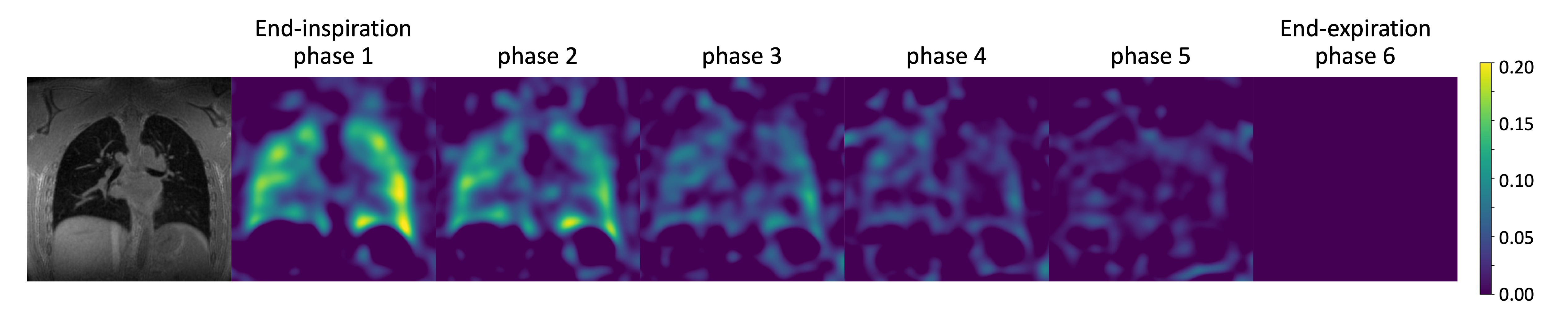
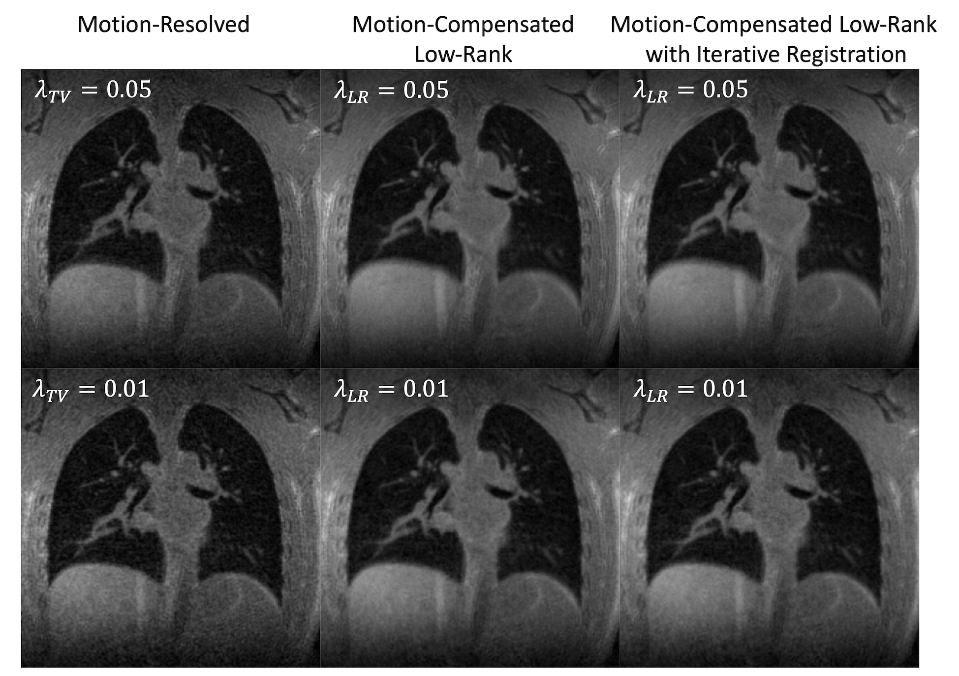
Figure 2 demonstrates ventilation maps calculated at six different respiratory states. The end-expiration state (phase 6) is used as the reference frame during registration. The ventilation inside the lung has a maximum value of 0.2, which represents 20% of volume change. The ventilation is higher at end inspiration and gradually becomes lower in the end-expiration state.Figure 3 is a comparison of the structural images using motion-resolved, motion-compensated with low-rank constraint, and motion-compensated low-rank with iterative registration methods. The signal-to-noise ratio is higher in the two motion-compensated images than the motion-resolved image. No significant difference is observed for the two motion-compensated approaches. We also compare images reconstructed using different regularization parameters. Stronger regularization parameters yield higher SNR and weak regularization keeps the sharp structures.
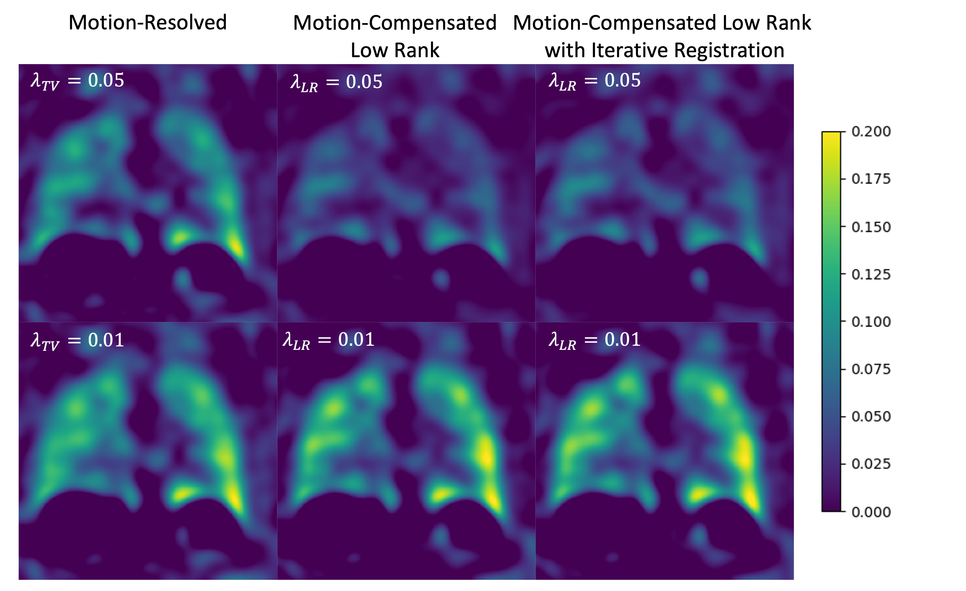
Figure 4 lists the regional ventilation maps calculated from different reconstruction methods. All reconstruction approaches provide ventilation maps with the same pattern. For example, there is higher ventilation in the left lung and in the lower lobes of the lung. However, a range difference exists across methods and regularization parameters. A larger regularization parameter provides lower ventilation for all reconstruction methods.
Discussion
The regularization parameter affects both the structural image and ventilation map. For structural images, it balances the trade-off between SNR and sharpness. For regional ventilation maps, stronger constraint suppresses the ventilation. Reconstruction regularization parameters should be considered in ventilation quantification. A minimum amount of regularization will potentially provide a more accurate functional analysis.We also observe that for ventilation analysis, the regularization parameter has a stronger effect for motion-compensated low-rank approaches than the motion-resolved approach. This could be caused by the characteristics of the low-rank assumption. It might average the areas with large motion and small motion, resulting in a smaller range of ventilation. On the other hand, however, the motion-compensated image provides a higher signal-to-noise ratio which will help reduce the registration error. And the iterative registration updates the motion field and can further improve the ventilation quantification.
Conclusion
In conclusion, the motion-compensated low-rank constrained with iterative registration methods is suitable for motion field-based regional ventilation quantification.Acknowledgements
This work is supported by NIH grant R01 HL136965.References
1. Wild JM, Marshall H, Bock M, et al. MRI of the lung (1/3): Methods. Insights Imaging 2012;3:345–353 doi: 10.1007/s13244-012-0176-x.
2. Jiang W, Ong F, Johnson KM, et al. Motion robust high resolution 3D free-breathing pulmonary MRI using dynamic 3D image self-navigator. Magn. Reson. Med. 2018;79:2954–2967 doi: 10.1002/mrm.26958.
3. Zhu X, Chan M, Lustig M, Johnson KM, Larson PEZ. Iterative motion-compensation reconstruction ultra-short TE (iMoCo UTE) for high-resolution free-breathing pulmonary MRI. Magn. Reson. Med. 2020;83:1208–1221 doi: 10.1002/mrm.27998.
4. Tan F, Zhu X, Larson PEZ. Pulmonary Ventilation Analysis Using Motion-Resolved Ultrashort Echo (UTE) MRI. In: Proc. Intl. Soc. Mag. Reson. Med. 28. ; 2020.
5. Johnson KM, Fain SB, Schiebler ML, Nagle S. Optimized 3D ultrashort echo time pulmonary MRI. Magn. Reson. Med. 2013;70:1241–1250 doi: 10.1002/mrm.24570.
6. Feng L, Axel L, Chandarana H, Block KT, Sodickson DK, Otazo R. XD-GRASP: Golden-angle radial MRI with reconstruction of extra motion-state dimensions using compressed sensing. Magn. Reson. Med. 2016;75:775–788 doi: 10.1002/mrm.25665.
7. Zhu X, Ong F, Lustig M, Larson P. Motion Compensated Low-Rank ( MoCoLoR ) constrained reconstruction with application to motion resolved lung MRI. In: Vol. 72. ; 2020. p. 2020.
8. Avants BB, Epstein CL, Grossman M, Gee JC. Symmetric diffeomorphic image registration with cross-correlation: Evaluating automated labeling of elderly and neurodegenerative brain. Med. Image Anal. 2008;12:26–41 doi: 10.1016/j.media.2007.06.004.
Figures