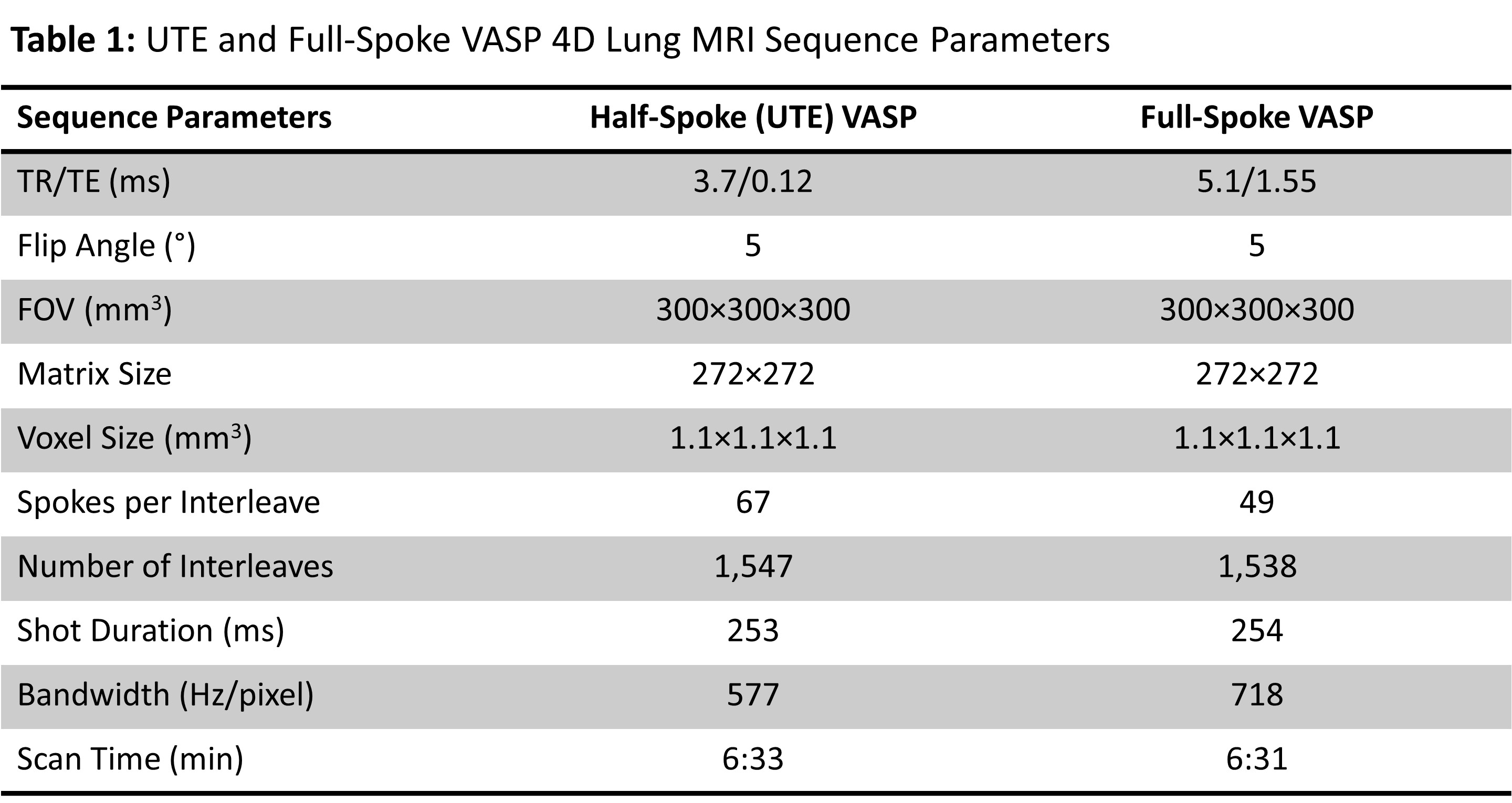
3373
High-Resolution Half-Spoke and Full-Spoke 4D Lung MRI with Golden-Angle 3D Radial Acquisition and Motion-Resolved Sparse Reconstruction1Department of Medical Physics, Memorial Sloan Kettering Cancer Center, New York, NY, United States, 2Philips Healthcare, MR R&D, Rochester, MN, United States, 3Department of Radiology, Memorial Sloan Kettering Cancer Center, New York, NY, United States
Synopsis
This work aims to develop high-resolution 4D lung MRI with golden-angle 3D radial (kooshball) acquisition and motion-resolved sparse reconstruction. The golden-angle radial k-space lines were continuously acquired during free-breathing and retrospectively binned to multiple respiratory phases with respect to the respiratory signal obtained from a built-in motion-sensing camera. The sparsity along the respiratory motion dimension was exploited using compressed sensing reconstruction to suppress artifacts and improve SNR. The feasibility of half-spoke ultrashort echo time (UTE) and full-spoke 4D lung MRI was demonstrated on healthy volunteers on a clinical 3T MRI scanner.
INTRODUCTION
Lung MRI remains challenging due to low proton density and short T2* of the lung parenchyma and motion artifacts. Ultrashort echo time (UTE) sequences have shown to provide decent imaging of lung parenchyma and can be promising in the diagnosis of lung diseases, such as lung nodules or cystic fibrosis.1-3 However, respiratory motion causes breathing artifacts that make diagnosis difficult. Breath hold and respiratory gating are common compensation techniques to reduce motion-induced artifacts, but they are either inefficient in data acquisition or cannot provide high-resolution 3D imaging. Recently, advanced UTE techniques were proposed for motion-resolved lung imaging using self-navigators, compressed sensing, and low-rank reconstructions.4-7 In addition, a VASP (Variable Anisotropic FOV for 3D radial imaging with Spiral Phyllotaxis) sequence was developed to reduce scan time or aliasing artifacts in 3D radial cardiac imaging,8 but has not been applied for UTE lung imaging. The purpose of this study was to develop high-resolution and motion-resolved half-spoke (UTE) and full-spoke 4D radial lung MRI using the VASP technique with golden angle spiral phyllotaxis acquisition and sparse reconstruction.METHODS
Figure 1 shows the k-space trajectory of half-spoke (UTE) VASP with golden angle spiral phyllotaxis (13 interleaves as an example). The X-Y projection (top view) demonstrates that the interleaves are spaced relatively uniform according to golden angle rotation. k-space trajectory for full-spoke VASP follows a similar pattern except that full spoke are used. The end of the spoke travels from the north pole to the south (UTE) or equator (full spoke) and then flies back to the north to minimize the impact of eddy currents.9VASP Data Acquisition: Free-breathing T1-weighted UTE and full-spoke VASP data acquisition was performed on two healthy volunteers (1 male, 1 female) on a clinical 3T MRI scanner (Ingenia Elition X, Philips Healthcare) using a combination of 16-channel anterior and 12-channel posterior coils. A camera-based respiratory sensing technology (VitalEye, Philips Healthcare) was used to provide real-time respiratory motion signals during data acquisition. k-space data and their associated 3D coordinates, density compensation weights, and respiratory signals were exported from the scanner for subsequent offline 4D lung image reconstruction. Table 1 summarizes the sequence parameters for UTE and full-spoke VASP acquisitions. The study was approved by local institutional review board and written informed consent was obtained from the subjects prior to MRI scans.
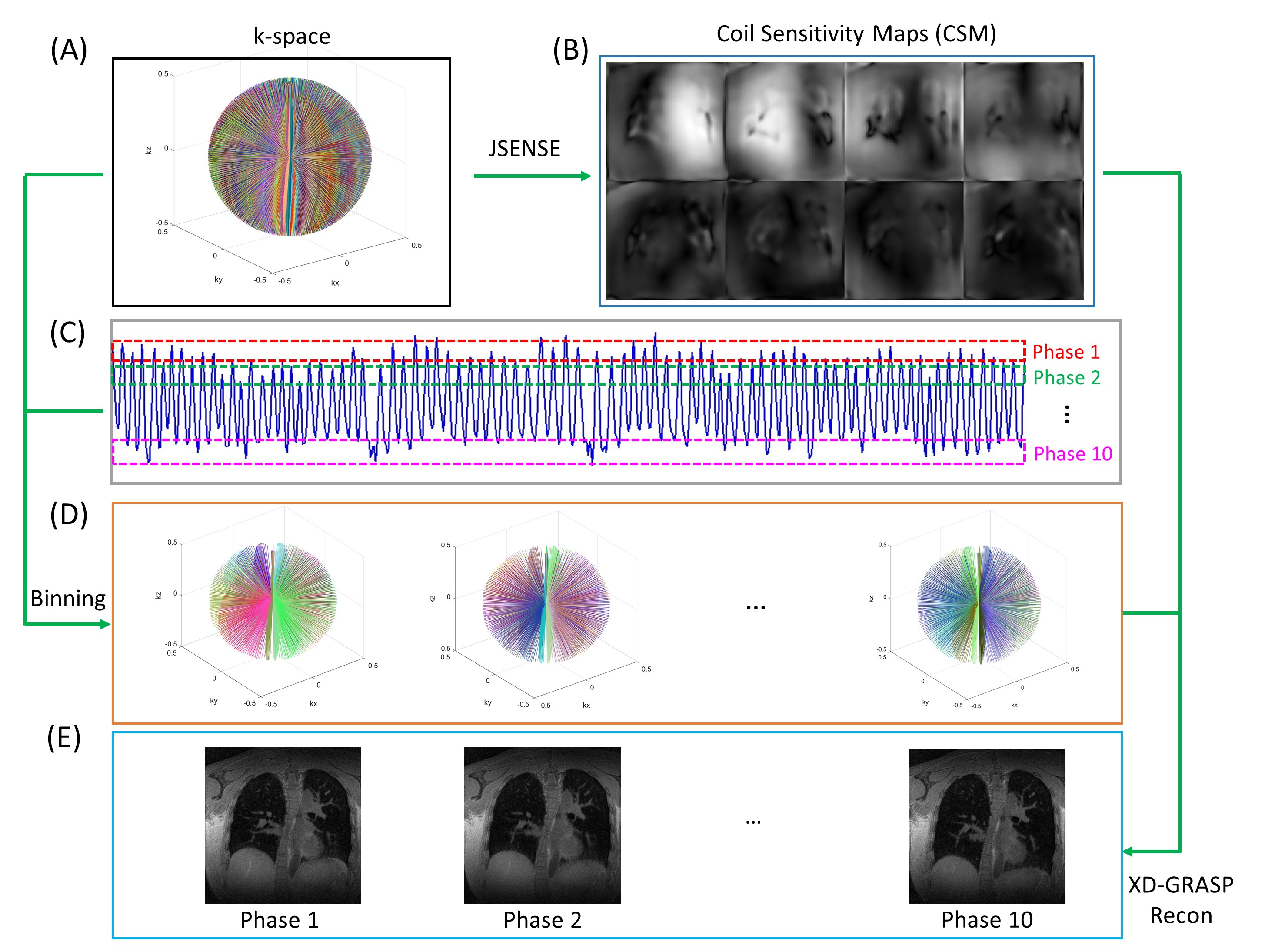
4D Image Reconstruction: Figure 2 illustrates the 4D lung MRI reconstruction workflow. Coil sensitivity maps (Figure 2B) were reconstructed from the complete k-space data (Figure 2A) using the joint image reconstruction and sensitivity estimation in SENSE (JSENSE) algorithm.10 K-space data was then sorted with respect to respiratory signals (Figure 2C) and subsequently binned into 10 motion states from expiration to inspiration (Figure 2D). Motion-resolved 4D lung image reconstruction (Figure 2E) was performed similar to the XD-GRASP framework11 by solving the following optimization problem:
$$d=\mathop\arg\min_{d}\frac{1}{2}\parallel Ed-u\parallel _2^2 + \lambda \parallel Td \parallel_{1}$$
where E is an encoding operator that incorporates coil sensitivities, nonuniform Fourier transform (NUFFT) and density compensation function, d is the 4D image target to reconstruct, u is the 4D under-sampled multi-coil k-space data binned in 10 motion states, λ is a regularization parameter to balance data consistency and temporal sparsity, and T is the sparsifying transform applied along the temporal domain. First-order finite difference was chosen as the sparsifying transform. The image reconstruction pipeline was implemented in Python and NUFFT operations were implemented using the SigPy package.12 All reconstructions were performed on a Linux server with 24GB NVIDIA GPU and 512GB RAM. The reconstruction time of each 4D dataset was about two hours.
RESULTS
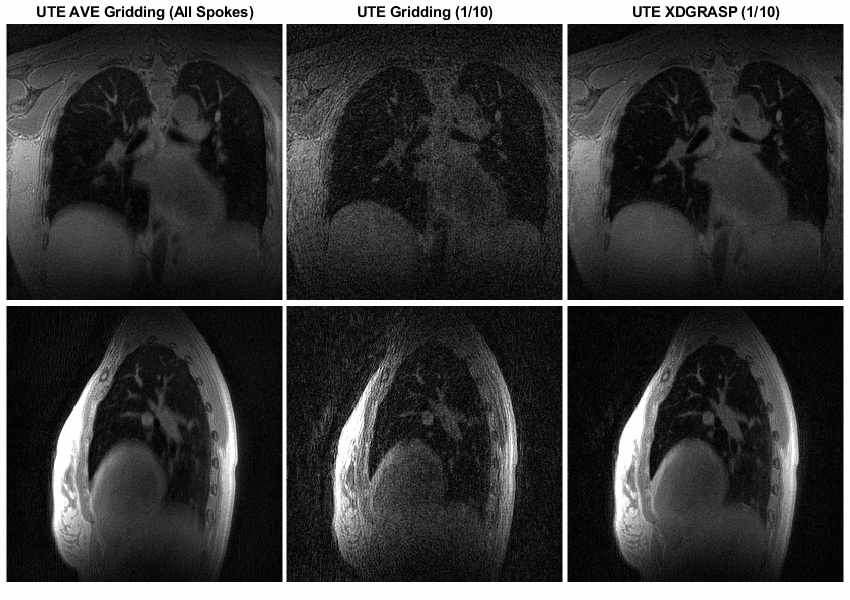
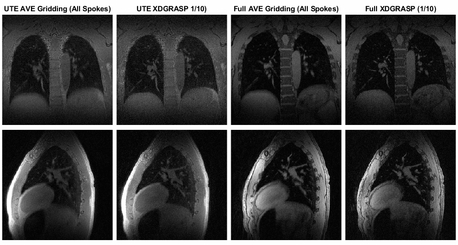
Data acquisition and image reconstruction were successfully performed for the subjects. Figure 3 shows a UTE VASP lung MRI scan from a healthy volunteer. The motion-averaged images from the gridding reconstruction of all spokes have good SNR and relatively well detection of the pulmonary vessels despite some blurring from respiratory motion. The gridding reconstruction of the binned data looks noisy and has artifacts due to substantial undersampling (only 1/10 data for each phase), which are significantly improved with XD-GRASP reconstruction. It is noted that the motion-resolved XD-GRASP reconstruction has comparable SNR to the motion-averaged images but appears sharper for the pulmonary vessels and the lung-liver diaphragm. Figure 4 demonstrates that both UTE and full-spoke VASP lung MRI were able to resolve respiratory motion, but UTE VASP provides more details of the lung parenchyma with slightly lower SNR compared to full-spoke VASP.DISCUSSION
4D UTE and full-spoke VASP allows high-resolution motion-resolved lung imaging. UTE VASP provides detailed information of the lung parenchyma for the evaluation of lung abnormalities. Full-spoke VASP shows superior SNR and could be useful in motion tracking of lung tumors and organs at risk in radiation therapy. Future study is warranted to demonstrate the clinical value of the proposed technique on patients with lung tumors and include metrics to quantitatively evaluate its performance.CONCLUSION
High-resolution UTE and full-spoke 4D radial lung MRI were successfully implemented on clinical MRI scanners and can be potentially useful for lung tumor evaluation and motion tracking in radiation therapy applications.Acknowledgements
None.References
1. Ohno Y, Koyama H, Yoshikawa T, et al. Standard-, Reduced-, and No-Dose Thin-Section Radiologic Examinations: Comparison of Capability for Nodule Detection and Nodule Type Assessment in Patients Suspected of Having Pulmonary Nodules. Radiology 2017;284(2);562-573.
2. Dournes G, Menut F, Macey J, et al. Lung morphology assessment of cystic fibrosis using MRI with ultra-short echo time at submillimeter spatial resolution. Eur Radiol 2016;26(11):3811-3820.
3. Heidenreich JF, Weng AM, Metz C, et al. Three-dimensional Ultrashort Echo Time MRI for Functional Lung Imaging in Cystic Fibrosis. Radiology 2020;296(1):191-199.
4. Jiang W, Ong F, Johnson KM. et al. Motion robust high resolution 3D free-breathing pulmonary MRI using dynamic 3D image self-navigator. Magn Reson Med 2018;79(6):3954-2967.
5. Zhu X, Chan M, Lustig M, et al. Iterative motion-compensation reconstruction ultra-short TE (iMoCo UTE) for high-resolution free-breathing pulmonary MRI. Magn Reson Med 2020;83(4):1208-1221.
6. Feng L, Delacoste J, Smith D, et al. Simultaneous Evaluation of Lung Anatomy and Ventilation Using 4D Respiratory-Motion-Resolved Ultrashort Echo Time Sparse MRI. J Magn Reson Imaging 2019;49(2):411-422.
7. Ong F, Zhu X, Cheng JY, et al. Extreme MRI: Large-scale volumetric dynamic imaging from continuous non-gated acquisitions. Magn Reson Med 2020;84(4):1763-1780.
8. Krishnamoorthy G, Smink J, Tourais J, et al. Variable anisotropic FOV for 3D radial imaging with spiral phyllotaxis (VASP). Magn Reson Med 2020;85(1):68-77.
9. Krishnamoorthy G, Beck G. B0 and eddy current optimized k-space profile ordering for 3D radial MRI. European Patent Application. EP3748385A1.
10. Ying L, Sheng J. Joint image reconstruction and sensitivity estimation in SENSE (JSENSE). Magn Reson Med 2007;57(6):1196-1202.
11. Feng L, Axel L, Chandarana H, et al. XD-GRASP: Golden-angle radial MRI with reconstruction of extra motion-state dimensions using compressed sensing. Magn Reson Med 2016;75(2):775-788.
12. Ong F, Lustig M. SigPy: a python package for high performance iterative reconstruction. Proc Int Soc Magn Reson Med 2019;27:4819.
Figures