3333
Maternal Obesity Alters Neuro-Connectivity in Baboon Offspring
Wei D Zhang1, Peter T Fox 1, Hillary F Huber2, Peter William Nathanielsz3, and Geoffrey D Clarke4
1Research Imaging Institute, UT Health San Antonio, San Antonio, TX, United States, 2Texas Pregnancy & Life-Course Health Research Center, Texas Biomedical Research Institute, San Antonio, TX, United States, 3Animal Science, University of Wyoming, Laramie, WY, United States, 4Research Imaging Insitute, UT Health San Antonio, San Antonio, TX, United States
1Research Imaging Institute, UT Health San Antonio, San Antonio, TX, United States, 2Texas Pregnancy & Life-Course Health Research Center, Texas Biomedical Research Institute, San Antonio, TX, United States, 3Animal Science, University of Wyoming, Laramie, WY, United States, 4Research Imaging Insitute, UT Health San Antonio, San Antonio, TX, United States
Synopsis
Maternal obesity status was found to affect neural functional connectivity of adult offspring in in a baboon model. Significant differences in connectivity between offspring of obese mothers and age/sex matched controls were found in regions of the left temporal lobe and frontal lobe using rs-fMRI.
Introduction
Maternal obesity (MO) has been linked to offspring’s brain development in neonates.1 We have developed a baboon model of developmental programming in maternal obesity to investigate the extent of perinatal malnutrition on subsequent development.2 This model can be used for finding MRI biomarkers of aging that indicate if MO drives the offspring on an irregular growth trajectory. The aim of the present study was to investigate the hypothesis that brain network connectivity differences exist in adult offspring of obese mothers, compared to a sex/age matched control group, using resting state functional magnetic resonance imaging (rs-fMRI) of whole brain.Methods
Females were randomly assigned prior to breeding to the control group (CTR) or the MO group. CTR mothers ate ad libitum SNPRC biscuits (Purina Monkey Diet and Monkey Diet Jumbo; Purina LabDiets, St Louis, MO, USA) containing 12% energy from fat, 0.29% from glucose, 0.32% from fructose, and a metabolizable energy content of 3.07 kcal/g. For at least 9 months prior to breeding, MO mothers ate ad lib CTR diet + ad lib Purina 5045-6 (Purina LabDiets, St Louis, MO, USA), a high-fat and high-energy diet containing 45% energy from fat, 4.6% from glucose, 5.6% from fructose, and a metabolizable energy content of 4.03 kcal/g. The MO group also had continuous access to a high fructose beverage. A total of 32 baboon offspring were studied, of which 19 were of MO dams (10 female /9 male), age 4.9±0.5 yrs, body weight 18.6±3.5 kg; and 13 control offspring (6 female /7 male), age 5.2 ± 1 yrs, body weight 17.7±2.6 kg. There were no age, sex, or body weight differences between the two groups. An rs-fMRI dataset of the whole brain was acquired using a BOLD sequence (TR/TE=3000/35, 1.9 mm3 voxel, 28 slices, EPI factor = 104, 200 measurements) for each animal under general anesthesia on a 3T scanner (TIM Trio, Siemens, Malvern, PA, USA). Each image was preprocessed for motion correction, slice timing correction, brain extraction, field-map distortion correction, high pass filtering, spatial smoothing, and registration to study template using FSL software. Group independent component analysis (ICA) was performed using GIFT software3 resulting in 40 components or networks which were, in turn, used to generate individual subject network maps and time courses using the GIG-ICA (group information guided ICA) method.4 A group comparison was conducted on the individual network maps using a two sample t-test controlling for age on GIFT.Results
Regions of left temporal lobe and frontal lobe including the insula, somatosensory area, perirhinal cortex, ventral premotor area, and area 45 showed significant functional connectivity change in the MO group. Two brain regions in one network showed significant functional connectivity change in the MO group. Cluster 1 (mean p = 0.000053 ) was 53 mm3 in size (246 voxels) and was located in the left auditory cortex and is displayed in Figure 1. Cluster 2 (mean p = 0.00025) was 12 mm3 in size (56 voxels) and was located in the left agranular frontal area F4 and is shown in Figure 2.Discussion
Previous studies in humans, from fetuses5 to newborns6,7 to young children8 have also found differences in brain connectivity in offspring of MO mothers and an association between these changes and maternal body mass index (BMI) occurring across multiple networks. However, the evaluation of offspring changes due to perinatal MO environment becomes more difficult as humans age since individuals can be exposed to multiple environmental factors. Nonhuman primates (NHP) offer an advantage in that their environment can be controlled. The current study is the first to investigate neural connectivity changes associated with MO in an NHP MO model and also the first in adult offspring. These data suggest that the disruption in neural connectivity seen in early life can persist into adulthood. Cognitive testing is being carried out on these subjects to determine behavioral correlates.Conclusion
Maternal obesity status changes the adult offspring’s neural functional connectivity in the regions of left temporal lobe and frontal lobe, which can be detected using rs-fMRI.Acknowledgements
This work was in part supported by the National Institutes of Health 5P01HD021350, 1U19AG057758, 5R24OD011183, OD P51 OD011133, 1R25EB016631, and EU FP 7/HEALTH/GA No.: 279281: BrainAge- Impact of Prenatal Stress on BRAINAGEing.References
- Li X, Andres A, Shankar K, Pivik RT, et al. Differences in brain functional connectivity at resting state in neonates born to healthy obese or normal-weight mothers. Intl J Obesity. 2016; 40:1931-1934.
- Li C, Jenkins S, Considine MM, Cox LA, Gerow KG, Huber HF, Nathanielsz PW. Effect of maternal obesity on fetal and postnatal baboon (Papio species) early life phenotype. Journal of medical primatology. 2019; 48:90-98.
- https://trendscenter.org/software/gift/
- Svensén M., Kruggel F., Benali H. ICA of fMRI group study data. 2002; Neuroimage 16, 551–563.
- Norr ME, Hect JL, Lenniger CJ, Van den Heuvel M, Thomason ME. An examination of maternal prenatal BMI and human fetal brain development. J Child Psychol Psychi. 2021; 62:458-469.
- Salzwedel AP, Gao W, Andres A, Badger TM, Glasier CM, Ramakrishnaiah RH, Rowell AC, Ou X. Maternal adiposity influences neonatal brain functional connectivity. Frontiers in human neuroscience. 2019; 12:514. https://doi.org/10.3389/fnhum.2018.00514
- Li X, Andres A, Shankar K, Pivik RT, Glasier CM, Ramakrishnaiah RH, Zhang Y, Badger TM, Ou X. Differences in brain functional connectivity at resting state in neonates born to healthy obese or normal-weight mothers. Intl J Obes. 2016; 40:1931-1934.
- Shapiro AL, Moore BF, Sutton B, Wilkening G, Stence N, Dabelea D, Tregellas JR. In utero exposure to maternal overweight or obesity is associated with altered offspring brain function in middle childhood. Obesity. 2020; 28:1718-1725.
Figures
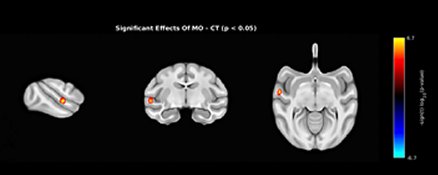
Figure 1. Overlay shows significant
spatial T-map (upper session) functional connectivity change (p = 0.00025) in
the MO offspring group, compared to the control group, in the in the left
agranular frontal area F4.
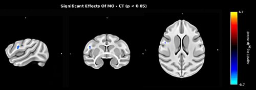
Figure 2. Overlay shows significant functional connectivity
change (p = 0.00025) in the MO offspring group, compared to the control group,
in the left auditory cortex. The spatial map of significant group effect: a
significant cluster with p < 0.05 shows as -sign(t)log (p-Value).
DOI: https://doi.org/10.58530/2022/3333