3324
Resting state functional connectivity subnetwork relates to prosocial behavior and compassion in adolescents
Benjamin Sipes1, Angela Jakary1, Melanie Morrison1, Tony T Yang2, and Olga Tymofiyeva1
1Radiology, University of California, San Francisco, San Francisco, CA, United States, 2Psychiatry and Behavioral Sciences, University of California, San Francisco, San Francisco, CA, United States
1Radiology, University of California, San Francisco, San Francisco, CA, United States, 2Psychiatry and Behavioral Sciences, University of California, San Francisco, San Francisco, CA, United States
Synopsis
In this study, we found that adolescent resting state functional brain networks contains a subnetwork significantly related to self-reported prosocial behavior and compassion. The subnetwork regions indicated connects many previously identified brain regions associated with prosocial behavioral tasks in adolescents, including the bilateral precuneus, left lateral prefrontal cortex, medial prefrontal cortex, inferior frontal gyri, temporal poles, left amygdala, right posterior cingulate cortex, occipital cortex, left supplementary motor area, and cerebellum.
Introduction
Prosocial behavior, defined as compassionate actions that benefit others, is associated with improved physical health of the giver [1-2], and while childhood prosocial behavior predicts adulthood prosociality [3], the brain network mechanisms of prosociality during development have yet to be elucidated. Functional magnetic resonance imaging (fMRI) studies investigating adolescent prosocial behavior have predominantly focused on identifying brain regions with significant activation during prosocial tasks [4-5] and one study analyzed seed-based resting state connectivity [6]. To our knowledge, no studies have yet investigated functional or structural brain networks in association with prosocial tendencies using a connectomics approach. Here, we sought to explore whether the resting state functional and diffusion MRI-based networks contain subnetworks related to prosocial behavior and compassion during adolescence.Methods
Ninety-five adolescents (16.0±1.3 yrs., range: 14-18 yrs., 49 females) underwent 3T MRI scans, which included a T1-weighted sequence, a T2*-weighted resting state fMRI sequence (TR=800ms, TE=30ms, 2.4mm isotropic resolution, 525 timepoints), and a 30-direction diffusion tensor imaging sequence (TR=7500ms, TE=60.7ms, 2mm isotropic resolution, b=1000). Functional connectomes were constructed using the default processing pipelines from the functional connectivity toolbox (CONN) [7], where connectivity was computed as the Fisher’s z transformed Pearson’s correlation of average BOLD timeseries from regions partitioned by the AAL atlas and thresholded to retain only positive correlations as network weights. Structural connectomes were constructed using deterministic tractography with the Diffusion Toolkit [8] with average fractional anisotropy as the edge weights between AAL atlas ROIs. Prosocial behavior and compassion were assessed by multiplying two complementary self-report measures: the Strengths and Difficulties Questionnaire (SDQ) prosocial behavior subscale and the Compassionate Engagement and Action Scales for Youth (CEASY) subscale measuring compassion for others [9-10]. Network-based statistics (NBS) (t-test; test-statistic=2.75; 10,000 permutations) were used to identify significant functional and structural subnetworks related to prosociality while controlling for age and sex and correcting for multiple comparisons [11]. Post-hoc analysis sought to verify this relationship by testing the correlation between each subject’s subnetwork weight (sum of all subnetwork edges) and their prosociality.Results
NBS revealed a statistically significant connected functional subnetwork associated with prosocial behavior and compassion in adolescents while controlling for age and sex as covariates (p = 0.021) (Figure 1A). The derived functional subnetwork’s highest degree nodes were the bilateral precuneus and the left lateral prefrontal cortex. Post-hoc analysis on the subnetwork’s weight confirmed a significant positive correlation with prosocial tendencies (r = 0.67, p < 0.0001) (Figure 1B). NBS revealed no significant structural subnetworks related to prosociality in adolescents.Conclusions
The present study provides preliminary evidence for a resting state functional connectivity subnetwork related to prosocial behavior and compassion in adolescents. We find that regions in this subnetwork exist across the brain and include the precuneus, left lateral prefrontal cortex, medial prefrontal cortex, inferior frontal gyri, temporal poles, left amygdala, right posterior cingulate cortex, occipital cortex, left supplementary motor area, and cerebellum. This work aligns with previous fMRI findings showing increased activation during prosocial tasks in many of these same regions [4-6]. Our results suggest that these disparate regions are a coordinated functional subnetwork that is related to prosocial behavior and compassion absent of any tasks. This finding could prove useful to guide interventions seeking to improve prosocial behavior and compassion in adolescents.Acknowledgements
This study was partially supported by the National Center for Complementary and Integrative Health (NCCIH) R21AT009173 and R61AT009864; NIH, through UCSF-CTSI UL1TR001872; the American Foundation for Suicide Prevention (AFSP) SRG-1-141-18; the UCSF Quantitative Biosciences Institute (QBI) Bold and Basic Grant.References
- Moieni et al. Exploring the role of gratitude and support-giving on inflammatory outcomes. Emotion. 2019;19(6):939-949.
- Inagaki & Eisenberger. Giving support to others reduces sympathetic nervous system-related responses to stress. Psychophysiology. 2016;53(4):427-435.
- Eisenberg et al. Brazilian adolescents' prosocial moral judgment and behavior: relations to sympathy, perspective taking, gender-role orientation, and demographic characteristics. Child Dev. 2001;72(2):518-534.
- Duell et al. Hormonal and neural correlates of prosocial conformity in adolescents. Developmental Cognitive Neuroscience. 2021;48:100936.
- Lemmers-Jansen et al. Giving others the option of choice: An fMRI study on low-cost cooperation. Neuropsychologia. 2018;109:1–9.
- Okada et al. Neurometabolic and functional connectivity basis of prosocial behavior in early adolescence. Scientific Reports. 2019;9(1):732.
- Whitfield-Gabrieli & Nieto-Castanon. Conn: a functional connectivity toolbox for correlated and anticorrelated brain networks. Brain connectivity. 2012;2(3):125-141.
- Wang, et al. Diffusion toolkit: a software package for diffusion imaging data processing and tractography. Proc Intl Soc Mag Reson Med. 2007;15(3720).
- Goodman, R. Psychometric properties of the strengths and difficulties questionnaire. Journal of the American Academy of Child & Adolescent Psychiatry. 2001;40(11):1337-1345.
- Gilbert et al. The development of compassionate engagement and action scales for self and others. J of Compassionate Health Care. 2017;4(4):1-24.
- Zalesky et al. Network-based statistic: identifying differences in brain networks. Neuroimage. 2010;53(4):1197-1207.
- Xia et al. BrainNet Viewer: a network visualization tool for human brain connectomics. PloS one. 2013;8(7):e68910.
Figures
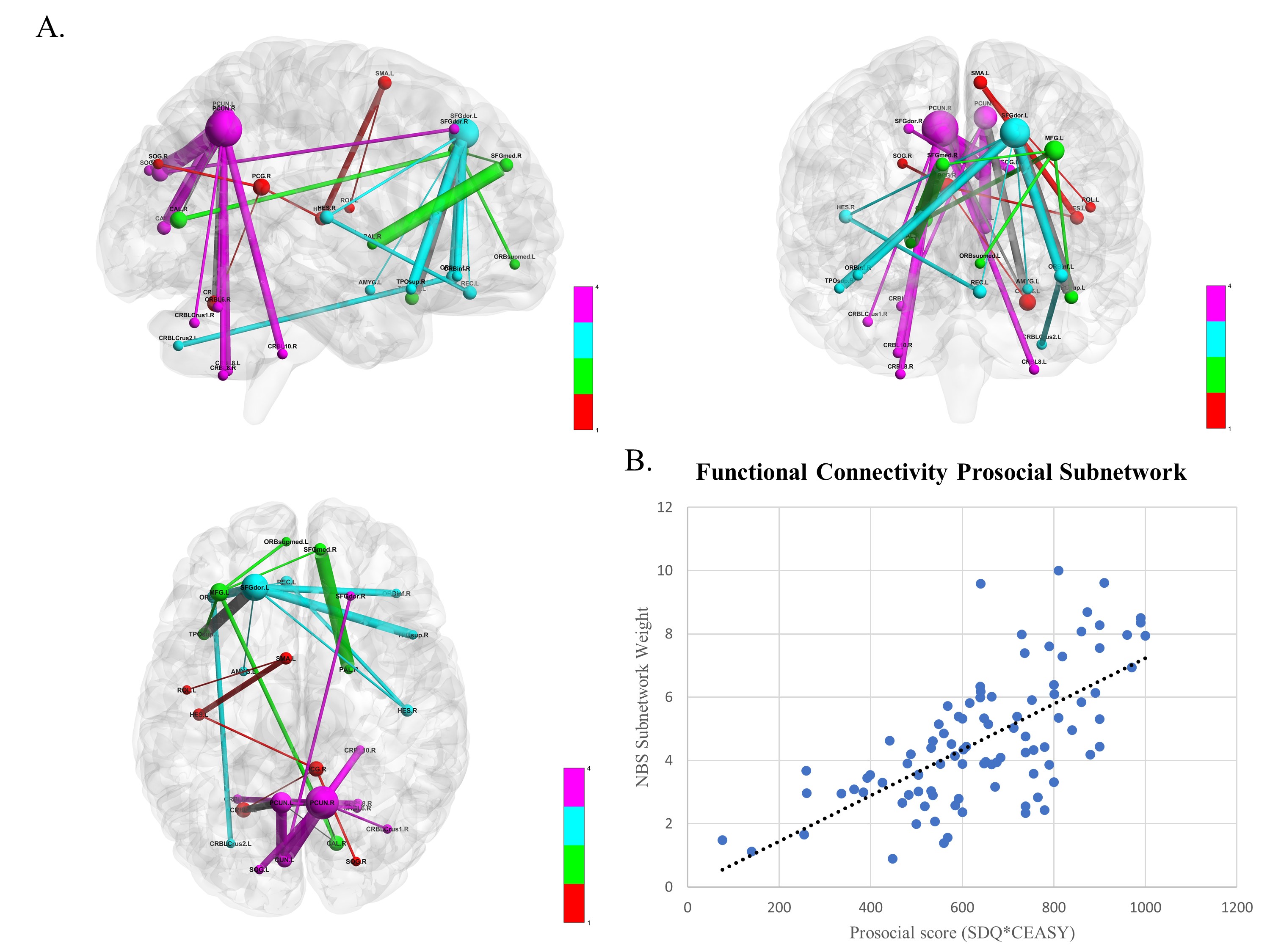
A) Network-Based Statistics subnetwork, controlled for age and sex and corrected for multiple comparisons (p=0.021). Louvain community detection (γ=0.8) revealed a precuneus module (purple), two left dorsolateral prefrontal cortex modules (blue & green), and a posterior cingulate to sensory cortices module (red). Edges here are visually weighted by their test statistic. Visualized with the BrainNet Viewer [12]. B) A scatter plot displaying the significant positive correlation (r = 0.67, p < 0.0001) between the subnetwork’s weight and the subject’s associated prosocial score.
DOI: https://doi.org/10.58530/2022/3324