3320
Evaluating a Volumetric High-Permittivity (HPM) Helmet for Brain Imaging at 3.0 T1Bernard and Irene Schwartz Center for Biomedical Imaging, Department of Radiology, NYU Grossman School of Medicine, Newyork, NY, United States, 2Center for Advanced Imaging Innovation and Research (CAI2R), Department of Radiology, NYU Grossman School of Medicine, New york, NY, United States, 3HyQRS, LLC, State College, PA, United States, 4Vilcek Institute of Graduate Biomedical Science, NYU Grossman School of Medicine, New york, NY, United States
Synopsis
A thin (8mm) helmet-shaped shell of high-permittivity material (HPM) is seen to double transmit efficiency and increase SNR by 40% at the center of tissue equivalent head phantom when compared to the body coil alone, with little effect on B1 homogeneity within the brain region. While evaluation of effects on receive arrays is ongoing, from the results shown here it seems an HPM helmet could be used at 3T in place of more electronically cumbersome head transmit coils.
Introduction
Materials with high electric permittivity have been used strategically to improve excitation efficiency and receive sensitivity1-5. Our recent work demonstrated that a RF receive coil array with an integrated volumetric high permittivity material (HPM) helmet can achieve significant SNR boost in whole brain volume at 7T when compared to an identical array without the helmet operating in a close-fitting receive array6. Towards translation of these effects to more widely-available field strengths, here we present a first evaluation of effects of an HPM helmet designed for use at 3T on the transmit efficiency and receive sensitivity of a whole-body coil with and without the HPM helmet.Methods
HPM Shell Construction: To investigate the benefit of a fully surrounding HPM insert at 3T a custom ceramic HPM helmet was designed and constructed. Briefly, numerical simulations of SNR in the human brain for a receive array at 3T were performed in a manner similar to those performed previously for 7T6. It was found that to translate effects like those seen at 7T to 3T, a permittivity of 650 should be effective, roughly matching the expectation of εrµ(1/f2)7. Methods similar to those to produce the HPM helmet for 7T6 were followed, but with different combination of powders to achieve specified properties for 3T (εr = 650 and σ = 0.004 S/m, thickness = 8mm) (Fig.1).Imaging: To quantify the performance of the HPM shell, imaging experiments were performed on a tissue equivalent head-shaped phantom8 with and without the HPM shell. Measurements were performed using a whole-body 3 T scanner (MAGNETOM, Siemens, Healthineers, Germany). Spin excitation and signal reception were evaluated using the whole body coil. B1+ and SNR maps were respectively acquired using TurboFLASH (TE-1.9ms, TR-10000ms) and GRE sequences (with and without RF).
Results
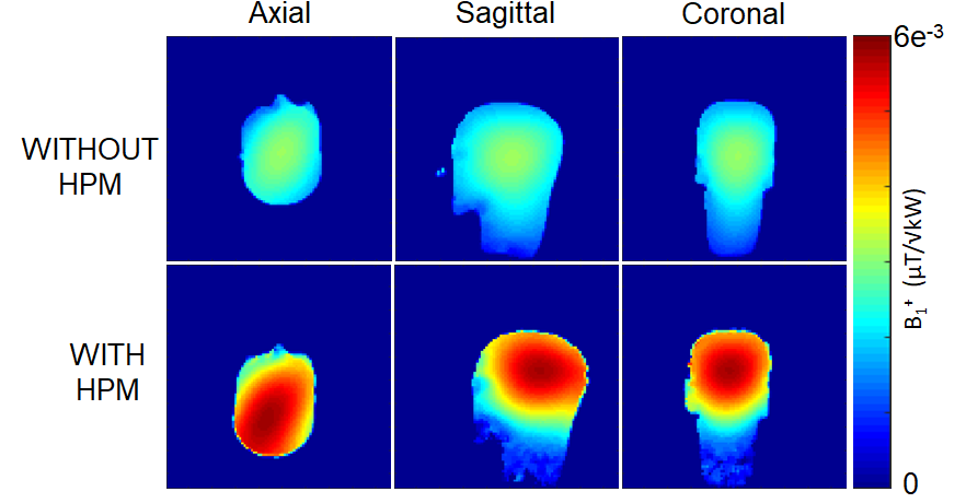
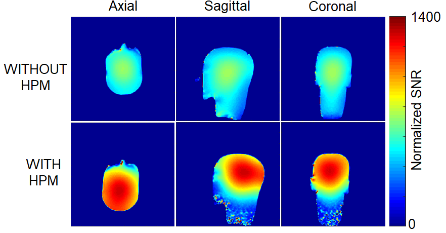
The HPM helmet provided a two-fold improvement in transmit efficiency at the phantom center over the setup without the helmet (Fig.2). The body coil with HPM helmet required a 103v, 500μs hard pulse to achieve 90° flip angle compared to 211v, 500μs hard pulse when the HPM shell was not used. The B1+ uniformity, quantified as standard deviation of the flip angle normalized by the mean on the transverse plane, was very similar with and without the helmet (0.631 and 0.635, respectively).Three-axis SNR maps (normalized to excitation flip angle) are presented in Figure 3. SNR maps show a 40% improvement at the center of the phantom with the HPM. Interestingly the sagittal and coronal B1+ maps with the HPM helmet show that the transmit field is focused in the brain volume, whereas the sagittal and coronal B1+ maps without the HPM shell show a more distributed B1+ profile with the transmit field into the extending into the neck area. Similar behavior is observed in SNR maps.
Discussion and Conclusions
A volumetric HPM shell was designed and implemented to demonstrate B1+ and SNR improvement in the brain at 3T when coupled to the body coil. Our encompassing HPM helmet provided a two-fold improvement in transmit efficiency and a SNR boost of 40% at the center of a tissue equivalent head phantom compared to the body coil alone.This approach can be seen as similar to that used recently to enhance transmit efficiency and SNR in breast imaging at 3T with specially-designed ceramic resonators9, but in our case the ceramic helmet was designed with receive sensitivity of a close-fitting receive array in mind6. Evaluations of close-fitting receive arrays with and without a helmet at 3T are ongoing at this time. The HPM helmet substantially focused the B1+ field in the brain volume increasing transmit efficiency and SNR. Although the B1+ homogeneity over the whole head and neck appears to be adversely affected with addition of the helmet (Fig. 2), evaluation of B1+ homogeneity in the region of brain shows little effect from the helmet. This is in contrast with a prior study using a smaller HPM cap at the top of the head with a much higher permittivity than used here, which had strong adverse effects on B1+ homogeneity in brain at 3T5. While evaluations of effects on SNR of a close-fitting receive array are ongoing, it appears that HPM materials coupled to the body coil could be used in place of more electronically cumbersome transmit head coils to achieve localized excitation in the head at 3T.
Acknowledgements
No acknowledgement found.References
1. Alsop et al., MRM 1998; 40:49-54.
2. Yang et al., JMRI 2006; 24:197-202.
3. Haines at al., JMRI 2010; 203:323-327.
4. Webb, Concepts Magn Reson Part A 2011; 38(4):148-84.
5. Sica C et al., MRM. 2020 83(3):1123-34.
6. Lakshmanan et al., MRM. 2021; 86: 1167– 1174.
7. Collins et al., Proc. ISMRM 2019, p. 1566
8. Ianniello et al., MRM. 2021; 80: 413-419
9. Shchelokova et al., Nature communications. 2020 Jul 31;11(1):1-7.
Figures
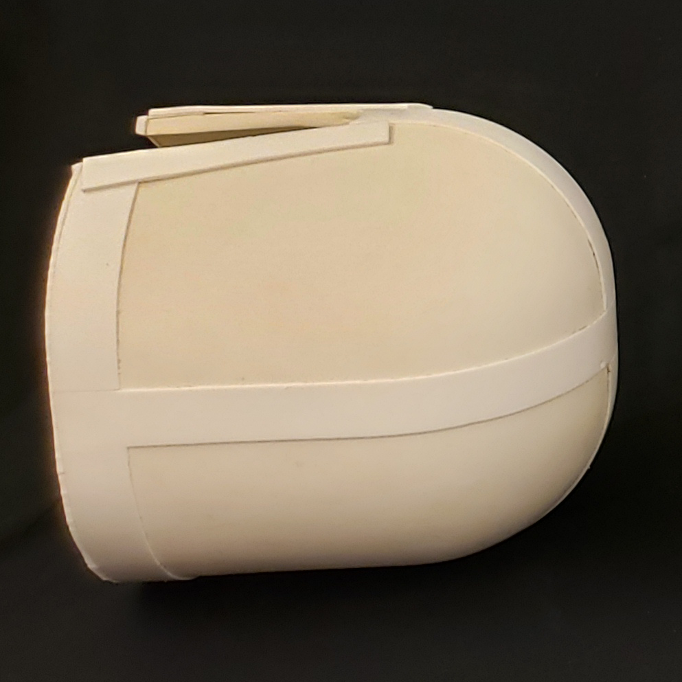
Figure 1. A photograph of the volumetric HPM shell (εr = 650, σ = 0.004 S/m, thickness = 8mm)