3303
Anatomically Informed Unsupervised Deep Learning for Fast and Effective White Matter Fiber Clustering1Harvard Medical School, Boston, MA, United States, 2The University of Sydney, Sydney, Australia, 3The University of New South Wales, Sydney, Australia
Synopsis
We propose a novel unsupervised deep learning framework for white matter fiber clustering. Self-supervised learning is adopted to enable joint deep embedding and cluster assignment. Anatomical information is incorporated into the neural network to improve anatomical coherence. In addition, outlier removal is performed to further improve cluster quality. Our method is evaluated on three datasets and showed superior performance in terms of cluster compactness, anatomical coherence and generalization across subjects compared to several state-of-the-art algorithms.
Introduction
Diffusion magnetic resonance imaging (dMRI)1 uniquely enables mapping of the brain’s white matter fiber tracts via tractography2, to study the brain’s connections in health and disease3. For clinical and research purposes, tractography parcellation is needed to divide whole brain tractography into anatomically meaningful fiber bundles. One widely used tractography method, white matter fiber clustering (WMFC), groups fibers with similar geometric trajectory into clusters4. Though it showed good performance in many applications5,6, key challenges remain such as expensive computation, sensitivity to fiber point order, existence of outlier fibers, and difficulty in utilizing both spatial and anatomical information, as well as inter-subject correspondence of fiber clusters7,8,9.Unsupervised clustering has been extensively studied in the computer vision community10,11,12. Auto-encoder-based methods are widely adopted to learn deep embeddings11,12. Besides, self-supervised learning is also a promising approach for unsupervised learning which shows good performance in many applications13.
Though attempts have been made to apply deep learning to tractography segmentation14,15,16, most of them are based on supervised learning. In this work, we propose a novel unsupervised deep learning framework for fast and effective fiber clustering. We adopt self-supervised learning by designing a novel pretext task, incorporate anatomical information into the network and perform outlier removal to further improve cluster quality. Our proposed method shows superior performance compared to several state-of-the-art methods.
Methods
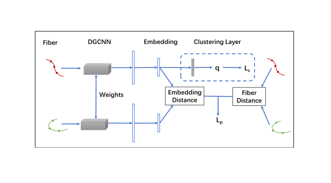
As shown in Figure-1, our training pipeline includes the pretraining stage and clustering stage. In the pretraining stage, self-supervised learning is performed to learn deep embeddings. A pretext task is designed to predict distance between the input pair of fibers with fiber distance calculated directly from fiber spatial coordinates8 as pseudo labels. After pretraining, k-means is performed to obtain initial clusters. In the clustering stage, the network is fine tuned in a self-training manner and cluster centroids are updated as parameters, as in 12.Intuitively, point clouds should be an efficient and discriminative representation of fibers. We adopt the DGCNN17 model, which improves PointNet18 by considering relations between nearby points. A Siamese Neural Network19 is adopted to learn deep embeddings and predict distances between pairs of input fibers.
In the clustering stage of training, we include anatomical information into the neural network by designing a new definition of soft label assignment probability adapted from13 to regularize that fibers within the same cluster pass through the same brain regions and cortical parcellations:
$$q_{ij}=\frac{1+\parallel z_{i}-\mu_{j}\parallel^{2}*(1-D_{aij})*(1-D_{sij}))^{-1}}{\sum_{j{'}}(1+\parallel z_i-\mu_{j{'}}\parallel^{2}*(1-D_{aij{'}})(1-D_{sij{'}}))^{-1}}$$
Where Daij is the Dice score between Freesurfer regions of fibers i and those of cluster j as defined in 8. Dsij is the percentage of cortical parcellations of fiber i in those of cluster j.
During inference, outlier removal is performed to filter anatomically implausible fibers. Fibers are removed if their soft label assignment probabilities are lower than the cluster mean probability for over n standard deviations. The parameter n is set to 0.7 so that our method removes the same percentage of fibers as WhiteMatterAnalysis (WMA)8.
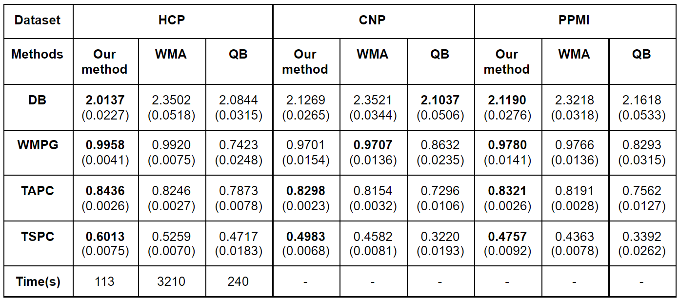
To evaluate our proposed method, we conduct experiments on three datasets: Human Connectome Project (HCP)20, Parkinson’s Progression Markers Initiative (PPMI)21 and Consortium for Neuropsychiatric Phenomics (CNP)22. Quantitative evaluations are performed in terms of the Davies-Douldin (DB) index23 (measuring within cluster scatter and separation between clusters), White Matter Parcellation Generalization (WMPG)8 (the percentage of successfully detected clusters (with more than 20 fibers)), Tract Anatomical Profile Coherence (TAPC)8 (measuring coherence of brain regions fibers within a cluster pass through) and Tract Surface Profile Coherence (TSPC) (measuring coherence of cortical parcellations fibers within a cluster end in).
We compared the performance of our method with two state-of-the-art (SOTA) fiber clustering methods: WMA8 and QuickBundles (QB)7.
Results
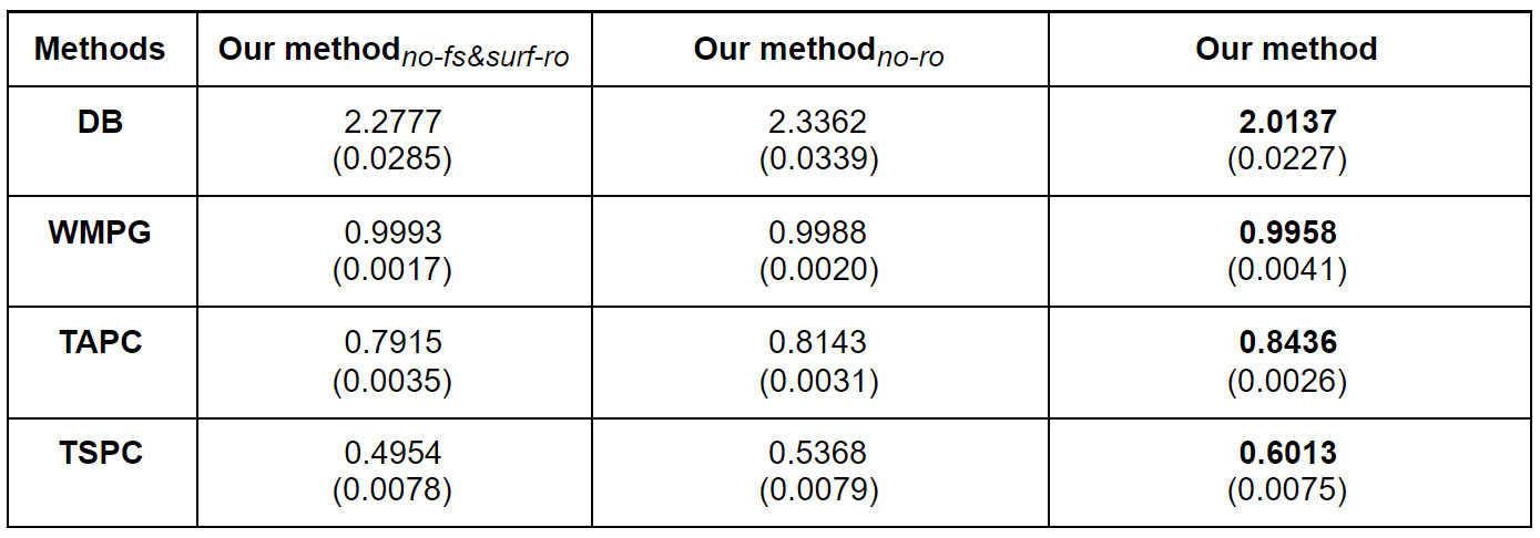
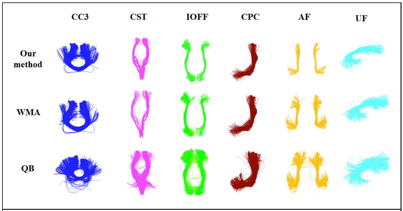
Figure-2 give the comparison results with the SOTA methods. As we can see, for HCP and PPMI data, our method shows the best performance for all evaluation metrics. For PPMI data, QB has slightly smaller DB score and WMA shows slightly better WMPG score than our method while our method has the highest TAPC and TSPC score. In addition, our method is the most efficient method. Results in Figure-3 shows that incorporation of anatomical and cortical parcellation information improves TAPC and TSPC score and outlier removal improves all evaluation metrics. Figure-4 gives a visualization of example clusters of three methods.Discussion
Our method successfully addresses several key challenges in WMFC and shows superior performance compared to SOTA methods. First, our deep learning-based method is computationally efficient owing to utilization of GPU acceleration. Second, by using the self-supervised learning strategy and point cloud as input, the clustering process is not sensitive to point order along fibers. Third, efficient outlier removal is performed to further improve cluster quality. Fourth, both spatial and anatomical information are utilized in our method which improves anatomical coherence. Finally, inter-subject correspondence is achieved by building a training model and demonstrated with high WMPG scores of our method.Conclusion
In this work, we propose a novel deep learning-based method for white matter fiber clustering by adopting self-supervised learning strategy. Our method was evaluated on three independently acquired datasets and showed superior performance compared to several SOTA algorithms.Acknowledgements
We acknowledge funding provided by the following National Institutes of Health(NIH) grants: R01MH125860, R01MH119222, R01MH074794, and P41EB015902.References
Basser, P.J., Mattiello, J., LeBihan, D.: MR diffusion tensor spectroscopy and imaging. Biophysical journal 66(1), 259-267 (1994)
Basser, P.J., Pajevic, S., Pierpaoli, C., Duda, J., Aldroubi, A.: In vivo fiber tractography using DT-MRI data. Mag. Res. Med 44(4), 625-632 (2000)
Zhang F, Daducci A, He Y, et al. Quantitative mapping of the brain's structural connectivity using diffusion MRI tractography: a review[J]. arXiv preprint arXiv:2104.11644, 2021.
O'Donnell, L.J., Golby, A.J.,Westin, C.F.: Fiber clustering versus the parcellation-based connectome. NeuroImage 80, 283-289 (2013)
Wu, Y., Hong, Y., Ahmad, S., Lin, W., et al.: Tract dictionary learning for fast and robust recognition of ber bundles. In: MICCAI. pp. 251-259 (2020)
Zhang, F., Savadjiev, P., Cai, W., Song, Y., Rathi, Y., Tunc, B., Parker, D., Kapur, T., Schultz, R.T., Makris, N., et al.: Whole brain white matter connectivity analysis using machine learning: an application to autism. NeuroImage 172, 826-837 (2018)
Garyfallidis, E., Brett, M., Correia, M.M., et al.: Quickbundles, a method for tractography simplification. Frontiers in neuroscience 6, 175 (2012)
Zhang, F., Wu, Y., Norton, I., Rigolo, L., Rathi, Y., Makris, N., O'Donnell, L.J.: An anatomically curated ber clustering white matter atlas for consistent white matter tract parcellation across the lifespan. NeuroImage 179, 429-447 (2018)
O'Donnell, L., Westin, C.F.: White matter tract clustering and correspondence in populations. In: MICCAI. pp. 140-147 (2005)
Caron, M., Bojanowski, P., Joulin, A., Douze, M.: Deep clustering for unsupervised learning of visual features. In: ECCV. pp. 132-149 (2018)
Guo, X., Liu, X., Zhu, E., Yin, J.: Deep clustering with convolutional autoencoders. In: ICNIP. pp. 373-382 (2017)
Xie, J., Girshick, R., Farhadi, A.: Unsupervised deep embedding for clustering analysis. In: ICML. pp. 478-487. PMLR (2016)
Kolesnikov, A., Zhai, X., Beyer, L.: Revisiting self-supervised visual representationlearning. In: CVPR. pp. 1920{1929 (2019)
Wasserthal, J., Neher, P., Maier-Hein, K.H.: Tractseg-fast and accurate white matter tract segmentation. NeuroImage 183, 239-253 (2018)
Zhang, F., Karayumak, S.C., Homann, N., Rathi, Y., Golby, A.J., O'Donnell, L.J.: Deep white matter analysis (DeepWMA): Fast and consistent tractography segmentation. Medical Image Analysis 65, 101761 (2020)
Chen Y, Zhang C, Song Y, et al. Deep Fiber Clustering: Anatomically Informed Unsupervised Deep Learning for Fast and Effective White Matter Parcellation[C]//International Conference on Medical Image Computing and Computer-Assisted Intervention. Springer, Cham, 2021: 497-507.
Wang Y, Sun Y, Liu Z, et al. Dynamic graph cnn for learning on point clouds[J]. Acm Transactions On Graphics (tog), 2019, 38(5): 1-12.
Qi, Charles R., et al. "Pointnet: Deep learning on point sets for 3d classification and segmentation." Proceedings of the IEEE conference on computer vision and pattern recognition. 2017.
Chopra, S., Hadsell, R., LeCun, Y.: Learning a similarity metric discriminatively, with application to face verification. In: CVPR. vol. 1, pp. 539-546 (20)
Van Essen, D.C., Smith, S.M., Barch, D.M., et al.: The WU-Minn human connectome project: an overview. Neuroimage 80, 62–79 (2013)
Marek K, Jennings D, Lasch S, et al. The Parkinson progression marker initiative (PPMI) [J]. Progress in neurobiology, 2011, 95(4): 629-635.
Poldrack R A, Congdon E, Triplett W, et al. A phenome-wide examination of neural and cognitive function[J]. Scientific data, 2016, 3(1): 1-12.
Xu, D., Tian, Y.: A comprehensive survey of clustering algorithms. Annals of Data Science 2(2), 165–193 (2015)
Norton, I. et al. SlicerDMRI: Open Source Diffusion MRI Software for Brain Cancer Research. Cancer Res. 77, e101–e103 (2017).
Zhang F, Noh T, Juvekar P, et al. SlicerDMRI: Diffusion MRI and tractography research software for brain cancer surgery planning and visualization[J]. JCO clinical cancer informatics, 2020, 4: 299-309.
Figures